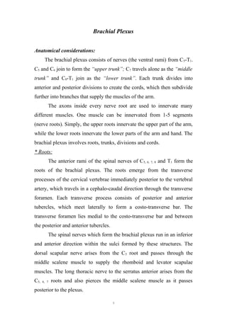
SlideShare a Scribd company logo
Brachial Plexus
Anatomical considerations:
The brachial plexus consists of nerves (the ventral rami) from C5-T1.
C5 and C6 join to form the “upper trunk”; C7 travels alone as the “middle
trunk” and C8-T1 join as the “lower trunk”. Each trunk divides into
anterior and posterior divisions to create the cords, which then subdivide
further into branches that supply the muscles of the arm.
The axons inside every nerve root are used to innervate many
different muscles. One muscle can be innervated from 1-5 segments
(nerve roots). Simply, the upper roots innervate the upper part of the arm,
while the lower roots innervate the lower parts of the arm and hand. The
brachial plexus involves roots, trunks, divisions and cords.
* Roots:
The anterior rami of the spinal nerves of C5, 6, 7, 8 and T1 form the
roots of the brachial plexus. The roots emerge from the transverse
processes of the cervical vertebrae immediately posterior to the vertebral
artery, which travels in a cephalo-caudal direction through the transverse
foramen. Each transverse process consists of posterior and anterior
tubercles, which meet laterally to form a costo-transverse bar. The
transverse foramen lies medial to the costo-transverse bar and between
the posterior and anterior tubercles.
The spinal nerves which form the brachial plexus run in an inferior
and anterior direction within the sulci formed by these structures. The
dorsal scapular nerve arises from the C5 root and passes through the
middle scalene muscle to supply the rhomboid and levator scapulae
muscles. The long thoracic nerve to the serratus anterior arises from the
C5, 6, 7 roots and also pierces the middle scalene muscle as it passes
posterior to the plexus.
1
* Trunks and divisions:
Trunks of the brachial plexus pass between the anterior and middle
scalene muscles. The superior trunk lies closest to the surface and is
formed by C5 and C6 roots. The suprascapular nerve and the nerve to
subclavius arise from the superior trunk. The suprascapular nerve
contributes sensory fibers to the shoulder joint and provides motor
innervation to supraspinatus and infraspinatus muscles. The C7 root
continues as the middle trunk, while C8 and T1 roots join to form the
inferior trunk. The trunks divide into anterior and posterior divisions,
separating the innervation of the ventral and dorsal halves of the upper
limb.
The phrenic nerve (C3, 4, 5) passes between the anterior and middle
scalenes and continues over the surface of the anterior scalene muscle.
Diaphragmatic twitch during interscalene brachial plexus, performed with
a nerve stimulator, indicates needle placement anterior to the plexus.
The spinal accessory nerve (cranial nerve XI) runs posterior to the
brachial plexus over the surface of middle and posterior scalenes. Contact
of spinal accessory nerve with a nerve stimulator (stimulating twitch in
trapezius) indicates placement of the needle posterior to the plexus.
* Cords and branches
The cords are named the lateral, posterior and medial cord,
according to their relationship to the axillary artery. The lateral cord
receives fibers from the anterior divisions of the superior and middle
trunks; it is the origin of the lateral pectoral nerve (C5, 6, 7). The posterior
divisions of the superior, middle and inferior trunks combine to form the
posterior cord. The inferior trunk continues as the medial cord and gives
off the median pectoral nerve (C8, T1), the medial brachial cutaneous
nerve (T1) and the medial ante-brachial cutaneous nerve (C8, T1).
2
The upper and lower subscapular nerves (C7, 8 and C5, 6) leave the
posterior cord and descend behind the axillary artery to supply the
subscapularis and teres major muscles, respectively. The thorcodorsal
nerve to the latissimus dorsi, also known as the middle subscapular nerve
(C6, 7, 8) also arises from the posterior cord.
The lateral cord divides into the lateral root of the median nerve
and the musculocutaneous nerve. The posterior cord gives off the axillary
nerve at the lower border of the subscapularis muscle and continues along
the inferior and posterior surface of the axillary artery as the radial nerve.
The axillary nerve supplies the teres minor muscle before ending as the
superior lateral brachial cutaneous nerve. The medial cord contributes the
medial root of the median nerve and continues as the ulnar nerve along
the medial and anterior surface of the axillary artery. The medial and
lateral roots join to form the median nerve which continues along the
posterior and lateral surface of the axillary artery.
Fig. (1): Overview of the brachial plexus
3
Brachial Plexus Palsy (BPP)
Brachial plexus injuries are more common in over-weight babies,
particularly where delivery is complicated by shoulder dystocia (difficult
labor). A brachial plexus lesion is revealed by lack of movement in the
arm; initially the arm is flaccid. After 48 hours, an upper palsy can be
distinguished from a complete palsy. In upper root palsy (C5,6 and
sometimes C7), the arm is internally rotated and pronated and there is no
active shoulder abduction or elbow flexion (Waiter’s tip position). In a
complete palsy of upper and lower roots, the arm is flail; there may be a
ptosis and a Horner’s syndrome due to damage to the stellate ganglion
(star-shaped collection of nerve cells in the sympathetic chain) adjacent to
C8 and T1. Phrenic nerve palsy should be considered in these cases.
Most infantile injuries to the brachial plexus predominantly involve
the upper trunk (C5,6); the classic Erb's palsy. However, many of these
infants also have impairment of the C7 nerve root, which postpones the
poorer prognosis. Far less frequently, the entire plexus (C5,6,7,8 and T1) or
the lower trunk (C8 and T1; Klumpke's palsy), may be involved.
Incidence:
With improvement in obstetrical care, the incidence of brachial
plexus injuries has decreased significantly. In countries in which
obstetrical care is poor, obstetric brachial plexus palsy (OBPP) is noted
more frequently. The incidence ranges globally from 0.2-4 % of live-
births. According to the World Health Organization (WHO), the
prevalence is generally 1-2 % worldwide, with the higher numbers in the
underdeveloped countries. In the United States, the prevalence is
approximately 0.2 %. Studies in France suggest an incidence of 1.09-1.19
cases per 1000 live-births.
4
Etiology:
Most neonatal BPP occurs in the birthing process. Risk factors for
this type of injury (OBPP) include:
- Over weight at birth (average vertex BPP is 3.8 - 5.0 kg, average breech
BPP is 1.8 - 3.7 kg, while the average unaffected is 2.8 - 4.5 kg).
- Breech presentation.
- Maternal diabetes.
- Second stage of labor that lasts more than 60 minutes.
- Assisted delivery (e.g. use of forceps or vacuum extraction).
- Previous child with OBPP.
- Intrauterine torticollis.
- Shoulder dystocia (difficult labor).
Other less common causes of neonatal BPP include:
- Neoplasms (e.g. neuromas, rhomboid tumors).
- Intrauterine compression.
- Humeral osteomyelitis.
- Hemangioma.
- Exostosis (overgrowth of bone tissue) of the first rib.
Although the traditional mechanism of injury is lateral neck
flexion, the upper rootlets (C5, 6, 7) are 25 % as likely to be avulsed as the
lower roots (C7, 8 and T1). The upper roots (C5, 6), however, are far more
likely to be ruptured (88 %) because of the anatomy of the transverse
processes and the degree of flexibility at that level.
Pathophysiology:
Brachial plexus palsy (BPP) results from excessive lateral traction
on the head away from the shoulder. This force on the brachial plexus can
cause varying degrees of injury to the nerves, including rupture of the
nerve roots or trunks, avulsion of the nerve roots from the spinal cord and
5
traction preserving the continuity of the nerve but causing excessive
scarring. Injury to the brachial plexus may result from demyelination,
axonal degeneration or avulsion. Clinically, this injury results in
disruption of the sensory and / or motor function of the injured nerve.
Spontaneous recovery of function occurs with remyelination, with or
without axonal regeneration and reinnervation of the sensory receptors,
muscle endplates or both. However, in some cases of severe nerve injury
and with avulsion injuries, spontaneous recovery does not occur and
surgical intervention is warranted.
Severity depends upon the number of nerves involved and the
degree to which each level is injured. The basic types of brachial plexus
palsies include:
- Duchenne-Erb palsy; affects nerves arising from C5 and C6.
- Upper-middle trunk BPP; involves nerve fibers from C5, 6, 7 levels.
- Klumpke palsy; results in deficits at levels of C8 and T1, although many
clinicians agree that pure C8 - T1 injuries do not occur in infants and may
be indicative of spinal cord injury (SCI).
- Total BPP; affects nerves at all levels (C5, 6, 7, 8 - T1).
When defining severity of a peripheral nerve injury, differentiation
between neuropraxic, axonotemetic, and neurotemetic lesions is helpful:
* Purely neuropraxic lesions do not affect the axon itself. These lesions
generally are reversible and do not leave sequelae.
* Axonotemetic lesions involve disruption of both the myelin sheath and
the axon, leading to degeneration of the axon distal to the injury. The
connective tissue across the lesion remains intact. These injuries improve
gradually over 4 - 6 months, depending on the level of the lesion.
* Neurotemetic lesions are the most severe and destroy not only the axon
and myelin but also the supporting structures across a nerve. As the
proximal end of the nerve attempts to regenerate without this supportive
6
connective tissue, a neuroma may develop. The extent of improvement in
the patient's condition depends upon the ultimate number of nerve fibers
that reconnect distal to the neuroma.
Depending on the degree of nerve injury, recovery may be noted
within a few days. In most cases, some degree of spontaneous recovery
occurs within 1 month, although in some injuries evidence of full
recovery may not appear for up to 3 months. Sunderland described a
classification for nerve injuries including 5 degrees of nerve injury:
- 1st
degree injury (neuropraxia): It involves a temporary conduction
block with demyelination of the nerve at the site of injury. Recovery may
take up to 12 weeks.
- 2nd
degree injury (axonotemesis): It results from a more severe trauma
or compression. This causes Wallarian degeneration distal to the level of
injury and proximal axonal degeneration to at least at the next node of
Ranvier. Axonal regeneration occurs at a rate of 1 mm / day or 1 inch /
month and can be followed with an advancing Tinel sign.
- 3rd
degree injury: It is more severe than a second-degree injury, as
regeneration occurs at a rate of 1 mm / day and progress may be followed
with an advancing Tinel sign. However, with the increased severity of the
injury, the endo-neural tubes are not intact. The pattern of recovery is
mixed and incomplete. Reinnervation occurs only if sensory fibers reach
their sensory end organs and motor fibers reach their muscle targets.
- 4th
degree injury: It results in a large area of scar at the site of nerve
injury, precluding any axons from advancing distal to the level of nerve
injury.
- 5th
degree injury: It is a complete transection of the nerve. Similar to a
fourth-degree injury, surgery is required to restore neural continuity.
7
Examination and diagnosis:
Diagnosis of brachial plexus injuries relies heavily upon the
clinical evaluation; children are first evaluated between birth and 6 weeks
of age with a physical therapist starting at 3 - 4 weeks then at 6 weeks of
age. Follow-up evaluations occur at three-month intervals.
Physical findings:
The newborn infants can have many of the following findings:
- In infants with complete BPP (C5 - T1), the arm is held limply at his side.
Deep tendon reflexes in the affected arm are absent and the Moro
response is asymmetric with no active abduction of the ipsilateral arm. In
children with total arm involvement, careful examination of the child's
eye often demonstrates Horner syndrome. Children with intrinsic hand
weakness, associated with BPP, generally have Horner syndrome and
vice versa. Respiratory status should be evaluated since the phrenic nerve
can be injured simultaneously.
- The infant with upper plexus palsy (C5, 6, 7) keeps the arm adducted and
internally rotated with the elbow extended, forearm pronated, wrist flexed
and the hand in a fist. In the first hours of life, the hand also may appear
flaccid but strength soon returns.
- The infant with a nerve injury to the lower plexus (C8 - T1) holds the arm
supinated with the elbow bent and wrist extended.
- Sensation should be assessed closely, with the clinician noting any
sensory loss in corresponding dermatomes.
- Reflexes typically absent in the affected limb and should be evaluated.
- In the newborn nursery, it is essential that the physician carefully
inspect the size of the hand and arm and bulk of the pectoralis major.
These children may have a smaller limb with asymmetric palmar creases,
pectoralis atrophy and / or joint contractures at the time of delivery.
8
The examination consists of a motor evaluation that scores both
individual muscle groups (using the five-point British Research Council
Grading System) and functional muscle group activities, including
abduction, external rotation, hand-to-head, hand-to-back and hand-to-
mouth movements as well as sensory and reflex examinations. Physicians
should compare clinical assessments on subsequent visits every three
months
The back should be inspected carefully, assessing muscle bulk
including the trapezius, rhomboids, supraspinatus, infraspinatus,
latissimus dorsi, teres major, and teres minor. Contracture of the
pectoralis major can be assessed by palpation of the anterior axillary fold
during external rotation.
Laboratory investigations:
Children who recover partial function between examinations
should continue to be followed at three-month intervals. Children, who
fail to improve on subsequent visits or who improve initially but then
plateau at a nonfunctional level, should be evaluated initially via
electromyography (EMG). This will assess the levels of injury and
determine if there is evidence of early reinnervation
Complications:
Children with BPP are at risk for developing complications such as
progressive contractures, bony deformities, scoliosis and posterior
shoulder dislocation. Children with chronic upper trunk plexopathy may
develop external rotation weakness and internal rotation contractures
about the shoulder. This muscle imbalance will progressively alter the
glenohumeral joint.
9
Prognosis:
Peripheral nerves remyelinate at a rate of 1 mm/day. Thus, if the
nerve is not transected, recovery can be expected by 4-5 months in Erb’s
palsy, 6-7 months for upper-middle trunk palsy and 14 months for a total
BPP. Some authors believe that infants who do not show signs of
spontaneous recovery by 3-5 months are usually left with residual deficits
if managed conservatively. Unfavorable functional outcome is related
more often to faulty reinnervation than to lack of reinnervation. Faulty
reinnervation is especially common in brachial plexus lesions, secondary
to the close proximity of the nerves involved.
Evaluation:
* Electrophysiological evaluation:
- Electromyography: Evidence of denervation is usually not seen before 1
week of age.
- Nerve conduction velocity.
* Physical evaluation:
Assessment is carried out with the infant undressed and in warm
room. Any broken bone, the extent of passive and active range of motion,
sensation, strength and achievement of developmental milestones should
be thoroughly checked. Asymmetrical position of the upper limb is
recorded as the newborn must be held in flexion. If there is scapular
winging, rhomboids are involved. The diaphragm may be also involved.
The following tests are conducted:
* Muscles tone.
* Range of motion (flexibility test) for all the joint include shoulder
internal rotators (subscpularis), pronators and wrist and finger flexors.
* Round measurement.
10
* Muscles test or functional test according to the infant’s age.
* Reflex testing according the age of the infant (e.g. Moro reflex, placing
reaction, neck righting reaction and grasp reflex).
* Sensory test to detect any sensory disturbance, which interferes with the
treatment progress.
* Activities of daily living (feeding, washing and grooming) with the
affected extremity.
* Mallet System:
Assessment of the function of the shoulder is based on five criteria.
The ability to actively abduct the arm, the ability to externally rotate the
arm, the ability to place the hand behind the neck as well as behind the
back and the ability to place the hand over the mouth.
- Grade I indicates a stiff shoulder or a flail arm.
- Grade II indicates active abduction of 30° or less.
- Grade III indicates active abduction of 30° to 90°, active external
rotation of 20° or less.
- Grade IV indicates active abduction of at least 90°, active external
rotation of more than 20°, and place the hand behind the neck and over
the thoraco-lumbar. The hand can be brought to the mouth without
abduction of the arm.
- Grade V indicates a clinically normal shoulder.
Physical treatment:
Goals:
- To facilitate weak muscles.
- To resume function as soon as neural regeneration has taken place.
- To train motor control.
- To maintain normal range of motion.
- To prevent soft tissue contracture and skeletal deformity.
11
Methods:
* Facilitatory stimuli such as scratch, painful stimuli, quick stretch,
brushing, brief icing and faradic stimulation.
* Functional strengthening exercises: Active use of involved extremity
using a variety of developmentally appropriate activities to increase
strength and coordination, beginning in gravity eliminated then advance
to against gravity.
* Passive range of motion exercises for all joints of the limb.
* Stretching exercises.
* Scapular mobilization.
* Manipulative exercises.
* Hydrotherapy.
* Sensory stimulation to prevent disuse muscle atrophy by encouraging
muscle growth.
* Motor training:
- It should start within the first month although there may be no activity
in the denervated muscles.
- Early training aims probably to elicit activity in temporarily
denervated muscles, to minimize soft tissue contraction and to set up a
regular pattern of behavior in the parents.
- Training should be direct to age-related actions with particular
emphasis on reaching to touch and eventually to grasping and
manipulate different objects.
- Specific games can be played with the older children, aiming to
improve sensory awareness of the affected limb.
12
13

More Related Content

What's hot

spinal cord and applied aspects of spine
spinal cord and applied aspects of spinespinal cord and applied aspects of spine
spinal cord and applied aspects of spine
mrinal joshi
 
Anatomy of brachial plexus
Anatomy of brachial plexusAnatomy of brachial plexus
Anatomy of brachial plexusananya nanda
 
5. Brainstem (2): Pons & Midbrain
5. Brainstem (2): Pons & Midbrain5. Brainstem (2): Pons & Midbrain
5. Brainstem (2): Pons & Midbrain
Ahmed Eljack
 
spinal cord
spinal cordspinal cord
spinal cord
zoheb khan
 
Brachial plexopathies
Brachial plexopathiesBrachial plexopathies
Brachial plexopathies
rzgar hamed
 
Neuroanatomy spinal cord
Neuroanatomy   spinal cordNeuroanatomy   spinal cord
Neuroanatomy spinal cordMBBS IMS MSU
 
Brachial plexus
Brachial plexusBrachial plexus
Brachial plexus
Neurology resident slides
 
Spinal cord Anatomy
Spinal cord AnatomySpinal cord Anatomy
Spinal cord Anatomy
CIMS
 
Brachial plexus
Brachial plexusBrachial plexus
Brachial plexusLucidante1
 
Anatomy of the upper limb – brachial plexus
Anatomy of the upper limb – brachial plexusAnatomy of the upper limb – brachial plexus
Anatomy of the upper limb – brachial plexus
ess_online
 
Brain stem dr.gosai
Brain stem dr.gosaiBrain stem dr.gosai
Brain stem dr.gosai
Dr.B.B. Gosai
 
Motor lesions : Neuroanatomy
Motor lesions : NeuroanatomyMotor lesions : Neuroanatomy
Motor lesions : Neuroanatomy
Medvizz institute of medical education
 
Anatomy of brachial plexus
Anatomy of brachial plexusAnatomy of brachial plexus
Anatomy of brachial plexus
Ankush Honda
 
Arterial supply to the shoulder and brachial plexus
Arterial supply to the shoulder and brachial plexusArterial supply to the shoulder and brachial plexus
Arterial supply to the shoulder and brachial plexus
محمد حسن
 
Brachial plexus
Brachial plexusBrachial plexus
Brachial plexus
Ali Faris
 
Anatomy & functions of the Brainstem & Cerebellum
Anatomy & functions of the Brainstem & CerebellumAnatomy & functions of the Brainstem & Cerebellum
Anatomy & functions of the Brainstem & Cerebellum
Rafid Rashid
 
Anatomy and blood supply of spinal cord
Anatomy and blood supply of spinal cordAnatomy and blood supply of spinal cord
Anatomy and blood supply of spinal cord
NeurologyKota
 
Brachial plexus by Dr. Rahul Singh Thakur
Brachial plexus by Dr. Rahul Singh ThakurBrachial plexus by Dr. Rahul Singh Thakur
Brachial plexus by Dr. Rahul Singh Thakur
DrRahul Thakur
 

What's hot (20)

spinal cord and applied aspects of spine
spinal cord and applied aspects of spinespinal cord and applied aspects of spine
spinal cord and applied aspects of spine
 
Anatomy of brachial plexus
Anatomy of brachial plexusAnatomy of brachial plexus
Anatomy of brachial plexus
 
Brachial plexus seminar dr saumya agarwal
Brachial plexus seminar dr saumya agarwalBrachial plexus seminar dr saumya agarwal
Brachial plexus seminar dr saumya agarwal
 
5. Brainstem (2): Pons & Midbrain
5. Brainstem (2): Pons & Midbrain5. Brainstem (2): Pons & Midbrain
5. Brainstem (2): Pons & Midbrain
 
spinal cord
spinal cordspinal cord
spinal cord
 
Brachial plexopathies
Brachial plexopathiesBrachial plexopathies
Brachial plexopathies
 
Neuroanatomy spinal cord
Neuroanatomy   spinal cordNeuroanatomy   spinal cord
Neuroanatomy spinal cord
 
Brachial plexus
Brachial plexusBrachial plexus
Brachial plexus
 
Spinal cord Anatomy
Spinal cord AnatomySpinal cord Anatomy
Spinal cord Anatomy
 
Brachial plexus
Brachial plexusBrachial plexus
Brachial plexus
 
Anatomy of the upper limb – brachial plexus
Anatomy of the upper limb – brachial plexusAnatomy of the upper limb – brachial plexus
Anatomy of the upper limb – brachial plexus
 
Brain stem dr.gosai
Brain stem dr.gosaiBrain stem dr.gosai
Brain stem dr.gosai
 
Motor lesions : Neuroanatomy
Motor lesions : NeuroanatomyMotor lesions : Neuroanatomy
Motor lesions : Neuroanatomy
 
Anatomy of brachial plexus
Anatomy of brachial plexusAnatomy of brachial plexus
Anatomy of brachial plexus
 
Arterial supply to the shoulder and brachial plexus
Arterial supply to the shoulder and brachial plexusArterial supply to the shoulder and brachial plexus
Arterial supply to the shoulder and brachial plexus
 
Brachial plexus
Brachial plexusBrachial plexus
Brachial plexus
 
The spine & spinal cord
The spine & spinal cordThe spine & spinal cord
The spine & spinal cord
 
Anatomy & functions of the Brainstem & Cerebellum
Anatomy & functions of the Brainstem & CerebellumAnatomy & functions of the Brainstem & Cerebellum
Anatomy & functions of the Brainstem & Cerebellum
 
Anatomy and blood supply of spinal cord
Anatomy and blood supply of spinal cordAnatomy and blood supply of spinal cord
Anatomy and blood supply of spinal cord
 
Brachial plexus by Dr. Rahul Singh Thakur
Brachial plexus by Dr. Rahul Singh ThakurBrachial plexus by Dr. Rahul Singh Thakur
Brachial plexus by Dr. Rahul Singh Thakur
 

Similar to Brachial plexus detailed lecture

Brachial plexus
Brachial plexusBrachial plexus
Brachial plexus
baibhav177
 
Brachial plexus - Made so Easy
Brachial plexus - Made so EasyBrachial plexus - Made so Easy
Brachial plexus - Made so Easy
Uthamalingam Murali
 
Brachial plexus injury
Brachial plexus injuryBrachial plexus injury
Brachial plexus injury
aditya romadhon
 
Brachial plexus in the Upper Extremity. By Dr. G Kamau
Brachial plexus in the Upper Extremity. By Dr. G KamauBrachial plexus in the Upper Extremity. By Dr. G Kamau
Brachial plexus in the Upper Extremity. By Dr. G Kamau
MathewJude
 
Brachial plexus.ppt
Brachial plexus.pptBrachial plexus.ppt
Brachial plexus.ppt
DrMohammed43
 
brachial plexus final.pptx
brachial plexus final.pptxbrachial plexus final.pptx
brachial plexus final.pptx
shyam sunder
 
cervical and brachial plexus
cervical and brachial plexuscervical and brachial plexus
cervical and brachial plexus
Sado Anatomist
 
6-Pectoral Region and Axilla pdf.pptx hj
6-Pectoral Region and Axilla pdf.pptx hj6-Pectoral Region and Axilla pdf.pptx hj
6-Pectoral Region and Axilla pdf.pptx hj
rk7424934
 
Bracial plexus injury localization and management
Bracial plexus injury localization and managementBracial plexus injury localization and management
Bracial plexus injury localization and management
drajay02
 
Anatomy of brachial plexus
Anatomy of brachial plexusAnatomy of brachial plexus
Anatomy of brachial plexus
AmeyDixit6
 
Brachial plexus block
Brachial plexus blockBrachial plexus block
Brachial plexus block
Sami Ur Rehman
 
4._Brachial_Plexus.pptx
4._Brachial_Plexus.pptx4._Brachial_Plexus.pptx
4._Brachial_Plexus.pptx
debbychikwe
 
Brachial Plexus - Julie Cornish
Brachial Plexus - Julie CornishBrachial Plexus - Julie Cornish
Brachial Plexus - Julie Cornish
welshbarbers
 
BRACHIAL PLEXUS INJURY: EVALUATION AND MANAGEMENT
BRACHIAL PLEXUS INJURY: EVALUATION AND MANAGEMENTBRACHIAL PLEXUS INJURY: EVALUATION AND MANAGEMENT
BRACHIAL PLEXUS INJURY: EVALUATION AND MANAGEMENT
ashupara
 
Brachial plexus by Dr. Nasir Mustafa
Brachial plexus by Dr. Nasir MustafaBrachial plexus by Dr. Nasir Mustafa
Brachial plexus by Dr. Nasir Mustafa
Dr. Nasir Mustafa
 
Anatomy Lect 5 Trunk & Spine
Anatomy Lect 5 Trunk & SpineAnatomy Lect 5 Trunk & Spine
Anatomy Lect 5 Trunk & SpineMiami Dade
 
165792 upper-extremity-muscle
165792 upper-extremity-muscle165792 upper-extremity-muscle
165792 upper-extremity-muscleYoAmoNYC
 
Muscles of Upper Extremities
Muscles of Upper ExtremitiesMuscles of Upper Extremities
Muscles of Upper Extremities
Examville.com LLC
 
165792 upper-extremity-muscle
165792 upper-extremity-muscle165792 upper-extremity-muscle
165792 upper-extremity-muscleabctutor
 
Brachial plexus anatomy and block
Brachial plexus anatomy and blockBrachial plexus anatomy and block
Brachial plexus anatomy and block
ZIKRULLAH MALLICK
 

Similar to Brachial plexus detailed lecture (20)

Brachial plexus
Brachial plexusBrachial plexus
Brachial plexus
 
Brachial plexus - Made so Easy
Brachial plexus - Made so EasyBrachial plexus - Made so Easy
Brachial plexus - Made so Easy
 
Brachial plexus injury
Brachial plexus injuryBrachial plexus injury
Brachial plexus injury
 
Brachial plexus in the Upper Extremity. By Dr. G Kamau
Brachial plexus in the Upper Extremity. By Dr. G KamauBrachial plexus in the Upper Extremity. By Dr. G Kamau
Brachial plexus in the Upper Extremity. By Dr. G Kamau
 
Brachial plexus.ppt
Brachial plexus.pptBrachial plexus.ppt
Brachial plexus.ppt
 
brachial plexus final.pptx
brachial plexus final.pptxbrachial plexus final.pptx
brachial plexus final.pptx
 
cervical and brachial plexus
cervical and brachial plexuscervical and brachial plexus
cervical and brachial plexus
 
6-Pectoral Region and Axilla pdf.pptx hj
6-Pectoral Region and Axilla pdf.pptx hj6-Pectoral Region and Axilla pdf.pptx hj
6-Pectoral Region and Axilla pdf.pptx hj
 
Bracial plexus injury localization and management
Bracial plexus injury localization and managementBracial plexus injury localization and management
Bracial plexus injury localization and management
 
Anatomy of brachial plexus
Anatomy of brachial plexusAnatomy of brachial plexus
Anatomy of brachial plexus
 
Brachial plexus block
Brachial plexus blockBrachial plexus block
Brachial plexus block
 
4._Brachial_Plexus.pptx
4._Brachial_Plexus.pptx4._Brachial_Plexus.pptx
4._Brachial_Plexus.pptx
 
Brachial Plexus - Julie Cornish
Brachial Plexus - Julie CornishBrachial Plexus - Julie Cornish
Brachial Plexus - Julie Cornish
 
BRACHIAL PLEXUS INJURY: EVALUATION AND MANAGEMENT
BRACHIAL PLEXUS INJURY: EVALUATION AND MANAGEMENTBRACHIAL PLEXUS INJURY: EVALUATION AND MANAGEMENT
BRACHIAL PLEXUS INJURY: EVALUATION AND MANAGEMENT
 
Brachial plexus by Dr. Nasir Mustafa
Brachial plexus by Dr. Nasir MustafaBrachial plexus by Dr. Nasir Mustafa
Brachial plexus by Dr. Nasir Mustafa
 
Anatomy Lect 5 Trunk & Spine
Anatomy Lect 5 Trunk & SpineAnatomy Lect 5 Trunk & Spine
Anatomy Lect 5 Trunk & Spine
 
165792 upper-extremity-muscle
165792 upper-extremity-muscle165792 upper-extremity-muscle
165792 upper-extremity-muscle
 
Muscles of Upper Extremities
Muscles of Upper ExtremitiesMuscles of Upper Extremities
Muscles of Upper Extremities
 
165792 upper-extremity-muscle
165792 upper-extremity-muscle165792 upper-extremity-muscle
165792 upper-extremity-muscle
 
Brachial plexus anatomy and block
Brachial plexus anatomy and blockBrachial plexus anatomy and block
Brachial plexus anatomy and block
 

More from Medvizz institute of medical education

Common injuries to the upper extremity
Common injuries to the upper extremityCommon injuries to the upper extremity
Common injuries to the upper extremity
Medvizz institute of medical education
 
arterial puncture procedure
arterial puncture procedure arterial puncture procedure
arterial puncture procedure
Medvizz institute of medical education
 
MEMBRANE POTENTIALS
MEMBRANE POTENTIALS MEMBRANE POTENTIALS
Cardiac cycle Detailed
Cardiac cycle DetailedCardiac cycle Detailed
CARDIOLOGY - ECG MADE SIMPLE - HIGH YIELD USMLE
CARDIOLOGY - ECG MADE SIMPLE - HIGH YIELD USMLECARDIOLOGY - ECG MADE SIMPLE - HIGH YIELD USMLE
CARDIOLOGY - ECG MADE SIMPLE - HIGH YIELD USMLE
Medvizz institute of medical education
 
Biochemistry chart dr.g.bhanu prakash
Biochemistry chart dr.g.bhanu prakashBiochemistry chart dr.g.bhanu prakash
Biochemistry chart dr.g.bhanu prakash
Medvizz institute of medical education
 
Guillain barre syndrome
Guillain  barre syndromeGuillain  barre syndrome
Ecg interpretation ; USMLE
Ecg interpretation ; USMLEEcg interpretation ; USMLE
Ecg interpretation ; USMLE
Medvizz institute of medical education
 
Atrial fibrillation
Atrial fibrillationAtrial fibrillation
Acute coronary syndrome
Acute coronary syndromeAcute coronary syndrome
Patent ductus arteriosus
Patent ductus arteriosusPatent ductus arteriosus
Patent ductus arteriosus
Medvizz institute of medical education
 
paediatric gastroenteritis : emergency medicine
paediatric gastroenteritis : emergency medicinepaediatric gastroenteritis : emergency medicine
paediatric gastroenteritis : emergency medicine
Medvizz institute of medical education
 
Placental development
Placental developmentPlacental development
Embryologic derivatives endoderm
Embryologic derivatives  endodermEmbryologic derivatives  endoderm
Embryologic derivatives endoderm
Medvizz institute of medical education
 
Long Case Template : Abdominal Pain and Vomiting 50 Year Old Male
Long Case Template   : Abdominal Pain and Vomiting 50 Year Old MaleLong Case Template   : Abdominal Pain and Vomiting 50 Year Old Male
Long Case Template : Abdominal Pain and Vomiting 50 Year Old Male
Medvizz institute of medical education
 
Biochemistry chart dr.g.bhanu prakash
Biochemistry chart dr.g.bhanu prakashBiochemistry chart dr.g.bhanu prakash
Biochemistry chart dr.g.bhanu prakash
Medvizz institute of medical education
 
Usmle complete gold collection
Usmle complete gold collectionUsmle complete gold collection
Usmle complete gold collection
Medvizz institute of medical education
 
ECG BASICS IN DETAIL
ECG BASICS IN DETAILECG BASICS IN DETAIL

More from Medvizz institute of medical education (20)

Common injuries to the upper extremity
Common injuries to the upper extremityCommon injuries to the upper extremity
Common injuries to the upper extremity
 
arterial puncture procedure
arterial puncture procedure arterial puncture procedure
arterial puncture procedure
 
MEMBRANE POTENTIALS
MEMBRANE POTENTIALS MEMBRANE POTENTIALS
MEMBRANE POTENTIALS
 
Cardiac cycle Detailed
Cardiac cycle DetailedCardiac cycle Detailed
Cardiac cycle Detailed
 
CARDIOLOGY - ECG MADE SIMPLE - HIGH YIELD USMLE
CARDIOLOGY - ECG MADE SIMPLE - HIGH YIELD USMLECARDIOLOGY - ECG MADE SIMPLE - HIGH YIELD USMLE
CARDIOLOGY - ECG MADE SIMPLE - HIGH YIELD USMLE
 
Biochemistry chart dr.g.bhanu prakash
Biochemistry chart dr.g.bhanu prakashBiochemistry chart dr.g.bhanu prakash
Biochemistry chart dr.g.bhanu prakash
 
Guillain barre syndrome
Guillain  barre syndromeGuillain  barre syndrome
Guillain barre syndrome
 
Ecg interpretation ; USMLE
Ecg interpretation ; USMLEEcg interpretation ; USMLE
Ecg interpretation ; USMLE
 
Atrial fibrillation
Atrial fibrillationAtrial fibrillation
Atrial fibrillation
 
Aortic stenosis
Aortic stenosisAortic stenosis
Aortic stenosis
 
Atrial flutter
Atrial flutterAtrial flutter
Atrial flutter
 
Acute coronary syndrome
Acute coronary syndromeAcute coronary syndrome
Acute coronary syndrome
 
Patent ductus arteriosus
Patent ductus arteriosusPatent ductus arteriosus
Patent ductus arteriosus
 
paediatric gastroenteritis : emergency medicine
paediatric gastroenteritis : emergency medicinepaediatric gastroenteritis : emergency medicine
paediatric gastroenteritis : emergency medicine
 
Placental development
Placental developmentPlacental development
Placental development
 
Embryologic derivatives endoderm
Embryologic derivatives  endodermEmbryologic derivatives  endoderm
Embryologic derivatives endoderm
 
Long Case Template : Abdominal Pain and Vomiting 50 Year Old Male
Long Case Template   : Abdominal Pain and Vomiting 50 Year Old MaleLong Case Template   : Abdominal Pain and Vomiting 50 Year Old Male
Long Case Template : Abdominal Pain and Vomiting 50 Year Old Male
 
Biochemistry chart dr.g.bhanu prakash
Biochemistry chart dr.g.bhanu prakashBiochemistry chart dr.g.bhanu prakash
Biochemistry chart dr.g.bhanu prakash
 
Usmle complete gold collection
Usmle complete gold collectionUsmle complete gold collection
Usmle complete gold collection
 
ECG BASICS IN DETAIL
ECG BASICS IN DETAILECG BASICS IN DETAIL
ECG BASICS IN DETAIL
 

Recently uploaded

A Strategic Approach: GenAI in Education
A Strategic Approach: GenAI in EducationA Strategic Approach: GenAI in Education
A Strategic Approach: GenAI in Education
Peter Windle
 
2024.06.01 Introducing a competency framework for languag learning materials ...
2024.06.01 Introducing a competency framework for languag learning materials ...2024.06.01 Introducing a competency framework for languag learning materials ...
2024.06.01 Introducing a competency framework for languag learning materials ...
Sandy Millin
 
678020731-Sumas-y-Restas-Para-Colorear.pdf
678020731-Sumas-y-Restas-Para-Colorear.pdf678020731-Sumas-y-Restas-Para-Colorear.pdf
678020731-Sumas-y-Restas-Para-Colorear.pdf
CarlosHernanMontoyab2
 
Language Across the Curriculm LAC B.Ed.
Language Across the  Curriculm LAC B.Ed.Language Across the  Curriculm LAC B.Ed.
Language Across the Curriculm LAC B.Ed.
Atul Kumar Singh
 
CACJapan - GROUP Presentation 1- Wk 4.pdf
CACJapan - GROUP Presentation 1- Wk 4.pdfCACJapan - GROUP Presentation 1- Wk 4.pdf
CACJapan - GROUP Presentation 1- Wk 4.pdf
camakaiclarkmusic
 
Introduction to AI for Nonprofits with Tapp Network
Introduction to AI for Nonprofits with Tapp NetworkIntroduction to AI for Nonprofits with Tapp Network
Introduction to AI for Nonprofits with Tapp Network
TechSoup
 
How libraries can support authors with open access requirements for UKRI fund...
How libraries can support authors with open access requirements for UKRI fund...How libraries can support authors with open access requirements for UKRI fund...
How libraries can support authors with open access requirements for UKRI fund...
Jisc
 
"Protectable subject matters, Protection in biotechnology, Protection of othe...
"Protectable subject matters, Protection in biotechnology, Protection of othe..."Protectable subject matters, Protection in biotechnology, Protection of othe...
"Protectable subject matters, Protection in biotechnology, Protection of othe...
SACHIN R KONDAGURI
 
Synthetic Fiber Construction in lab .pptx
Synthetic Fiber Construction in lab .pptxSynthetic Fiber Construction in lab .pptx
Synthetic Fiber Construction in lab .pptx
Pavel ( NSTU)
 
Overview on Edible Vaccine: Pros & Cons with Mechanism
Overview on Edible Vaccine: Pros & Cons with MechanismOverview on Edible Vaccine: Pros & Cons with Mechanism
Overview on Edible Vaccine: Pros & Cons with Mechanism
DeeptiGupta154
 
Digital Tools and AI for Teaching Learning and Research
Digital Tools and AI for Teaching Learning and ResearchDigital Tools and AI for Teaching Learning and Research
Digital Tools and AI for Teaching Learning and Research
Vikramjit Singh
 
Thesis Statement for students diagnonsed withADHD.ppt
Thesis Statement for students diagnonsed withADHD.pptThesis Statement for students diagnonsed withADHD.ppt
Thesis Statement for students diagnonsed withADHD.ppt
EverAndrsGuerraGuerr
 
Adversarial Attention Modeling for Multi-dimensional Emotion Regression.pdf
Adversarial Attention Modeling for Multi-dimensional Emotion Regression.pdfAdversarial Attention Modeling for Multi-dimensional Emotion Regression.pdf
Adversarial Attention Modeling for Multi-dimensional Emotion Regression.pdf
Po-Chuan Chen
 
Phrasal Verbs.XXXXXXXXXXXXXXXXXXXXXXXXXX
Phrasal Verbs.XXXXXXXXXXXXXXXXXXXXXXXXXXPhrasal Verbs.XXXXXXXXXXXXXXXXXXXXXXXXXX
Phrasal Verbs.XXXXXXXXXXXXXXXXXXXXXXXXXX
MIRIAMSALINAS13
 
Mule 4.6 & Java 17 Upgrade | MuleSoft Mysore Meetup #46
Mule 4.6 & Java 17 Upgrade | MuleSoft Mysore Meetup #46Mule 4.6 & Java 17 Upgrade | MuleSoft Mysore Meetup #46
Mule 4.6 & Java 17 Upgrade | MuleSoft Mysore Meetup #46
MysoreMuleSoftMeetup
 
How to Make a Field invisible in Odoo 17
How to Make a Field invisible in Odoo 17How to Make a Field invisible in Odoo 17
How to Make a Field invisible in Odoo 17
Celine George
 
aaaaaaaaaaaaaaaaaaaaaaaaaaaaaaaaaaaaaaaaa
aaaaaaaaaaaaaaaaaaaaaaaaaaaaaaaaaaaaaaaaaaaaaaaaaaaaaaaaaaaaaaaaaaaaaaaaaaaaaaaaaa
aaaaaaaaaaaaaaaaaaaaaaaaaaaaaaaaaaaaaaaaa
siemaillard
 
1.4 modern child centered education - mahatma gandhi-2.pptx
1.4 modern child centered education - mahatma gandhi-2.pptx1.4 modern child centered education - mahatma gandhi-2.pptx
1.4 modern child centered education - mahatma gandhi-2.pptx
JosvitaDsouza2
 
The Challenger.pdf DNHS Official Publication
The Challenger.pdf DNHS Official PublicationThe Challenger.pdf DNHS Official Publication
The Challenger.pdf DNHS Official Publication
Delapenabediema
 
Supporting (UKRI) OA monographs at Salford.pptx
Supporting (UKRI) OA monographs at Salford.pptxSupporting (UKRI) OA monographs at Salford.pptx
Supporting (UKRI) OA monographs at Salford.pptx
Jisc
 

Recently uploaded (20)

A Strategic Approach: GenAI in Education
A Strategic Approach: GenAI in EducationA Strategic Approach: GenAI in Education
A Strategic Approach: GenAI in Education
 
2024.06.01 Introducing a competency framework for languag learning materials ...
2024.06.01 Introducing a competency framework for languag learning materials ...2024.06.01 Introducing a competency framework for languag learning materials ...
2024.06.01 Introducing a competency framework for languag learning materials ...
 
678020731-Sumas-y-Restas-Para-Colorear.pdf
678020731-Sumas-y-Restas-Para-Colorear.pdf678020731-Sumas-y-Restas-Para-Colorear.pdf
678020731-Sumas-y-Restas-Para-Colorear.pdf
 
Language Across the Curriculm LAC B.Ed.
Language Across the  Curriculm LAC B.Ed.Language Across the  Curriculm LAC B.Ed.
Language Across the Curriculm LAC B.Ed.
 
CACJapan - GROUP Presentation 1- Wk 4.pdf
CACJapan - GROUP Presentation 1- Wk 4.pdfCACJapan - GROUP Presentation 1- Wk 4.pdf
CACJapan - GROUP Presentation 1- Wk 4.pdf
 
Introduction to AI for Nonprofits with Tapp Network
Introduction to AI for Nonprofits with Tapp NetworkIntroduction to AI for Nonprofits with Tapp Network
Introduction to AI for Nonprofits with Tapp Network
 
How libraries can support authors with open access requirements for UKRI fund...
How libraries can support authors with open access requirements for UKRI fund...How libraries can support authors with open access requirements for UKRI fund...
How libraries can support authors with open access requirements for UKRI fund...
 
"Protectable subject matters, Protection in biotechnology, Protection of othe...
"Protectable subject matters, Protection in biotechnology, Protection of othe..."Protectable subject matters, Protection in biotechnology, Protection of othe...
"Protectable subject matters, Protection in biotechnology, Protection of othe...
 
Synthetic Fiber Construction in lab .pptx
Synthetic Fiber Construction in lab .pptxSynthetic Fiber Construction in lab .pptx
Synthetic Fiber Construction in lab .pptx
 
Overview on Edible Vaccine: Pros & Cons with Mechanism
Overview on Edible Vaccine: Pros & Cons with MechanismOverview on Edible Vaccine: Pros & Cons with Mechanism
Overview on Edible Vaccine: Pros & Cons with Mechanism
 
Digital Tools and AI for Teaching Learning and Research
Digital Tools and AI for Teaching Learning and ResearchDigital Tools and AI for Teaching Learning and Research
Digital Tools and AI for Teaching Learning and Research
 
Thesis Statement for students diagnonsed withADHD.ppt
Thesis Statement for students diagnonsed withADHD.pptThesis Statement for students diagnonsed withADHD.ppt
Thesis Statement for students diagnonsed withADHD.ppt
 
Adversarial Attention Modeling for Multi-dimensional Emotion Regression.pdf
Adversarial Attention Modeling for Multi-dimensional Emotion Regression.pdfAdversarial Attention Modeling for Multi-dimensional Emotion Regression.pdf
Adversarial Attention Modeling for Multi-dimensional Emotion Regression.pdf
 
Phrasal Verbs.XXXXXXXXXXXXXXXXXXXXXXXXXX
Phrasal Verbs.XXXXXXXXXXXXXXXXXXXXXXXXXXPhrasal Verbs.XXXXXXXXXXXXXXXXXXXXXXXXXX
Phrasal Verbs.XXXXXXXXXXXXXXXXXXXXXXXXXX
 
Mule 4.6 & Java 17 Upgrade | MuleSoft Mysore Meetup #46
Mule 4.6 & Java 17 Upgrade | MuleSoft Mysore Meetup #46Mule 4.6 & Java 17 Upgrade | MuleSoft Mysore Meetup #46
Mule 4.6 & Java 17 Upgrade | MuleSoft Mysore Meetup #46
 
How to Make a Field invisible in Odoo 17
How to Make a Field invisible in Odoo 17How to Make a Field invisible in Odoo 17
How to Make a Field invisible in Odoo 17
 
aaaaaaaaaaaaaaaaaaaaaaaaaaaaaaaaaaaaaaaaa
aaaaaaaaaaaaaaaaaaaaaaaaaaaaaaaaaaaaaaaaaaaaaaaaaaaaaaaaaaaaaaaaaaaaaaaaaaaaaaaaaa
aaaaaaaaaaaaaaaaaaaaaaaaaaaaaaaaaaaaaaaaa
 
1.4 modern child centered education - mahatma gandhi-2.pptx
1.4 modern child centered education - mahatma gandhi-2.pptx1.4 modern child centered education - mahatma gandhi-2.pptx
1.4 modern child centered education - mahatma gandhi-2.pptx
 
The Challenger.pdf DNHS Official Publication
The Challenger.pdf DNHS Official PublicationThe Challenger.pdf DNHS Official Publication
The Challenger.pdf DNHS Official Publication
 
Supporting (UKRI) OA monographs at Salford.pptx
Supporting (UKRI) OA monographs at Salford.pptxSupporting (UKRI) OA monographs at Salford.pptx
Supporting (UKRI) OA monographs at Salford.pptx
 

Brachial plexus detailed lecture

  • 1. Brachial Plexus Anatomical considerations: The brachial plexus consists of nerves (the ventral rami) from C5-T1. C5 and C6 join to form the “upper trunk”; C7 travels alone as the “middle trunk” and C8-T1 join as the “lower trunk”. Each trunk divides into anterior and posterior divisions to create the cords, which then subdivide further into branches that supply the muscles of the arm. The axons inside every nerve root are used to innervate many different muscles. One muscle can be innervated from 1-5 segments (nerve roots). Simply, the upper roots innervate the upper part of the arm, while the lower roots innervate the lower parts of the arm and hand. The brachial plexus involves roots, trunks, divisions and cords. * Roots: The anterior rami of the spinal nerves of C5, 6, 7, 8 and T1 form the roots of the brachial plexus. The roots emerge from the transverse processes of the cervical vertebrae immediately posterior to the vertebral artery, which travels in a cephalo-caudal direction through the transverse foramen. Each transverse process consists of posterior and anterior tubercles, which meet laterally to form a costo-transverse bar. The transverse foramen lies medial to the costo-transverse bar and between the posterior and anterior tubercles. The spinal nerves which form the brachial plexus run in an inferior and anterior direction within the sulci formed by these structures. The dorsal scapular nerve arises from the C5 root and passes through the middle scalene muscle to supply the rhomboid and levator scapulae muscles. The long thoracic nerve to the serratus anterior arises from the C5, 6, 7 roots and also pierces the middle scalene muscle as it passes posterior to the plexus. 1
  • 2. * Trunks and divisions: Trunks of the brachial plexus pass between the anterior and middle scalene muscles. The superior trunk lies closest to the surface and is formed by C5 and C6 roots. The suprascapular nerve and the nerve to subclavius arise from the superior trunk. The suprascapular nerve contributes sensory fibers to the shoulder joint and provides motor innervation to supraspinatus and infraspinatus muscles. The C7 root continues as the middle trunk, while C8 and T1 roots join to form the inferior trunk. The trunks divide into anterior and posterior divisions, separating the innervation of the ventral and dorsal halves of the upper limb. The phrenic nerve (C3, 4, 5) passes between the anterior and middle scalenes and continues over the surface of the anterior scalene muscle. Diaphragmatic twitch during interscalene brachial plexus, performed with a nerve stimulator, indicates needle placement anterior to the plexus. The spinal accessory nerve (cranial nerve XI) runs posterior to the brachial plexus over the surface of middle and posterior scalenes. Contact of spinal accessory nerve with a nerve stimulator (stimulating twitch in trapezius) indicates placement of the needle posterior to the plexus. * Cords and branches The cords are named the lateral, posterior and medial cord, according to their relationship to the axillary artery. The lateral cord receives fibers from the anterior divisions of the superior and middle trunks; it is the origin of the lateral pectoral nerve (C5, 6, 7). The posterior divisions of the superior, middle and inferior trunks combine to form the posterior cord. The inferior trunk continues as the medial cord and gives off the median pectoral nerve (C8, T1), the medial brachial cutaneous nerve (T1) and the medial ante-brachial cutaneous nerve (C8, T1). 2
  • 3. The upper and lower subscapular nerves (C7, 8 and C5, 6) leave the posterior cord and descend behind the axillary artery to supply the subscapularis and teres major muscles, respectively. The thorcodorsal nerve to the latissimus dorsi, also known as the middle subscapular nerve (C6, 7, 8) also arises from the posterior cord. The lateral cord divides into the lateral root of the median nerve and the musculocutaneous nerve. The posterior cord gives off the axillary nerve at the lower border of the subscapularis muscle and continues along the inferior and posterior surface of the axillary artery as the radial nerve. The axillary nerve supplies the teres minor muscle before ending as the superior lateral brachial cutaneous nerve. The medial cord contributes the medial root of the median nerve and continues as the ulnar nerve along the medial and anterior surface of the axillary artery. The medial and lateral roots join to form the median nerve which continues along the posterior and lateral surface of the axillary artery. Fig. (1): Overview of the brachial plexus 3
  • 4. Brachial Plexus Palsy (BPP) Brachial plexus injuries are more common in over-weight babies, particularly where delivery is complicated by shoulder dystocia (difficult labor). A brachial plexus lesion is revealed by lack of movement in the arm; initially the arm is flaccid. After 48 hours, an upper palsy can be distinguished from a complete palsy. In upper root palsy (C5,6 and sometimes C7), the arm is internally rotated and pronated and there is no active shoulder abduction or elbow flexion (Waiter’s tip position). In a complete palsy of upper and lower roots, the arm is flail; there may be a ptosis and a Horner’s syndrome due to damage to the stellate ganglion (star-shaped collection of nerve cells in the sympathetic chain) adjacent to C8 and T1. Phrenic nerve palsy should be considered in these cases. Most infantile injuries to the brachial plexus predominantly involve the upper trunk (C5,6); the classic Erb's palsy. However, many of these infants also have impairment of the C7 nerve root, which postpones the poorer prognosis. Far less frequently, the entire plexus (C5,6,7,8 and T1) or the lower trunk (C8 and T1; Klumpke's palsy), may be involved. Incidence: With improvement in obstetrical care, the incidence of brachial plexus injuries has decreased significantly. In countries in which obstetrical care is poor, obstetric brachial plexus palsy (OBPP) is noted more frequently. The incidence ranges globally from 0.2-4 % of live- births. According to the World Health Organization (WHO), the prevalence is generally 1-2 % worldwide, with the higher numbers in the underdeveloped countries. In the United States, the prevalence is approximately 0.2 %. Studies in France suggest an incidence of 1.09-1.19 cases per 1000 live-births. 4
  • 5. Etiology: Most neonatal BPP occurs in the birthing process. Risk factors for this type of injury (OBPP) include: - Over weight at birth (average vertex BPP is 3.8 - 5.0 kg, average breech BPP is 1.8 - 3.7 kg, while the average unaffected is 2.8 - 4.5 kg). - Breech presentation. - Maternal diabetes. - Second stage of labor that lasts more than 60 minutes. - Assisted delivery (e.g. use of forceps or vacuum extraction). - Previous child with OBPP. - Intrauterine torticollis. - Shoulder dystocia (difficult labor). Other less common causes of neonatal BPP include: - Neoplasms (e.g. neuromas, rhomboid tumors). - Intrauterine compression. - Humeral osteomyelitis. - Hemangioma. - Exostosis (overgrowth of bone tissue) of the first rib. Although the traditional mechanism of injury is lateral neck flexion, the upper rootlets (C5, 6, 7) are 25 % as likely to be avulsed as the lower roots (C7, 8 and T1). The upper roots (C5, 6), however, are far more likely to be ruptured (88 %) because of the anatomy of the transverse processes and the degree of flexibility at that level. Pathophysiology: Brachial plexus palsy (BPP) results from excessive lateral traction on the head away from the shoulder. This force on the brachial plexus can cause varying degrees of injury to the nerves, including rupture of the nerve roots or trunks, avulsion of the nerve roots from the spinal cord and 5
  • 6. traction preserving the continuity of the nerve but causing excessive scarring. Injury to the brachial plexus may result from demyelination, axonal degeneration or avulsion. Clinically, this injury results in disruption of the sensory and / or motor function of the injured nerve. Spontaneous recovery of function occurs with remyelination, with or without axonal regeneration and reinnervation of the sensory receptors, muscle endplates or both. However, in some cases of severe nerve injury and with avulsion injuries, spontaneous recovery does not occur and surgical intervention is warranted. Severity depends upon the number of nerves involved and the degree to which each level is injured. The basic types of brachial plexus palsies include: - Duchenne-Erb palsy; affects nerves arising from C5 and C6. - Upper-middle trunk BPP; involves nerve fibers from C5, 6, 7 levels. - Klumpke palsy; results in deficits at levels of C8 and T1, although many clinicians agree that pure C8 - T1 injuries do not occur in infants and may be indicative of spinal cord injury (SCI). - Total BPP; affects nerves at all levels (C5, 6, 7, 8 - T1). When defining severity of a peripheral nerve injury, differentiation between neuropraxic, axonotemetic, and neurotemetic lesions is helpful: * Purely neuropraxic lesions do not affect the axon itself. These lesions generally are reversible and do not leave sequelae. * Axonotemetic lesions involve disruption of both the myelin sheath and the axon, leading to degeneration of the axon distal to the injury. The connective tissue across the lesion remains intact. These injuries improve gradually over 4 - 6 months, depending on the level of the lesion. * Neurotemetic lesions are the most severe and destroy not only the axon and myelin but also the supporting structures across a nerve. As the proximal end of the nerve attempts to regenerate without this supportive 6
  • 7. connective tissue, a neuroma may develop. The extent of improvement in the patient's condition depends upon the ultimate number of nerve fibers that reconnect distal to the neuroma. Depending on the degree of nerve injury, recovery may be noted within a few days. In most cases, some degree of spontaneous recovery occurs within 1 month, although in some injuries evidence of full recovery may not appear for up to 3 months. Sunderland described a classification for nerve injuries including 5 degrees of nerve injury: - 1st degree injury (neuropraxia): It involves a temporary conduction block with demyelination of the nerve at the site of injury. Recovery may take up to 12 weeks. - 2nd degree injury (axonotemesis): It results from a more severe trauma or compression. This causes Wallarian degeneration distal to the level of injury and proximal axonal degeneration to at least at the next node of Ranvier. Axonal regeneration occurs at a rate of 1 mm / day or 1 inch / month and can be followed with an advancing Tinel sign. - 3rd degree injury: It is more severe than a second-degree injury, as regeneration occurs at a rate of 1 mm / day and progress may be followed with an advancing Tinel sign. However, with the increased severity of the injury, the endo-neural tubes are not intact. The pattern of recovery is mixed and incomplete. Reinnervation occurs only if sensory fibers reach their sensory end organs and motor fibers reach their muscle targets. - 4th degree injury: It results in a large area of scar at the site of nerve injury, precluding any axons from advancing distal to the level of nerve injury. - 5th degree injury: It is a complete transection of the nerve. Similar to a fourth-degree injury, surgery is required to restore neural continuity. 7
  • 8. Examination and diagnosis: Diagnosis of brachial plexus injuries relies heavily upon the clinical evaluation; children are first evaluated between birth and 6 weeks of age with a physical therapist starting at 3 - 4 weeks then at 6 weeks of age. Follow-up evaluations occur at three-month intervals. Physical findings: The newborn infants can have many of the following findings: - In infants with complete BPP (C5 - T1), the arm is held limply at his side. Deep tendon reflexes in the affected arm are absent and the Moro response is asymmetric with no active abduction of the ipsilateral arm. In children with total arm involvement, careful examination of the child's eye often demonstrates Horner syndrome. Children with intrinsic hand weakness, associated with BPP, generally have Horner syndrome and vice versa. Respiratory status should be evaluated since the phrenic nerve can be injured simultaneously. - The infant with upper plexus palsy (C5, 6, 7) keeps the arm adducted and internally rotated with the elbow extended, forearm pronated, wrist flexed and the hand in a fist. In the first hours of life, the hand also may appear flaccid but strength soon returns. - The infant with a nerve injury to the lower plexus (C8 - T1) holds the arm supinated with the elbow bent and wrist extended. - Sensation should be assessed closely, with the clinician noting any sensory loss in corresponding dermatomes. - Reflexes typically absent in the affected limb and should be evaluated. - In the newborn nursery, it is essential that the physician carefully inspect the size of the hand and arm and bulk of the pectoralis major. These children may have a smaller limb with asymmetric palmar creases, pectoralis atrophy and / or joint contractures at the time of delivery. 8
  • 9. The examination consists of a motor evaluation that scores both individual muscle groups (using the five-point British Research Council Grading System) and functional muscle group activities, including abduction, external rotation, hand-to-head, hand-to-back and hand-to- mouth movements as well as sensory and reflex examinations. Physicians should compare clinical assessments on subsequent visits every three months The back should be inspected carefully, assessing muscle bulk including the trapezius, rhomboids, supraspinatus, infraspinatus, latissimus dorsi, teres major, and teres minor. Contracture of the pectoralis major can be assessed by palpation of the anterior axillary fold during external rotation. Laboratory investigations: Children who recover partial function between examinations should continue to be followed at three-month intervals. Children, who fail to improve on subsequent visits or who improve initially but then plateau at a nonfunctional level, should be evaluated initially via electromyography (EMG). This will assess the levels of injury and determine if there is evidence of early reinnervation Complications: Children with BPP are at risk for developing complications such as progressive contractures, bony deformities, scoliosis and posterior shoulder dislocation. Children with chronic upper trunk plexopathy may develop external rotation weakness and internal rotation contractures about the shoulder. This muscle imbalance will progressively alter the glenohumeral joint. 9
  • 10. Prognosis: Peripheral nerves remyelinate at a rate of 1 mm/day. Thus, if the nerve is not transected, recovery can be expected by 4-5 months in Erb’s palsy, 6-7 months for upper-middle trunk palsy and 14 months for a total BPP. Some authors believe that infants who do not show signs of spontaneous recovery by 3-5 months are usually left with residual deficits if managed conservatively. Unfavorable functional outcome is related more often to faulty reinnervation than to lack of reinnervation. Faulty reinnervation is especially common in brachial plexus lesions, secondary to the close proximity of the nerves involved. Evaluation: * Electrophysiological evaluation: - Electromyography: Evidence of denervation is usually not seen before 1 week of age. - Nerve conduction velocity. * Physical evaluation: Assessment is carried out with the infant undressed and in warm room. Any broken bone, the extent of passive and active range of motion, sensation, strength and achievement of developmental milestones should be thoroughly checked. Asymmetrical position of the upper limb is recorded as the newborn must be held in flexion. If there is scapular winging, rhomboids are involved. The diaphragm may be also involved. The following tests are conducted: * Muscles tone. * Range of motion (flexibility test) for all the joint include shoulder internal rotators (subscpularis), pronators and wrist and finger flexors. * Round measurement. 10
  • 11. * Muscles test or functional test according to the infant’s age. * Reflex testing according the age of the infant (e.g. Moro reflex, placing reaction, neck righting reaction and grasp reflex). * Sensory test to detect any sensory disturbance, which interferes with the treatment progress. * Activities of daily living (feeding, washing and grooming) with the affected extremity. * Mallet System: Assessment of the function of the shoulder is based on five criteria. The ability to actively abduct the arm, the ability to externally rotate the arm, the ability to place the hand behind the neck as well as behind the back and the ability to place the hand over the mouth. - Grade I indicates a stiff shoulder or a flail arm. - Grade II indicates active abduction of 30° or less. - Grade III indicates active abduction of 30° to 90°, active external rotation of 20° or less. - Grade IV indicates active abduction of at least 90°, active external rotation of more than 20°, and place the hand behind the neck and over the thoraco-lumbar. The hand can be brought to the mouth without abduction of the arm. - Grade V indicates a clinically normal shoulder. Physical treatment: Goals: - To facilitate weak muscles. - To resume function as soon as neural regeneration has taken place. - To train motor control. - To maintain normal range of motion. - To prevent soft tissue contracture and skeletal deformity. 11
  • 12. Methods: * Facilitatory stimuli such as scratch, painful stimuli, quick stretch, brushing, brief icing and faradic stimulation. * Functional strengthening exercises: Active use of involved extremity using a variety of developmentally appropriate activities to increase strength and coordination, beginning in gravity eliminated then advance to against gravity. * Passive range of motion exercises for all joints of the limb. * Stretching exercises. * Scapular mobilization. * Manipulative exercises. * Hydrotherapy. * Sensory stimulation to prevent disuse muscle atrophy by encouraging muscle growth. * Motor training: - It should start within the first month although there may be no activity in the denervated muscles. - Early training aims probably to elicit activity in temporarily denervated muscles, to minimize soft tissue contraction and to set up a regular pattern of behavior in the parents. - Training should be direct to age-related actions with particular emphasis on reaching to touch and eventually to grasping and manipulate different objects. - Specific games can be played with the older children, aiming to improve sensory awareness of the affected limb. 12
  • 13. 13