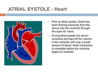
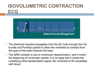
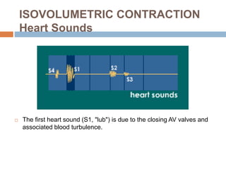
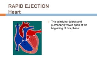
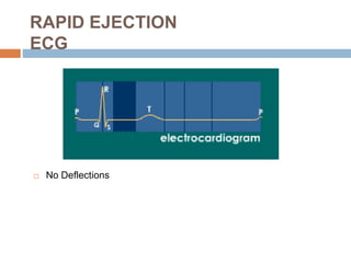
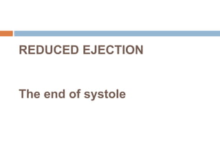
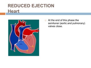
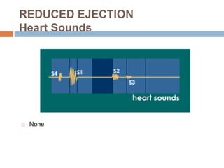
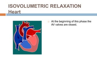
THE CARDIAC CYCLE
Nursing Study Contents
ATRIAL SYSTOLE
The end of diastole
ATRIAL SYSTOLE - Heart
 Prior to atrial systole, blood has
been flowing passively from the
atrium into the ventricle through
the open AV valve.
 During atrial systole the atrium
contracts and tops off the volume
in the ventricle with only a small
amount of blood. Atrial contraction
is complete before the ventricle
begins to contract.
ATRIAL SYSTOLE
Pressures & Volumes
 The "a" wave occurs when the
atrium contracts, increasing atrial
pressure (yellow).
 Blood arriving at the heart cannot
enter the atrium so it flows back
up the jugular vein, causing the
first discernible wave in the
jugular venous pulse.
 Atrial pressure drops when the
atria stop contracting.
 During atrial systole the atrium
contracts and tops off the volume
in the ventricle with only a small
amount of blood.
 Atrial contraction is complete
before the ventricle begins to
contract.
ATRIAL SYSTOLE
ECG
 An impulse arising from the SA node results in depolarization and
contraction of the atria (the right atrium contracts slightly before the
left atrium).
 The P wave is due to this atrial depolarization.
 The PR segment is electrically quiet as the depolarization proceeds
to the AV node.
 This brief pause before contraction allows the ventricles to fill
completely with blood.
ATRIAL SYSTOLE
Heart Sounds
 A fourth heart sound (S4) is abnormal and is associated with the end
of atrial emptying after atrial contraction.
 It occurs with hypertrophic congestive heart failure, massive
pulmonary embolism, tricuspid incompetence, or cor pulmonale.
ISOVOLUMETRIC
CONTRACTION
The Beginning of systole
ISOVOLUMETRIC CONTRACTION
Heart
 The atrioventricular (AV) valves
close at the beginning of this
phase.
 Electrically, ventricular systole is
defined as the interval between
the QRS complex and the end of
the T wave (the Q-T interval).
 Mechanically, ventricular systole
is defined as the interval between
the closing of the AV valves and
the opening of the semilunar
valves (aortic and pulmonary
valves).
ISOVOLUMETRIC CONTRACTION
Pressures & Volumes
 The AV valves close when the
pressure in the ventricles (red)
exceeds the pressure in the atria
(yellow).
 As the ventricles contract
isovolumetrically -- their volume
does not change (white) -- the
pressure inside increases,
approaching the pressure in the
aorta and pulmonary arteries
(green).
ISOVOLUMETRIC CONTRACTION
ECG
 The electrical impulse propagates from the AV node through the His
bundle and Purkinje system to allow the ventricles to contract from
the apex of the heart towards the base.
 The QRS complex is due to ventricular depolarization, and it marks
the beginning of ventricular systole. It is so large that it masks the
underlying atrial repolarization signal. the ventricles to fill completely
with blood.
ISOVOLUMETRIC CONTRACTION
Heart Sounds
 The first heart sound (S1, "lub") is due to the closing AV valves and
associated blood turbulence.
RAPID EJECTION
RAPID EJECTION
Heart
 The semilunar (aortic and
pulmonary) valves open at the
beginning of this phase.
RAPID EJECTION
Pressures & Volumes
 While the ventricles continue
contracting, the pressure in the
ventricles (red) exceeds the pressure
in the aorta and pulmonary arteries
(green); the semilunar valves open,
blood exits the ventricles, and the
volume in the ventricles decreases
rapidly (white).
 As more blood enters the arteries,
pressure there builds until the flow of
blood reaches a peak.
 The "c" wave of atrial pressure is not
normally discernible in the jugular
venous pulse. Right ventricular
contraction pushes the tricuspid valve
into the atrium and increases atrial
pressure, creating a small wave into
the jugular vein. It is normally
simultaneous with the carotid pulse.
RAPID EJECTION
ECG
 No Deflections
RAPID EJECTION
Heart Sounds
 None
REDUCED EJECTION
The end of systole
REDUCED EJECTION
Heart
 At the end of this phase the
semilunar (aortic and pulmonary)
valves close.
REDUCED EJECTION
Pressures & Volumes
 After the peak in ventricular and
arterial pressures (red and
green), blood flow out of the
ventricles decreases and
ventricular volume decreases
more slowly (white).
 When the pressure in the
ventricles falls below the pressure
in the arteries, blood in the
arteries begins to flow back
toward the ventricles and causes
the semilunar valves to close.
This marks the end of ventricular
systole mechanically.
REDUCED EJECTION
ECG
 The T wave is due to ventricular repolarization. The end of the T
wave marks the end of ventricular systole electrically.
REDUCED EJECTION
Heart Sounds
 None
ISOVOLUMETRIC
RELAXATION
The beginning of Diastole
ISOVOLUMETRIC RELAXATION
Heart
 At the beginning of this phase the
AV valves are closed.
ISOVOLUMETRIC RELAXATION
Pressures & Volumes
 Throughout this and the previous
two phases, the atrium in diastole
has been filling with blood on top
of the closed AV valve, causing
atrial pressure to rise gradually
(yellow).
 The "v" wave is due to the back
flow of blood after it hits the
closed AV valve. It is the second
discernible wave of the jugular
venous pulse.
 The pressure in the ventricles
(red) continues to drop.
 Ventricular volume (white) is at a
minimum and is ready to be filled
again with blood.
ISOVOLUMETRIC RELAXATION
ECG
 No Deflections
ISOVOLUMETRIC RELAXATION
Heart Sounds
 The second heart sound (S2, "dup") occurs when the semilunar
(aortic and pulmonary) valves close. S2 is normally split
because the aortic valve closes slightly earlier than the
pulmonary valve.
RAPID VENTRICULAR
FILLING
RAPID VENTRICULAR FILLING
Heart
 Once the AV valves open, blood
that has accumulated in the atria
flows rapidly into the ventricles.
RAPID VENTRICULAR FILLING
Pressures & Volumes
 Ventricular volume (white)
increases rapidly as blood flows
from the atria into the ventricles.
RAPID VENTRICULAR FILLING
ECG
 No Deflections
RAPID VENTRICULAR FILLING
Heart Sounds
 A third heart sound (S3) is usually abnormal and is due to rapid
passive ventricular filling. It occurs in dilated congestive heart
failure, severe hypertension, myocardial infarction, or mitral
incompetence.
REDUCED VENTRICULAR
FILLING
(Diastasis)
REDUCED VENTRICULAR FILLING
Heart
 Rest of blood that has
accumulated in the atria flows
slowly into the ventricles.
REDUCED VENTRICULAR FILLING
Pressures & Volumes
 Ventricular volume (white)
increases more slowly now. The
ventricles continue to fill with
blood until they are nearly full.
REDUCED VENTRICULAR FILLING
ECG
 No Deflections
REDUCED VENTRICULAR FILLING
Heart Sounds
0
THANKS
LIKE &
SHARE
Nursing Study Contents

Cardiac cycle

  • 1.
  • 2.
  • 3.
    ATRIAL SYSTOLE -Heart  Prior to atrial systole, blood has been flowing passively from the atrium into the ventricle through the open AV valve.  During atrial systole the atrium contracts and tops off the volume in the ventricle with only a small amount of blood. Atrial contraction is complete before the ventricle begins to contract.
  • 4.
    ATRIAL SYSTOLE Pressures &Volumes  The "a" wave occurs when the atrium contracts, increasing atrial pressure (yellow).  Blood arriving at the heart cannot enter the atrium so it flows back up the jugular vein, causing the first discernible wave in the jugular venous pulse.  Atrial pressure drops when the atria stop contracting.  During atrial systole the atrium contracts and tops off the volume in the ventricle with only a small amount of blood.  Atrial contraction is complete before the ventricle begins to contract.
  • 5.
    ATRIAL SYSTOLE ECG  Animpulse arising from the SA node results in depolarization and contraction of the atria (the right atrium contracts slightly before the left atrium).  The P wave is due to this atrial depolarization.  The PR segment is electrically quiet as the depolarization proceeds to the AV node.  This brief pause before contraction allows the ventricles to fill completely with blood.
  • 6.
    ATRIAL SYSTOLE Heart Sounds A fourth heart sound (S4) is abnormal and is associated with the end of atrial emptying after atrial contraction.  It occurs with hypertrophic congestive heart failure, massive pulmonary embolism, tricuspid incompetence, or cor pulmonale.
  • 7.
  • 8.
    ISOVOLUMETRIC CONTRACTION Heart  Theatrioventricular (AV) valves close at the beginning of this phase.  Electrically, ventricular systole is defined as the interval between the QRS complex and the end of the T wave (the Q-T interval).  Mechanically, ventricular systole is defined as the interval between the closing of the AV valves and the opening of the semilunar valves (aortic and pulmonary valves).
  • 9.
    ISOVOLUMETRIC CONTRACTION Pressures &Volumes  The AV valves close when the pressure in the ventricles (red) exceeds the pressure in the atria (yellow).  As the ventricles contract isovolumetrically -- their volume does not change (white) -- the pressure inside increases, approaching the pressure in the aorta and pulmonary arteries (green).
  • 10.
    ISOVOLUMETRIC CONTRACTION ECG  Theelectrical impulse propagates from the AV node through the His bundle and Purkinje system to allow the ventricles to contract from the apex of the heart towards the base.  The QRS complex is due to ventricular depolarization, and it marks the beginning of ventricular systole. It is so large that it masks the underlying atrial repolarization signal. the ventricles to fill completely with blood.
  • 11.
    ISOVOLUMETRIC CONTRACTION Heart Sounds The first heart sound (S1, "lub") is due to the closing AV valves and associated blood turbulence.
  • 12.
  • 13.
    RAPID EJECTION Heart  Thesemilunar (aortic and pulmonary) valves open at the beginning of this phase.
  • 14.
    RAPID EJECTION Pressures &Volumes  While the ventricles continue contracting, the pressure in the ventricles (red) exceeds the pressure in the aorta and pulmonary arteries (green); the semilunar valves open, blood exits the ventricles, and the volume in the ventricles decreases rapidly (white).  As more blood enters the arteries, pressure there builds until the flow of blood reaches a peak.  The "c" wave of atrial pressure is not normally discernible in the jugular venous pulse. Right ventricular contraction pushes the tricuspid valve into the atrium and increases atrial pressure, creating a small wave into the jugular vein. It is normally simultaneous with the carotid pulse.
  • 15.
  • 16.
  • 17.
  • 18.
    REDUCED EJECTION Heart  Atthe end of this phase the semilunar (aortic and pulmonary) valves close.
  • 19.
    REDUCED EJECTION Pressures &Volumes  After the peak in ventricular and arterial pressures (red and green), blood flow out of the ventricles decreases and ventricular volume decreases more slowly (white).  When the pressure in the ventricles falls below the pressure in the arteries, blood in the arteries begins to flow back toward the ventricles and causes the semilunar valves to close. This marks the end of ventricular systole mechanically.
  • 20.
    REDUCED EJECTION ECG  TheT wave is due to ventricular repolarization. The end of the T wave marks the end of ventricular systole electrically.
  • 21.
  • 22.
  • 23.
    ISOVOLUMETRIC RELAXATION Heart  Atthe beginning of this phase the AV valves are closed.
  • 24.
    ISOVOLUMETRIC RELAXATION Pressures &Volumes  Throughout this and the previous two phases, the atrium in diastole has been filling with blood on top of the closed AV valve, causing atrial pressure to rise gradually (yellow).  The "v" wave is due to the back flow of blood after it hits the closed AV valve. It is the second discernible wave of the jugular venous pulse.  The pressure in the ventricles (red) continues to drop.  Ventricular volume (white) is at a minimum and is ready to be filled again with blood.
  • 25.
  • 26.
    ISOVOLUMETRIC RELAXATION Heart Sounds The second heart sound (S2, "dup") occurs when the semilunar (aortic and pulmonary) valves close. S2 is normally split because the aortic valve closes slightly earlier than the pulmonary valve.
  • 27.
  • 28.
    RAPID VENTRICULAR FILLING Heart Once the AV valves open, blood that has accumulated in the atria flows rapidly into the ventricles.
  • 29.
    RAPID VENTRICULAR FILLING Pressures& Volumes  Ventricular volume (white) increases rapidly as blood flows from the atria into the ventricles.
  • 30.
  • 31.
    RAPID VENTRICULAR FILLING HeartSounds  A third heart sound (S3) is usually abnormal and is due to rapid passive ventricular filling. It occurs in dilated congestive heart failure, severe hypertension, myocardial infarction, or mitral incompetence.
  • 32.
  • 33.
    REDUCED VENTRICULAR FILLING Heart Rest of blood that has accumulated in the atria flows slowly into the ventricles.
  • 34.
    REDUCED VENTRICULAR FILLING Pressures& Volumes  Ventricular volume (white) increases more slowly now. The ventricles continue to fill with blood until they are nearly full.
  • 35.
  • 36.
  • 37.