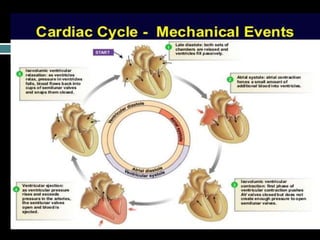
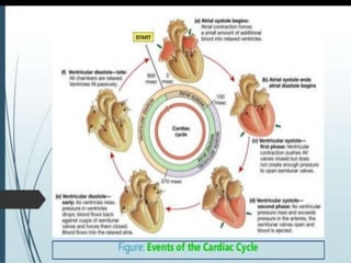
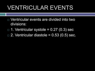
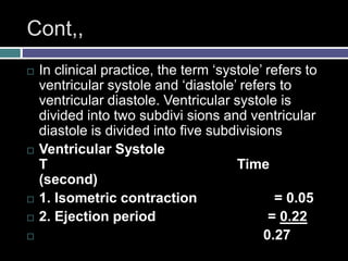
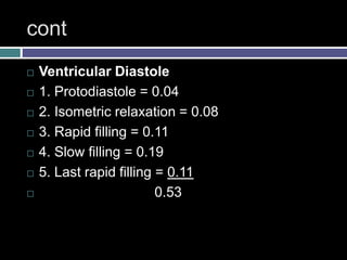
CARDIAC CYCLE
Abdus salam
DEFINITION
 Cardiac cycle is defined as the succession of
(sequence of) coordinated events taking place
in the heart during each beat. Each heartbeat
consists of two major periods called systole
and diastole. During systole, heart contracts
and pumps the blood through arteries. During
diastole, heart relaxes and blood is filled in the
heart. All these changes are repeated during
every heartbeat, in a cyclic manner
EVENTS OF CARDIAC CYCLE
 Events of cardiac cycle are classified into two
 1. Atrial events
 2. Ventricular events.
 In every event discuss what happening in the
ventricle atrium and aorta.
ATRIAL SYSTOL
 Durning artrial contraction there is
cummunication between the artium and
ventricle not with arota.
 Before artrial contraction start about 80% (
passive) filling already done
 They add just 20% of blood filling ( valuable
during exercise
 The pr in the left atrium will increased valve
open so pr easily communicate with ventricle
 Blood move to ventricle pr transfer to ventricle
 Before the contraction the pr in the ventricle is
0
 Arotic valve closed
 Mitral valve open
Ventricular contraction (systole)
 Depolarized current come to ventricle
 Onset of ventricular contraction arotic valve
already closed
 When ventricular start contraction pr is rise very
begnining round to 5 mmhg and more than artria
so mitral and tricuspid valve closed (S2)
 Ventricle continue to be contraction and contract
is closed chamber b/c valve are closed
 Now pr goes up upto 80 mmhg but not open the
arotic valve called iso volumeic contraction
isovolumic contraction
 Blood entering to ventricle no blood is leaving
so amount of blood is same so called
isovolumic contraction ventricle contracted is
closed chamber . In this phase PR increased
 Blood coming into the atrium work is a tank
and PR in artria gradually increased bcz Blood
is coming from the lungs and venous return.
 When PR increased in ventricle arotic vlave
will open termination of iso volumeic
contraction

EJECTION PERIOD
 Due to the opening of semilunar valves and
isotonic contraction of ventricles, blood is ejected
out of both the ventricles. Hence, this period is
called ejection period. Duration of this period is
0.22 second. Ejection period is of two stages: now
arotic chamber and ventricular work is a single
chamber pr transfer into arota pr go to 120
 First Stage or Rapid Ejection Period
 First stage starts immediately after the opening of
semilunar valves. During this stage, a large
amount of blood is rapidly ejected from both the
ventricles. It lasts for 0.13 second.
 Second Stage or Slow Ejection Period
 During this stage, the blood is ejected slowly
with much less force. Duration of this period is
0.09 second.
 Ventricular is a still contractated but pr star
fallingpr in the arota also fall
End-systolic Volume
 Ventricles are not emptied at the end of
ejection period and some amount of blood
remains in each ventricle. Amount of blood
remaining in ventricles at the end of ejection
period (i.e. at the end of systole) is called
endsystolic volume. It is 60 to 80 mL per
ventricle.
Ventricular relaxation
 PROTODIASTOLE
 Protodiastole is the first stage of ventricular
diastole, hence the name protodiastole. Duration
of this period is 0.04 second. Due to the ejection
of blood, the pressure in aorta and pulmonary
artery increases and pressure in ventricles drops.
When intraventricular pressure becomes less than
the pressure in aorta and pulmonary artery, the
semilunar valves close. Atrioventricular valves are
already closed . No other change occurs in the
heart during this period. Thus, protodiastole
indicates only the end of systole and beginning of
diastole.
Cont ….
 Ventricular start relaxing pr start to fall
 Pr rapidly start falling blood from the arota try
to comeback into ventricle which has low pr
chamber in this phase pr closure of seminular
valve so connection is disconnected
 Pr in the arota is more than ventricle so arotic
valve closed and also more than atria
 There is a isovolumeic relaxation .
ISOMETRIC RELAXATION
PERIOD
 Isometric relaxation is the type of muscular
relaxation, characterized by decrease in tension
without any change in the length of muscle fibers.
Isometric relaxation of ventricular muscle is also
called isovolumetric relaxation. During isometric
relaxation period, once again all the valves of the
heart are closed . Now, both the ventricles relax
as closed cavities without any change in volume
or length of the muscle fiber. Intraventricular
pressure decreases during this period. Duration of
isometric relaxation period is 0.08 second
Significance of Isometric
Relaxation
 During isometric relaxation period, the
ventricular pressure decreases greatly. When
the ventricular pressure becomes less than the
pressure in the atria, the atrioventricular valves
open. Thus, the fall in pressure in the
ventricles, caused by isometric relaxation is
responsible for the opening of atrioventricular
valves, resulting in filling of ventricles.
End systolic volume
 The volume of blood present in the ventricle at
the end of systole
 At this movment when the pr in the ventricle is
less than artrium the mitral valve open and
artia ane ventricel are connected and rapid
filling of blood occurs.
RAPID FILLING PHASE
 When atrionventricular valves are opened,
there is a sudden rush of blood (which is
accumulated in atria during atrial diastole)
from atria into ventricles. So, this period is
called the first rapid filling period. Ventricles
also relax isotonically. About 70% of filling
takes place during this phase, which lasts for
0.11 second.
 Third heart sound
SLOW FILLING PHASE
 After the sudden rush of blood, the ventricular
filling becomes slow. Now, it is called the slow
filling. It is also called diastasis. About 20% of
filling occurs in this phase. Duration of slow
filling phase is 0.19 second.
LAST RAPID FILLING PHASE
 Last rapid filling phase occurs because of
atrial systole. After slow filling period, the atria
contract and push a small amount of blood into
ventricles. About 10% of ventricular filling
takes place during this period. Flow of
additional amount of blood into ventricle due to
atrial systole is called atrial kick.
DIVISIONS AND DURATION OF
CARDIAC CYCLE
 When the heart beats at a normal rate of
72/minute, duration of each cardiac cycle is
about 0.8 second.
 ATRIAL EVENTS
 Atrial events are divided into two divisions:
 1. Atrial systole = 0.11 (0.1) sec
 2. Atrial diastole = 0.69 (0.7) sec
VENTRICULAR EVENTS
 Ventricular events are divided into two
divisions:
 1. Ventricular systole = 0.27 (0.3) sec
 2. Ventricular diastole = 0.53 (0.5) sec.
Cont,,
 In clinical practice, the term ‘systole’ refers to
ventricular systole and ‘diastole’ refers to
ventricular diastole. Ventricular systole is
divided into two subdivi sions and ventricular
diastole is divided into five subdivisions
 Ventricular Systole
T Time
(second)
 1. Isometric contraction = 0.05
 2. Ejection period = 0.22
 0.27
cont
 Ventricular Diastole
 1. Protodiastole = 0.04
 2. Isometric relaxation = 0.08
 3. Rapid filling = 0.11
 4. Slow filling = 0.19
 5. Last rapid filling = 0.11
 0.53
Cardiac cycle

Cardiac cycle

  • 1.
  • 2.
    DEFINITION  Cardiac cycleis defined as the succession of (sequence of) coordinated events taking place in the heart during each beat. Each heartbeat consists of two major periods called systole and diastole. During systole, heart contracts and pumps the blood through arteries. During diastole, heart relaxes and blood is filled in the heart. All these changes are repeated during every heartbeat, in a cyclic manner
  • 3.
    EVENTS OF CARDIACCYCLE  Events of cardiac cycle are classified into two  1. Atrial events  2. Ventricular events.  In every event discuss what happening in the ventricle atrium and aorta.
  • 4.
    ATRIAL SYSTOL  Durningartrial contraction there is cummunication between the artium and ventricle not with arota.  Before artrial contraction start about 80% ( passive) filling already done  They add just 20% of blood filling ( valuable during exercise  The pr in the left atrium will increased valve open so pr easily communicate with ventricle
  • 5.
     Blood moveto ventricle pr transfer to ventricle  Before the contraction the pr in the ventricle is 0  Arotic valve closed  Mitral valve open
  • 6.
    Ventricular contraction (systole) Depolarized current come to ventricle  Onset of ventricular contraction arotic valve already closed  When ventricular start contraction pr is rise very begnining round to 5 mmhg and more than artria so mitral and tricuspid valve closed (S2)  Ventricle continue to be contraction and contract is closed chamber b/c valve are closed  Now pr goes up upto 80 mmhg but not open the arotic valve called iso volumeic contraction
  • 7.
    isovolumic contraction  Bloodentering to ventricle no blood is leaving so amount of blood is same so called isovolumic contraction ventricle contracted is closed chamber . In this phase PR increased  Blood coming into the atrium work is a tank and PR in artria gradually increased bcz Blood is coming from the lungs and venous return.  When PR increased in ventricle arotic vlave will open termination of iso volumeic contraction 
  • 8.
    EJECTION PERIOD  Dueto the opening of semilunar valves and isotonic contraction of ventricles, blood is ejected out of both the ventricles. Hence, this period is called ejection period. Duration of this period is 0.22 second. Ejection period is of two stages: now arotic chamber and ventricular work is a single chamber pr transfer into arota pr go to 120  First Stage or Rapid Ejection Period  First stage starts immediately after the opening of semilunar valves. During this stage, a large amount of blood is rapidly ejected from both the ventricles. It lasts for 0.13 second.
  • 9.
     Second Stageor Slow Ejection Period  During this stage, the blood is ejected slowly with much less force. Duration of this period is 0.09 second.  Ventricular is a still contractated but pr star fallingpr in the arota also fall
  • 10.
    End-systolic Volume  Ventriclesare not emptied at the end of ejection period and some amount of blood remains in each ventricle. Amount of blood remaining in ventricles at the end of ejection period (i.e. at the end of systole) is called endsystolic volume. It is 60 to 80 mL per ventricle.
  • 11.
    Ventricular relaxation  PROTODIASTOLE Protodiastole is the first stage of ventricular diastole, hence the name protodiastole. Duration of this period is 0.04 second. Due to the ejection of blood, the pressure in aorta and pulmonary artery increases and pressure in ventricles drops. When intraventricular pressure becomes less than the pressure in aorta and pulmonary artery, the semilunar valves close. Atrioventricular valves are already closed . No other change occurs in the heart during this period. Thus, protodiastole indicates only the end of systole and beginning of diastole.
  • 12.
    Cont ….  Ventricularstart relaxing pr start to fall  Pr rapidly start falling blood from the arota try to comeback into ventricle which has low pr chamber in this phase pr closure of seminular valve so connection is disconnected  Pr in the arota is more than ventricle so arotic valve closed and also more than atria  There is a isovolumeic relaxation .
  • 13.
    ISOMETRIC RELAXATION PERIOD  Isometricrelaxation is the type of muscular relaxation, characterized by decrease in tension without any change in the length of muscle fibers. Isometric relaxation of ventricular muscle is also called isovolumetric relaxation. During isometric relaxation period, once again all the valves of the heart are closed . Now, both the ventricles relax as closed cavities without any change in volume or length of the muscle fiber. Intraventricular pressure decreases during this period. Duration of isometric relaxation period is 0.08 second
  • 14.
    Significance of Isometric Relaxation During isometric relaxation period, the ventricular pressure decreases greatly. When the ventricular pressure becomes less than the pressure in the atria, the atrioventricular valves open. Thus, the fall in pressure in the ventricles, caused by isometric relaxation is responsible for the opening of atrioventricular valves, resulting in filling of ventricles.
  • 15.
    End systolic volume The volume of blood present in the ventricle at the end of systole  At this movment when the pr in the ventricle is less than artrium the mitral valve open and artia ane ventricel are connected and rapid filling of blood occurs.
  • 16.
    RAPID FILLING PHASE When atrionventricular valves are opened, there is a sudden rush of blood (which is accumulated in atria during atrial diastole) from atria into ventricles. So, this period is called the first rapid filling period. Ventricles also relax isotonically. About 70% of filling takes place during this phase, which lasts for 0.11 second.  Third heart sound
  • 17.
    SLOW FILLING PHASE After the sudden rush of blood, the ventricular filling becomes slow. Now, it is called the slow filling. It is also called diastasis. About 20% of filling occurs in this phase. Duration of slow filling phase is 0.19 second.
  • 18.
    LAST RAPID FILLINGPHASE  Last rapid filling phase occurs because of atrial systole. After slow filling period, the atria contract and push a small amount of blood into ventricles. About 10% of ventricular filling takes place during this period. Flow of additional amount of blood into ventricle due to atrial systole is called atrial kick.
  • 22.
    DIVISIONS AND DURATIONOF CARDIAC CYCLE  When the heart beats at a normal rate of 72/minute, duration of each cardiac cycle is about 0.8 second.  ATRIAL EVENTS  Atrial events are divided into two divisions:  1. Atrial systole = 0.11 (0.1) sec  2. Atrial diastole = 0.69 (0.7) sec
  • 23.
    VENTRICULAR EVENTS  Ventricularevents are divided into two divisions:  1. Ventricular systole = 0.27 (0.3) sec  2. Ventricular diastole = 0.53 (0.5) sec.
  • 24.
    Cont,,  In clinicalpractice, the term ‘systole’ refers to ventricular systole and ‘diastole’ refers to ventricular diastole. Ventricular systole is divided into two subdivi sions and ventricular diastole is divided into five subdivisions  Ventricular Systole T Time (second)  1. Isometric contraction = 0.05  2. Ejection period = 0.22  0.27
  • 25.
    cont  Ventricular Diastole 1. Protodiastole = 0.04  2. Isometric relaxation = 0.08  3. Rapid filling = 0.11  4. Slow filling = 0.19  5. Last rapid filling = 0.11  0.53