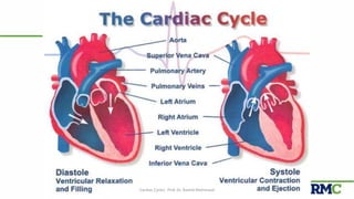
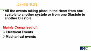
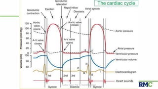
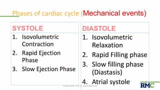
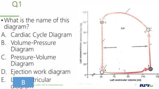
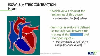
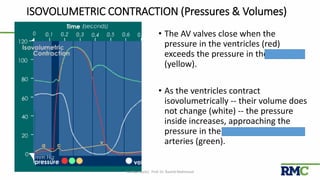
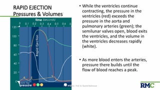
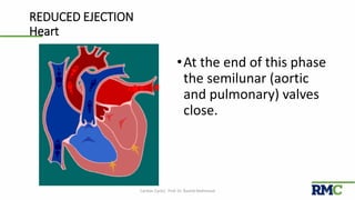
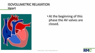
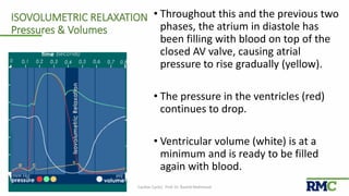
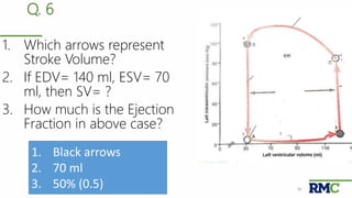
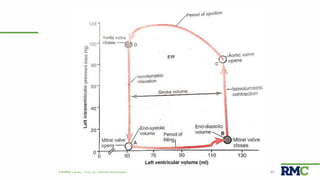
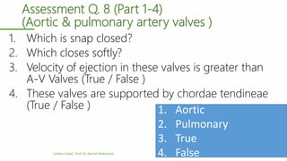
Cardiac Cycle
Prof. Dr. Rashid Mahmood
Cardiac Cycle| Prof. Dr. Rashid Mahmood 2
CourtesyCardiac Cycle| Prof. Dr. Rashid Mahmood 3
Cardiac Cycle
Cardiac Cycle| Prof. Dr. Rashid Mahmood 4
DEFINITION
•All the events taking place in the Heart from one
systole to another systole or from one Diastole to
another Diastole.
Mainly Comprised of:
Electrical Events
Mechanical events
Electrical & Mechanical events
Electrical Events
• ECG
Mechanical events
• Pressure Changes
• Volume Changes
• Production of Heart Sounds
Cardiac Cycle| Prof. Dr. Rashid Mahmood 6
Cardiac Cycle| Prof. Dr. Rashid Mahmood 7
Phases of cardiac cycle (Mechanical events)
SYSTOLE
1. Isovolumetric
Contraction
2. Rapid Ejection
Phase
3. Slow Ejection Phase
DIASTOLE
1. Isovolumetric
Relaxation
2. Rapid Filling phase
3. Slow filling phase
(Diastasis)
4. Atrial systoleCardiac Cycle| Prof. Dr. Rashid Mahmood 8
Q.1
• What is the name of this
diagram?
A. Cardiac Cycle Diagram
B. Volume-Pressure
Diagram
C. Pressure-Volume
Diagram
D. Ejection work diagram
E. Left-ventricular
diagramCardiac Cycle| Prof. Dr. Rashid Mahmood 9B
ISOVOLUMETRIC CONTRACTION
The Beginning of systole
Cardiac Cycle| Prof. Dr. Rashid Mahmood 10
ISOVOLUMETRIC CONTRACTION
Heart
• Which valves close at the
beginning of this phase
• atrioventricular (AV) valves
• Ventricular systole is defined
as the interval between the
closing of the AV valves and
the opening of ?
• the semilunar valves (aortic
and pulmonary valves).
Cardiac Cycle| Prof. Dr. Rashid Mahmood 11
ISOVOLUMETRIC CONTRACTION (Pressures & Volumes)
• The AV valves close when the
pressure in the ventricles (red)
exceeds the pressure in the atria
(yellow).
• As the ventricles contract
isovolumetrically -- their volume does
not change (white) -- the pressure
inside increases, approaching the
pressure in the aorta and pulmonary
arteries (green).
Cardiac Cycle| Prof. Dr. Rashid Mahmood 12
Q. 2
1. What happens at Point
A?
2. What volume is
represented at Point A?
3. What period is
represented by AB?
Cardiac Cycle| Prof. Dr. Rashid Mahmood 13
1. Mitral valve opens
2. End-Systolic Volume
3. Period of filling
RAPID EJECTION
Cardiac Cycle| Prof. Dr. Rashid Mahmood 14
RAPID EJECTION
Heart
•The semilunar (aortic and
pulmonary) valves open at
the beginning of this
phase.
Cardiac Cycle| Prof. Dr. Rashid Mahmood 15
RAPID EJECTION
Pressures & Volumes
• While the ventricles continue
contracting, the pressure in the
ventricles (red) exceeds the
pressure in the aorta and
pulmonary arteries (green); the
semilunar valves open, blood exits
the ventricles, and the volume in
the ventricles decreases rapidly
(white).
• As more blood enters the arteries,
pressure there builds until the
flow of blood reaches a peak.
Cardiac Cycle| Prof. Dr. Rashid Mahmood 16
REDUCED EJECTION
The end of systole
Cardiac Cycle| Prof. Dr. Rashid Mahmood 17
REDUCED EJECTION
Heart
•At the end of this phase
the semilunar (aortic
and pulmonary) valves
close.
Cardiac Cycle| Prof. Dr. Rashid Mahmood 18
REDUCED EJECTION
Pressures & Volumes
• After the peak in ventricular and
arterial pressures (red and green),
blood flow out of the ventricles
decreases and ventricular volume
decreases more slowly (white).
• When the pressure in the ventricles
falls below the pressure in the
arteries, blood in the arteries
begins to flow back toward the
ventricles and causes the semilunar
valves to close. This marks the end
of ventricular systole mechanically.
Cardiac Cycle| Prof. Dr. Rashid Mahmood 19
Q. 3
1. What happens at Point
B?
2. What volume is
represented at Point B?
3. What period is
represented by BC?
Cardiac Cycle| Prof. Dr. Rashid Mahmood 20
1. Mitral valve closes
2. End-Diastolic Volume
3. Isovolumic contraction
ISOVOLUMETRIC RELAXATION
The beginning of Diastole
Cardiac Cycle| Prof. Dr. Rashid Mahmood 21
ISOVOLUMETRIC RELAXATION
Heart
•At the beginning of this
phase the AV valves are
closed.
Cardiac Cycle| Prof. Dr. Rashid Mahmood 22
ISOVOLUMETRIC RELAXATION
Pressures & Volumes
• Throughout this and the previous two
phases, the atrium in diastole has
been filling with blood on top of the
closed AV valve, causing atrial
pressure to rise gradually (yellow).
• The pressure in the ventricles (red)
continues to drop.
• Ventricular volume (white) is at a
minimum and is ready to be filled
again with blood.
Cardiac Cycle| Prof. Dr. Rashid Mahmood 23
RAPID VENTRICULAR FILLING
Cardiac Cycle| Prof. Dr. Rashid Mahmood 24
RAPID VENTRICULAR FILLING
Heart
•Once the AV valves open,
blood that has
accumulated in the atria
flows rapidly into the
ventricles.
Cardiac Cycle| Prof. Dr. Rashid Mahmood 25
RAPID VENTRICULAR FILLING Pressures & Volumes
•Ventricular volume (white)
increases rapidly as blood flows
from the atria into the ventricles.
Cardiac Cycle| Prof. Dr. Rashid Mahmood 26
Q. 4
1. What happens at Point
C?
2. What volume is
represented at Point C?
3. What period is
represented by CD?
Cardiac Cycle| Prof. Dr. Rashid Mahmood 27
1. Aortic valve opens
2. End-Diastolic Volume
3. Period of Ejection
REDUCED VENTRICULAR FILLING
(Diastasis)
Cardiac Cycle| Prof. Dr. Rashid Mahmood 28
REDUCED VENTRICULAR FILLING
Heart
•Rest of blood that has
accumulated in the atria
flows slowly into the
ventricles.
Cardiac Cycle| Prof. Dr. Rashid Mahmood 29
REDUCED VENTRICULAR FILLING Pressures
& Volumes
•Ventricular volume
(white) increases more
slowly now.
•The ventricles continue
to fill with blood until
they are nearly full.
Cardiac Cycle| Prof. Dr. Rashid Mahmood 30
ATRIAL SYSTOLE
The end of diastole
Cardiac Cycle| Prof. Dr. Rashid Mahmood 31
ATRIAL SYSTOLE - Heart
• Prior to atrial systole, blood has been
flowing passively from the atrium into
the ventricle through the open AV valve.
• During atrial systole the atrium contracts
and tops off the volume in the ventricle
with only a small amount of blood.
• Atrial contraction is complete before the
ventricle begins to contract.
Cardiac Cycle| Prof. Dr. Rashid Mahmood 32
Pressure Changes in
the Atria
. ‘a Wave: Produced due to atrial contraction
Rt. Atrial Pressure ( 4-6mmHg <
Lt Atrial Pressure (7-8mmHg)
. ‘C’ Wave. Mainly due to bulging of A-V valve in to the
atrium during ventricular contraction.
. ‘V’ Wave: produced due to slow flow of blood into the
atrium when the A-V valves are still closed during
ventricular contraction
a c v
Atrial Pressure Curve (Blue)
Q. 5
1. What happens at Point
D?
2. What volume is
represented at Point C?
3. What period is
represented by DA?
Cardiac Cycle| Prof. Dr. Rashid Mahmood 34
1. Aortic valve closes
2. End-Systolic Volume
3. Isovolumic relaxation
Applied aspects
Stroke Volume
•Volume of blood pumped
by each ventricular
contraction
•Normal value
• 70 ml
Ejection Fraction
•Fraction of end-
diastolic volume that is
ejected
•Normal value
•60% (0.6)
Cardiac Cycle| Prof. Dr. Rashid Mahmood 35
Q. 6
1. Which arrows represent
Stroke Volume?
2. If EDV= 140 ml, ESV= 70
ml, then SV= ?
3. How much is the Ejection
Fraction in above case?
Cardiac Cycle| Prof. Dr. Rashid Mahmood 36
1. Black arrows
2. 70 ml
3. 50% (0.5)
THE COMPLETE
PICTURE
Cardiac Cycle| Prof. Dr. Rashid Mahmood 37
Cardiac Cycle| Prof. Dr. Rashid Mahmood 38
Cardiac Cycle| Prof. Dr. Rashid Mahmood 39
Cardiac Cycle| Prof. Dr. Rashid Mahmood 40
Assessment Q 7 (Part 1-4) (Yes/ No)
1. AV valves open and close passively
2. Stroke vol can be doubled by increasing ESV &
decreasing EDV
3. Calculations of ejection fraction
4. Increasing HR decreases heart filling
Cardiac Cycle| Prof. Dr. Rashid Mahmood 41
All: Yes
Assessment Q. 8 (Part 1-4)
(Aortic & pulmonary artery valves )
1. Which is snap closed?
2. Which closes softly?
3. Velocity of ejection in these valves is greater than
A-V Valves (True / False )
4. These valves are supported by chordae tendineae
(True / False )
Cardiac Cycle| Prof. Dr. Rashid Mahmood 42
1. Aortic
2. Pulmonary
3. True
4. False
Summary (Slide ½)
•Events that occur from the beginning of one heartbeat
to next are called the Cardiac Cycle
•Cardiac cycle consists of a period of contraction
(Systole) followed by relaxation (Diastole)
•Systole consists of isovolumic contraction, followed by
period of rapid & then slow ejection.
•Diastole consists of isovolumic relaxation, followed by
Rapid Filling phase, Slow filling phase (Diastasis), &
then Atrial systole
Cardiac Cycle| Prof. Dr. Rashid Mahmood 43
Summary (Slide 2/2)
•Normally ESV is 40-50 ml, EDV is 110-120 ml, and
stroke volume output 40-50 ml; with an ejection
fraction of 60% (0.6)
•Left ventricular pressure during rapid filling stage is 2-
3 mmHg, at the end of isovolumic contraction 80
mmHg, and 120 mmHg at the peak of the Ejection
Phase.
•‘a’ wave of Atrial systole is due to Atrial contraction, ‘c’
wave due to bulging of A-V valves backwards, and ‘v’
wave due to slow flow of blood from veins to atria.Cardiac Cycle| Prof. Dr. Rashid Mahmood 44
Thank You
ppt available on slideshare.com
Questions/ Comments:
drrashid62@gmail.com
rashid.mahmood@rmi.edu.pk
Cardiac Cycle| Prof. Dr. Rashid Mahmood 45

Cardiac cycle

  • 1.
  • 2.
    Cardiac Cycle| Prof.Dr. Rashid Mahmood 2
  • 3.
    CourtesyCardiac Cycle| Prof.Dr. Rashid Mahmood 3 Cardiac Cycle
  • 4.
    Cardiac Cycle| Prof.Dr. Rashid Mahmood 4
  • 5.
    DEFINITION •All the eventstaking place in the Heart from one systole to another systole or from one Diastole to another Diastole. Mainly Comprised of: Electrical Events Mechanical events
  • 6.
    Electrical & Mechanicalevents Electrical Events • ECG Mechanical events • Pressure Changes • Volume Changes • Production of Heart Sounds Cardiac Cycle| Prof. Dr. Rashid Mahmood 6
  • 7.
    Cardiac Cycle| Prof.Dr. Rashid Mahmood 7
  • 8.
    Phases of cardiaccycle (Mechanical events) SYSTOLE 1. Isovolumetric Contraction 2. Rapid Ejection Phase 3. Slow Ejection Phase DIASTOLE 1. Isovolumetric Relaxation 2. Rapid Filling phase 3. Slow filling phase (Diastasis) 4. Atrial systoleCardiac Cycle| Prof. Dr. Rashid Mahmood 8
  • 9.
    Q.1 • What isthe name of this diagram? A. Cardiac Cycle Diagram B. Volume-Pressure Diagram C. Pressure-Volume Diagram D. Ejection work diagram E. Left-ventricular diagramCardiac Cycle| Prof. Dr. Rashid Mahmood 9B
  • 10.
    ISOVOLUMETRIC CONTRACTION The Beginningof systole Cardiac Cycle| Prof. Dr. Rashid Mahmood 10
  • 11.
    ISOVOLUMETRIC CONTRACTION Heart • Whichvalves close at the beginning of this phase • atrioventricular (AV) valves • Ventricular systole is defined as the interval between the closing of the AV valves and the opening of ? • the semilunar valves (aortic and pulmonary valves). Cardiac Cycle| Prof. Dr. Rashid Mahmood 11
  • 12.
    ISOVOLUMETRIC CONTRACTION (Pressures& Volumes) • The AV valves close when the pressure in the ventricles (red) exceeds the pressure in the atria (yellow). • As the ventricles contract isovolumetrically -- their volume does not change (white) -- the pressure inside increases, approaching the pressure in the aorta and pulmonary arteries (green). Cardiac Cycle| Prof. Dr. Rashid Mahmood 12
  • 13.
    Q. 2 1. Whathappens at Point A? 2. What volume is represented at Point A? 3. What period is represented by AB? Cardiac Cycle| Prof. Dr. Rashid Mahmood 13 1. Mitral valve opens 2. End-Systolic Volume 3. Period of filling
  • 14.
    RAPID EJECTION Cardiac Cycle|Prof. Dr. Rashid Mahmood 14
  • 15.
    RAPID EJECTION Heart •The semilunar(aortic and pulmonary) valves open at the beginning of this phase. Cardiac Cycle| Prof. Dr. Rashid Mahmood 15
  • 16.
    RAPID EJECTION Pressures &Volumes • While the ventricles continue contracting, the pressure in the ventricles (red) exceeds the pressure in the aorta and pulmonary arteries (green); the semilunar valves open, blood exits the ventricles, and the volume in the ventricles decreases rapidly (white). • As more blood enters the arteries, pressure there builds until the flow of blood reaches a peak. Cardiac Cycle| Prof. Dr. Rashid Mahmood 16
  • 17.
    REDUCED EJECTION The endof systole Cardiac Cycle| Prof. Dr. Rashid Mahmood 17
  • 18.
    REDUCED EJECTION Heart •At theend of this phase the semilunar (aortic and pulmonary) valves close. Cardiac Cycle| Prof. Dr. Rashid Mahmood 18
  • 19.
    REDUCED EJECTION Pressures &Volumes • After the peak in ventricular and arterial pressures (red and green), blood flow out of the ventricles decreases and ventricular volume decreases more slowly (white). • When the pressure in the ventricles falls below the pressure in the arteries, blood in the arteries begins to flow back toward the ventricles and causes the semilunar valves to close. This marks the end of ventricular systole mechanically. Cardiac Cycle| Prof. Dr. Rashid Mahmood 19
  • 20.
    Q. 3 1. Whathappens at Point B? 2. What volume is represented at Point B? 3. What period is represented by BC? Cardiac Cycle| Prof. Dr. Rashid Mahmood 20 1. Mitral valve closes 2. End-Diastolic Volume 3. Isovolumic contraction
  • 21.
    ISOVOLUMETRIC RELAXATION The beginningof Diastole Cardiac Cycle| Prof. Dr. Rashid Mahmood 21
  • 22.
    ISOVOLUMETRIC RELAXATION Heart •At thebeginning of this phase the AV valves are closed. Cardiac Cycle| Prof. Dr. Rashid Mahmood 22
  • 23.
    ISOVOLUMETRIC RELAXATION Pressures &Volumes • Throughout this and the previous two phases, the atrium in diastole has been filling with blood on top of the closed AV valve, causing atrial pressure to rise gradually (yellow). • The pressure in the ventricles (red) continues to drop. • Ventricular volume (white) is at a minimum and is ready to be filled again with blood. Cardiac Cycle| Prof. Dr. Rashid Mahmood 23
  • 24.
    RAPID VENTRICULAR FILLING CardiacCycle| Prof. Dr. Rashid Mahmood 24
  • 25.
    RAPID VENTRICULAR FILLING Heart •Oncethe AV valves open, blood that has accumulated in the atria flows rapidly into the ventricles. Cardiac Cycle| Prof. Dr. Rashid Mahmood 25
  • 26.
    RAPID VENTRICULAR FILLINGPressures & Volumes •Ventricular volume (white) increases rapidly as blood flows from the atria into the ventricles. Cardiac Cycle| Prof. Dr. Rashid Mahmood 26
  • 27.
    Q. 4 1. Whathappens at Point C? 2. What volume is represented at Point C? 3. What period is represented by CD? Cardiac Cycle| Prof. Dr. Rashid Mahmood 27 1. Aortic valve opens 2. End-Diastolic Volume 3. Period of Ejection
  • 28.
    REDUCED VENTRICULAR FILLING (Diastasis) CardiacCycle| Prof. Dr. Rashid Mahmood 28
  • 29.
    REDUCED VENTRICULAR FILLING Heart •Restof blood that has accumulated in the atria flows slowly into the ventricles. Cardiac Cycle| Prof. Dr. Rashid Mahmood 29
  • 30.
    REDUCED VENTRICULAR FILLINGPressures & Volumes •Ventricular volume (white) increases more slowly now. •The ventricles continue to fill with blood until they are nearly full. Cardiac Cycle| Prof. Dr. Rashid Mahmood 30
  • 31.
    ATRIAL SYSTOLE The endof diastole Cardiac Cycle| Prof. Dr. Rashid Mahmood 31
  • 32.
    ATRIAL SYSTOLE -Heart • Prior to atrial systole, blood has been flowing passively from the atrium into the ventricle through the open AV valve. • During atrial systole the atrium contracts and tops off the volume in the ventricle with only a small amount of blood. • Atrial contraction is complete before the ventricle begins to contract. Cardiac Cycle| Prof. Dr. Rashid Mahmood 32
  • 33.
    Pressure Changes in theAtria . ‘a Wave: Produced due to atrial contraction Rt. Atrial Pressure ( 4-6mmHg < Lt Atrial Pressure (7-8mmHg) . ‘C’ Wave. Mainly due to bulging of A-V valve in to the atrium during ventricular contraction. . ‘V’ Wave: produced due to slow flow of blood into the atrium when the A-V valves are still closed during ventricular contraction a c v Atrial Pressure Curve (Blue)
  • 34.
    Q. 5 1. Whathappens at Point D? 2. What volume is represented at Point C? 3. What period is represented by DA? Cardiac Cycle| Prof. Dr. Rashid Mahmood 34 1. Aortic valve closes 2. End-Systolic Volume 3. Isovolumic relaxation
  • 35.
    Applied aspects Stroke Volume •Volumeof blood pumped by each ventricular contraction •Normal value • 70 ml Ejection Fraction •Fraction of end- diastolic volume that is ejected •Normal value •60% (0.6) Cardiac Cycle| Prof. Dr. Rashid Mahmood 35
  • 36.
    Q. 6 1. Whicharrows represent Stroke Volume? 2. If EDV= 140 ml, ESV= 70 ml, then SV= ? 3. How much is the Ejection Fraction in above case? Cardiac Cycle| Prof. Dr. Rashid Mahmood 36 1. Black arrows 2. 70 ml 3. 50% (0.5)
  • 37.
    THE COMPLETE PICTURE Cardiac Cycle|Prof. Dr. Rashid Mahmood 37
  • 38.
    Cardiac Cycle| Prof.Dr. Rashid Mahmood 38
  • 39.
    Cardiac Cycle| Prof.Dr. Rashid Mahmood 39
  • 40.
    Cardiac Cycle| Prof.Dr. Rashid Mahmood 40
  • 41.
    Assessment Q 7(Part 1-4) (Yes/ No) 1. AV valves open and close passively 2. Stroke vol can be doubled by increasing ESV & decreasing EDV 3. Calculations of ejection fraction 4. Increasing HR decreases heart filling Cardiac Cycle| Prof. Dr. Rashid Mahmood 41 All: Yes
  • 42.
    Assessment Q. 8(Part 1-4) (Aortic & pulmonary artery valves ) 1. Which is snap closed? 2. Which closes softly? 3. Velocity of ejection in these valves is greater than A-V Valves (True / False ) 4. These valves are supported by chordae tendineae (True / False ) Cardiac Cycle| Prof. Dr. Rashid Mahmood 42 1. Aortic 2. Pulmonary 3. True 4. False
  • 43.
    Summary (Slide ½) •Eventsthat occur from the beginning of one heartbeat to next are called the Cardiac Cycle •Cardiac cycle consists of a period of contraction (Systole) followed by relaxation (Diastole) •Systole consists of isovolumic contraction, followed by period of rapid & then slow ejection. •Diastole consists of isovolumic relaxation, followed by Rapid Filling phase, Slow filling phase (Diastasis), & then Atrial systole Cardiac Cycle| Prof. Dr. Rashid Mahmood 43
  • 44.
    Summary (Slide 2/2) •NormallyESV is 40-50 ml, EDV is 110-120 ml, and stroke volume output 40-50 ml; with an ejection fraction of 60% (0.6) •Left ventricular pressure during rapid filling stage is 2- 3 mmHg, at the end of isovolumic contraction 80 mmHg, and 120 mmHg at the peak of the Ejection Phase. •‘a’ wave of Atrial systole is due to Atrial contraction, ‘c’ wave due to bulging of A-V valves backwards, and ‘v’ wave due to slow flow of blood from veins to atria.Cardiac Cycle| Prof. Dr. Rashid Mahmood 44
  • 45.
    Thank You ppt availableon slideshare.com Questions/ Comments: drrashid62@gmail.com rashid.mahmood@rmi.edu.pk Cardiac Cycle| Prof. Dr. Rashid Mahmood 45