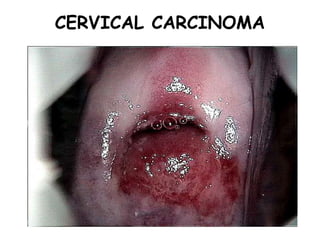
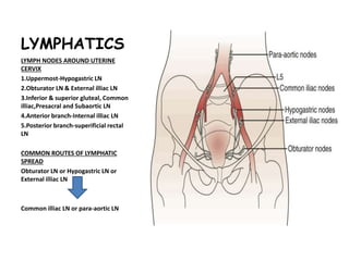
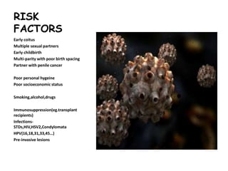
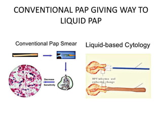
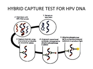
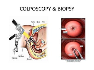
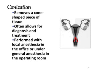
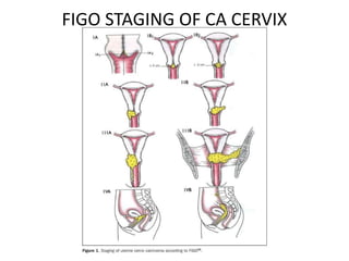
CERVICAL CARCINOMA
ANATOMY
Parts of Uterus
1.Body/Corpus
2.Isthmus
3.cervix-a)supra-vaginal
part b)vaginal part
Ligaments
1.Broad ligament
2.Round ligament
3.Uterosacral ligament
LYMPHATICS
LYMPH NODES AROUND UTERINE
CERVIX
1.Uppermost-Hypogastric LN
2.Obturator LN & External illiac LN
3.Inferior & superior gluteal, Common
illiac,Presacral and Subaortic LN
4.Anterior branch-Internal illiac LN
5.Posterior branch-superificial rectal
LN
COMMON ROUTES OF LYMPHATIC
SPREAD
Obturator LN or Hypogastric LN or
External illiac LN
Common illiac LN or para-aortic LN
ANATOMIC SITES OF FIRST
METASTASIS
HISTOLOGY
• Squamocolumner
junction
• Transformation
zone
RISK
FACTORS
Early coitus
Multiple sexual partners
Early childbirth
Multi-parity with poor birth spacing
Partner with penile cancer
Poor personal hygeine
Poor socioeconomic status
Smoking,alcohol,drugs
Immunosuppression(eg.transplant
recipients)
Infections-
STDs,HIV,HSV2,Condylomata
HPV(16,18,31,33,45…)
Pre-invasive lesions
HPV-an essential criteria for CA Cervix
HPV infection & proliferation
Molecular biology of HPV
infection
HPV vaccination
•GARDASIL-Quadrivalent(VLPs for HPV
6,11,16,18)
•CERVERIX-Bivalent(VLPs for HPV16,18)
Efficacy 100% for seronegative or
seropositive but with negative HPV
DNA
•ACS GUIDELINES
Reccomended for girls @ 11 to 12
years(9-18years)
Cervical Screening programme to
remain unaffected
PRE-INVASIVE LESIONS of CERVIX
COMPARISON B/W VARIOUS CLASSIFICATION
SYSTEMS
Pre-invasive to invasive
% of CIN REACH INVASIVE STAGE IN YEAR
4% 1 Y
11% 3 Y
22% 5 Y
30% 10 Y
screening
ACS NCCN ACOG
Start Within 3 years of 1st sexual
activity, no later than 21
year
Age 21 year
Upto 30
Y
Conventional Pap-annually
Liquid Pap-2yearly
Every 2 years
>30Y Every 2-3 years after 3
consecutive normal pap
smears
Every 2-3 years after 3
consecutive normal pap
smears
Or every 3 years when
cytology + HPV Test
Every 3 years after 3 consecutive
normal pap smears
Stop Age 70 years after 3
consecutive normal Paps &
no abnormal results within
10 years
Age 70 years after 3
consecutive normal Paps & no
abnormal results within 10
years
Age 65-70 years after 3
consecutive normal Paps & no
abnormal results within 10 years
Post
hystere
ctomy
none none None.except H/O CIN2-CIN3
HPV
DNA
>30 years,every 3 years with
cytology
DIAGNOSIS
• Clinical features
 Mostly asymptomatic
 Post coital bleeding
 Post menopausal bleeding
 On Inspection- cervicitis/erosion - bleeds on touch
• investigations
 Pap smear test
 Liquid based cytology
 DNA study
 Colposcopy
 Cone biopsy
CONVENTIONAL PAP GIVING WAY TO
LIQUID PAP
HYBRID CAPTURE TEST FOR HPV DNA
COLPOSCOPY & BIOPSY
29
Conization
•Removes a cone-
shaped piece of
tissue
•Often allows for
diagnosis and
treatment
•Performed with
local anesthesia in
the office or under
general anesthesia in
the operating room
CONE BIOPSY
TREATMENT
LOCAL DESTRUCTION LOCAL EXCISION RADICAL TREATMENT
Ceauterisation
Cryo-surgery
Laser ablation
Conisation
LLETZ
LEEP
NETZ
Trachelectomy
Hysterectomy with
removal of vaginal cuff
Local excision methods
RADICAL SURGICAL TREATMENT
INVASIVE CERVICLE CANCER
PATHOLOGY
•Invasive squamous carcinoma
Small cell
Large cell
Keratinising
Non-keratinising
Rare varients
Papillary
Verrucous
sarcomatoid
Adenocarcinoma
•Adenocarcinoma in situ
•Invasive adenocarcinoma
Mucinous
Minimal deviation adenocarcinoma
Glassy cell adenocarcinoma
Adenoid basal adenocarcinoma
Adenoid cystic adenocarcinoma
Serous
Other types
• Neuroendocrine tumors-Anaplastic small cell tumors
• Rare neoplasms
 Metastasis-from colon , breast
 Sarcomas - Embryonal Rhabdomyosarcoma,
Leiomyosarcoma,Adenosarcoma
 Lymphoma
 melanoma
38
CLINICAL FEATURES
1. BLEEDING --- postmenopausal, metrorrhagia, menorrhagia, post coital bleeding.
2. PAIN in the pelvis or hypogastrium
3. URINARY Symptoms
4. RECTAL Symptoms
5. DISTANT SITE SPECIFIC METASTATIC MANIFESTATIONS
a. LYMPHATIC SPREAD --- to supraclavicular LN, para-aortic lymphadenopathy (non
specific abdominal symptoms)
b. HEMATOGENOUS SPREAD---- to lungs (cough, respiratory distress, in 21% of patients
in metastatic setting)
---- bone pain
INTERNAL EXAMINATION (EXAMINATION UNDER ANAESTHESIA –
ADVISED)
1. INSPECTION ---
Cauliflower like growth --- exophytic nature
Bleeding from the growth
Serosanguineous vaginal discharge
2. PALPATION ----
1) uterus ---- size, shape, position
2) cervix ---- bulky
3) growth might obliterate the vaginal fornices
4) friable growth, ulcerated , which bleeds to touch ---- blood present
on finger tips
5) parametrium – nodular thickening extending upto the lateral pelvic
wall (by per –rectal examination)
39
CLINICAL FEATURES -EXAMINATION - DIAGNOSIS
40
41
STAGING --- FIGO STAGING
1) FIGO staging was based on
anatomical compartmental spread of
cervical cancer.
2) No inclusion of lymph nodal status
3) LVI not included because
pathologists do not agree on status
on presence of LVI
4) MRI, CT and PET Scan – not included
in formal staging.
FIGO STAGING OF CA CERVIX
Staging & survival
IMAGING STUDIES
CT scan
 Detects para-aortic metastasis(Sp-100%)
MRI scan
 Assessment of extracervical tumor extension
 Assessment of local tumor control
 Early prediction of tumor regression
 Can differentiate reccurant tumors from fibrosis
FDG PET scan
 detects para-aortic metastasis(Sn-72% & Sp-92%)
 Detects metabolically active recurrence or residual

Carcinoma cervix

  • 1.
  • 7.
    ANATOMY Parts of Uterus 1.Body/Corpus 2.Isthmus 3.cervix-a)supra-vaginal partb)vaginal part Ligaments 1.Broad ligament 2.Round ligament 3.Uterosacral ligament
  • 8.
    LYMPHATICS LYMPH NODES AROUNDUTERINE CERVIX 1.Uppermost-Hypogastric LN 2.Obturator LN & External illiac LN 3.Inferior & superior gluteal, Common illiac,Presacral and Subaortic LN 4.Anterior branch-Internal illiac LN 5.Posterior branch-superificial rectal LN COMMON ROUTES OF LYMPHATIC SPREAD Obturator LN or Hypogastric LN or External illiac LN Common illiac LN or para-aortic LN
  • 9.
    ANATOMIC SITES OFFIRST METASTASIS
  • 10.
  • 11.
    RISK FACTORS Early coitus Multiple sexualpartners Early childbirth Multi-parity with poor birth spacing Partner with penile cancer Poor personal hygeine Poor socioeconomic status Smoking,alcohol,drugs Immunosuppression(eg.transplant recipients) Infections- STDs,HIV,HSV2,Condylomata HPV(16,18,31,33,45…) Pre-invasive lesions
  • 13.
  • 15.
    HPV infection &proliferation
  • 16.
    Molecular biology ofHPV infection
  • 19.
    HPV vaccination •GARDASIL-Quadrivalent(VLPs forHPV 6,11,16,18) •CERVERIX-Bivalent(VLPs for HPV16,18) Efficacy 100% for seronegative or seropositive but with negative HPV DNA •ACS GUIDELINES Reccomended for girls @ 11 to 12 years(9-18years) Cervical Screening programme to remain unaffected
  • 20.
  • 21.
    COMPARISON B/W VARIOUSCLASSIFICATION SYSTEMS
  • 23.
    Pre-invasive to invasive %of CIN REACH INVASIVE STAGE IN YEAR 4% 1 Y 11% 3 Y 22% 5 Y 30% 10 Y
  • 24.
    screening ACS NCCN ACOG StartWithin 3 years of 1st sexual activity, no later than 21 year Age 21 year Upto 30 Y Conventional Pap-annually Liquid Pap-2yearly Every 2 years >30Y Every 2-3 years after 3 consecutive normal pap smears Every 2-3 years after 3 consecutive normal pap smears Or every 3 years when cytology + HPV Test Every 3 years after 3 consecutive normal pap smears Stop Age 70 years after 3 consecutive normal Paps & no abnormal results within 10 years Age 70 years after 3 consecutive normal Paps & no abnormal results within 10 years Age 65-70 years after 3 consecutive normal Paps & no abnormal results within 10 years Post hystere ctomy none none None.except H/O CIN2-CIN3 HPV DNA >30 years,every 3 years with cytology
  • 25.
    DIAGNOSIS • Clinical features Mostly asymptomatic  Post coital bleeding  Post menopausal bleeding  On Inspection- cervicitis/erosion - bleeds on touch • investigations  Pap smear test  Liquid based cytology  DNA study  Colposcopy  Cone biopsy
  • 26.
    CONVENTIONAL PAP GIVINGWAY TO LIQUID PAP
  • 27.
  • 28.
  • 29.
    29 Conization •Removes a cone- shapedpiece of tissue •Often allows for diagnosis and treatment •Performed with local anesthesia in the office or under general anesthesia in the operating room
  • 30.
  • 31.
    TREATMENT LOCAL DESTRUCTION LOCALEXCISION RADICAL TREATMENT Ceauterisation Cryo-surgery Laser ablation Conisation LLETZ LEEP NETZ Trachelectomy Hysterectomy with removal of vaginal cuff
  • 32.
  • 33.
  • 34.
  • 35.
    PATHOLOGY •Invasive squamous carcinoma Smallcell Large cell Keratinising Non-keratinising Rare varients Papillary Verrucous sarcomatoid
  • 36.
    Adenocarcinoma •Adenocarcinoma in situ •Invasiveadenocarcinoma Mucinous Minimal deviation adenocarcinoma Glassy cell adenocarcinoma Adenoid basal adenocarcinoma Adenoid cystic adenocarcinoma Serous
  • 37.
    Other types • Neuroendocrinetumors-Anaplastic small cell tumors • Rare neoplasms  Metastasis-from colon , breast  Sarcomas - Embryonal Rhabdomyosarcoma, Leiomyosarcoma,Adenosarcoma  Lymphoma  melanoma
  • 38.
    38 CLINICAL FEATURES 1. BLEEDING--- postmenopausal, metrorrhagia, menorrhagia, post coital bleeding. 2. PAIN in the pelvis or hypogastrium 3. URINARY Symptoms 4. RECTAL Symptoms 5. DISTANT SITE SPECIFIC METASTATIC MANIFESTATIONS a. LYMPHATIC SPREAD --- to supraclavicular LN, para-aortic lymphadenopathy (non specific abdominal symptoms) b. HEMATOGENOUS SPREAD---- to lungs (cough, respiratory distress, in 21% of patients in metastatic setting) ---- bone pain
  • 39.
    INTERNAL EXAMINATION (EXAMINATIONUNDER ANAESTHESIA – ADVISED) 1. INSPECTION --- Cauliflower like growth --- exophytic nature Bleeding from the growth Serosanguineous vaginal discharge 2. PALPATION ---- 1) uterus ---- size, shape, position 2) cervix ---- bulky 3) growth might obliterate the vaginal fornices 4) friable growth, ulcerated , which bleeds to touch ---- blood present on finger tips 5) parametrium – nodular thickening extending upto the lateral pelvic wall (by per –rectal examination) 39 CLINICAL FEATURES -EXAMINATION - DIAGNOSIS
  • 40.
  • 41.
    41 STAGING --- FIGOSTAGING 1) FIGO staging was based on anatomical compartmental spread of cervical cancer. 2) No inclusion of lymph nodal status 3) LVI not included because pathologists do not agree on status on presence of LVI 4) MRI, CT and PET Scan – not included in formal staging.
  • 42.
    FIGO STAGING OFCA CERVIX
  • 43.
  • 44.
    IMAGING STUDIES CT scan Detects para-aortic metastasis(Sp-100%) MRI scan  Assessment of extracervical tumor extension  Assessment of local tumor control  Early prediction of tumor regression  Can differentiate reccurant tumors from fibrosis FDG PET scan  detects para-aortic metastasis(Sn-72% & Sp-92%)  Detects metabolically active recurrence or residual