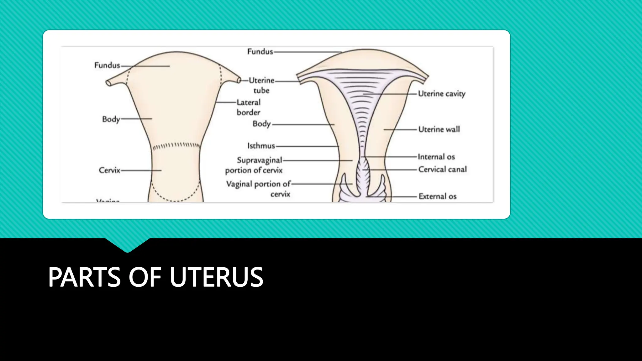
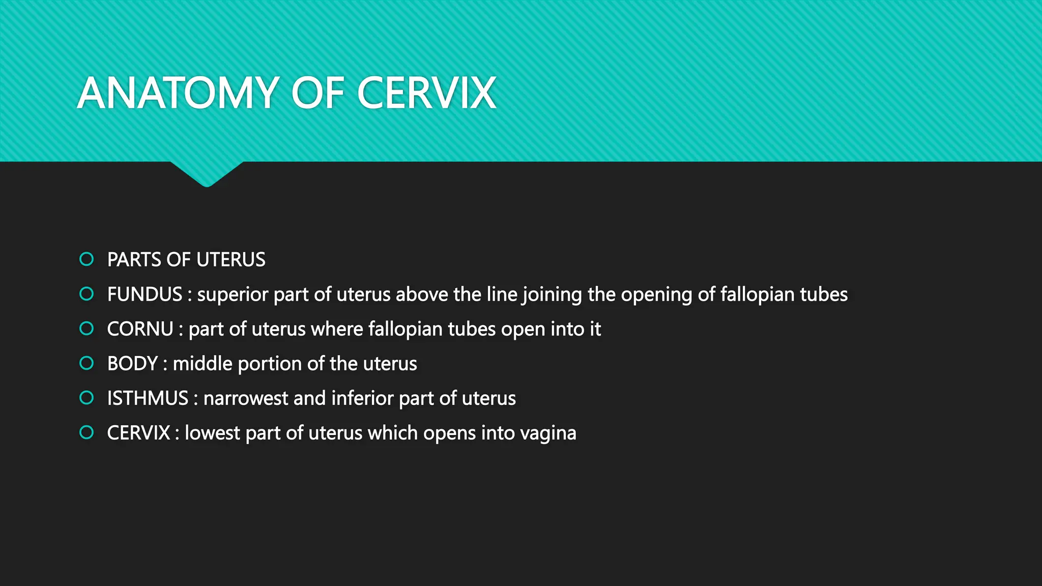
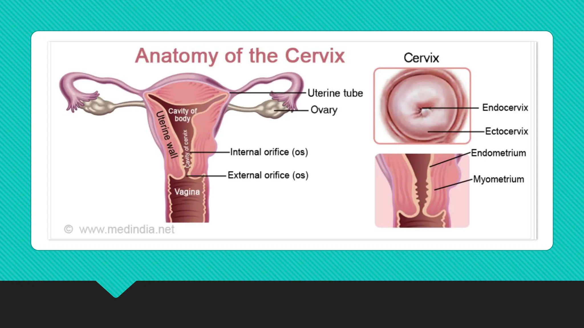
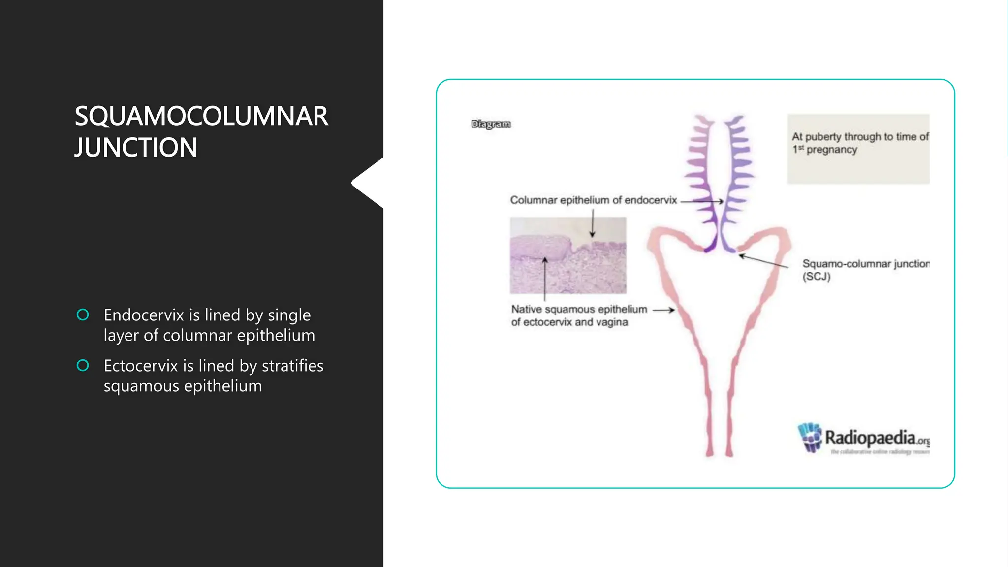
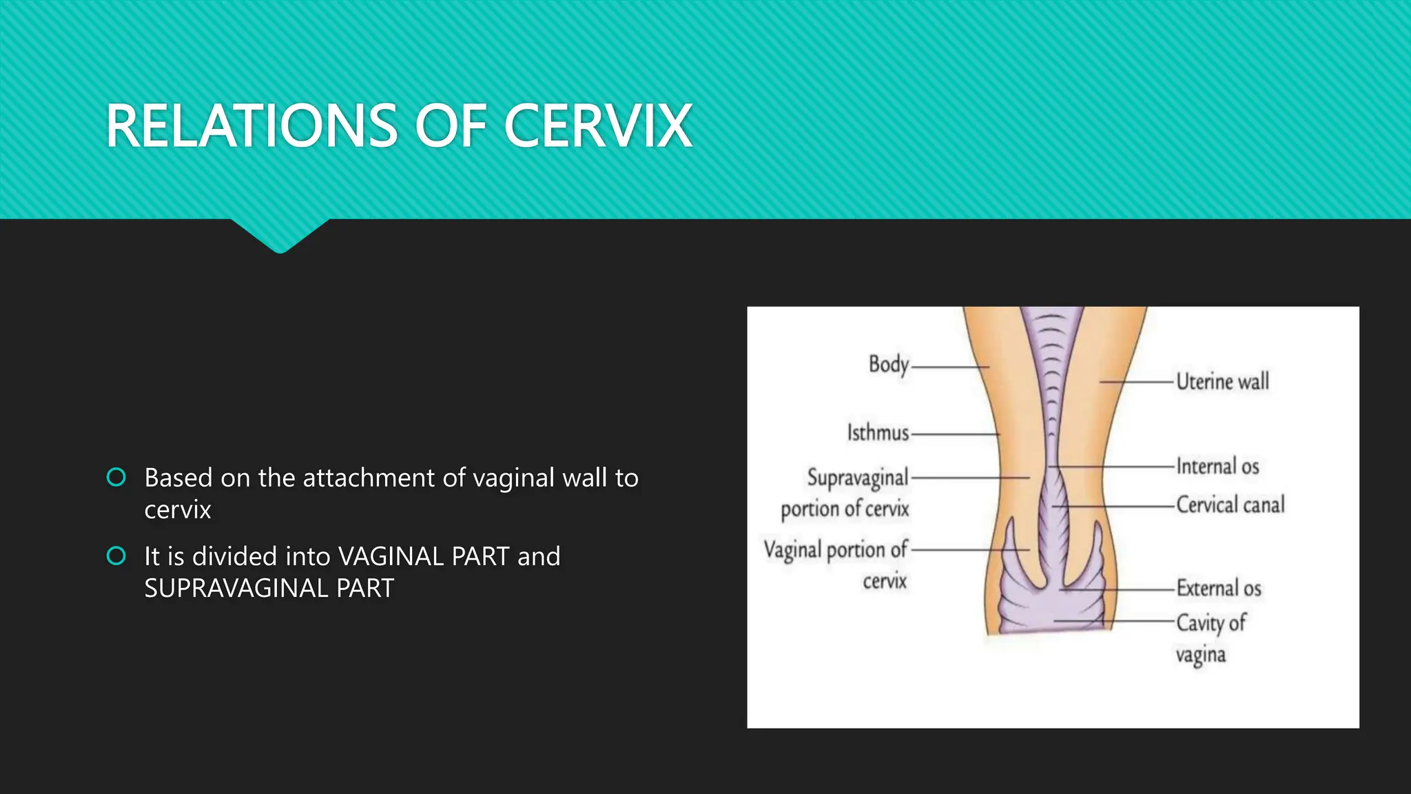
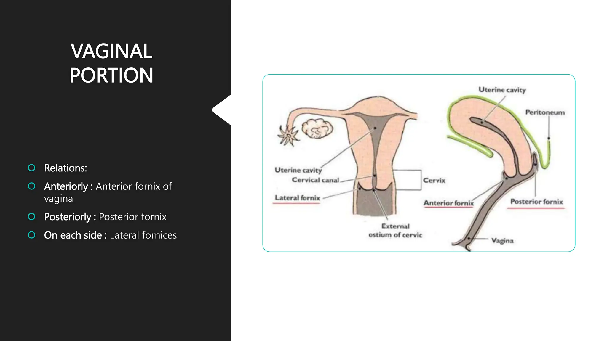
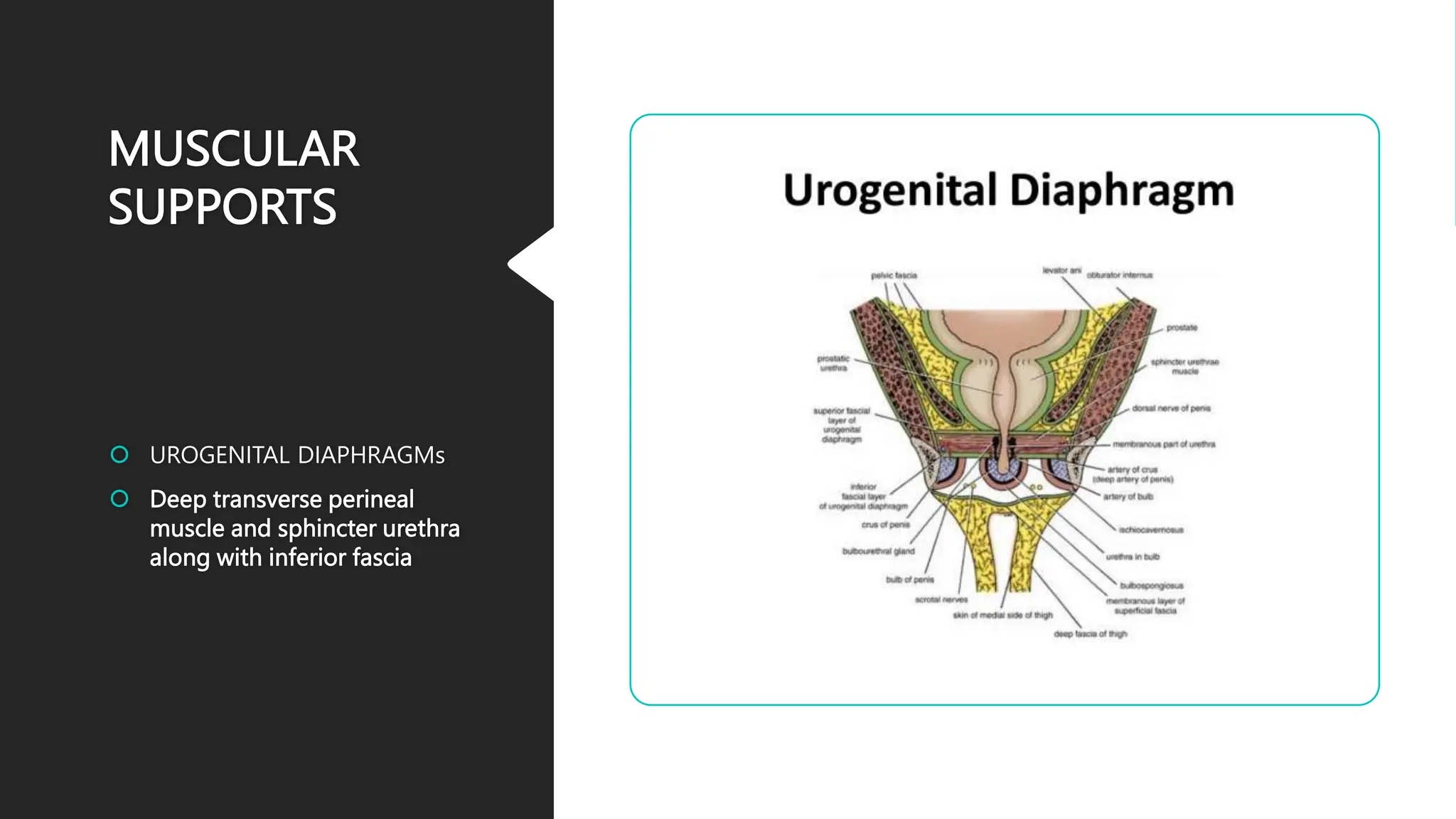
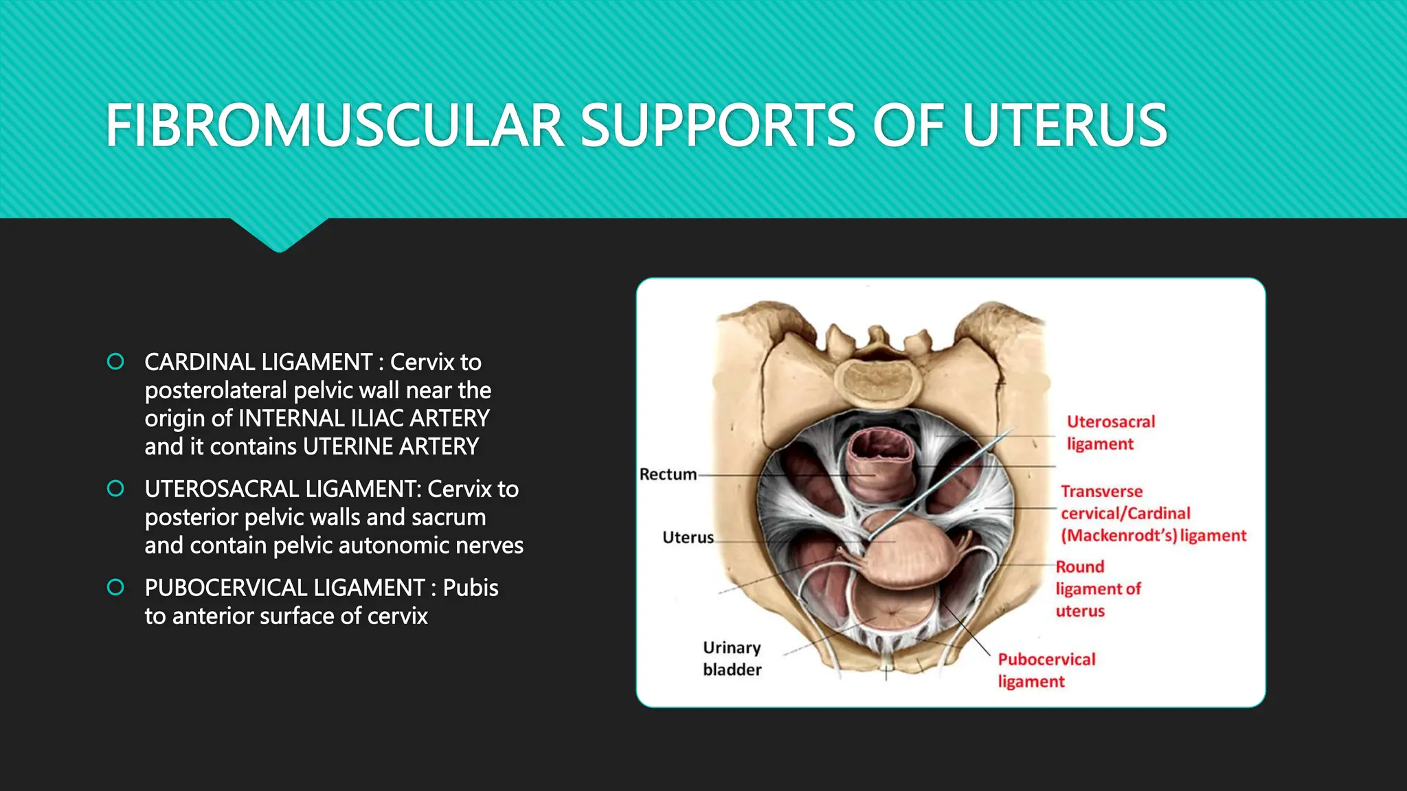
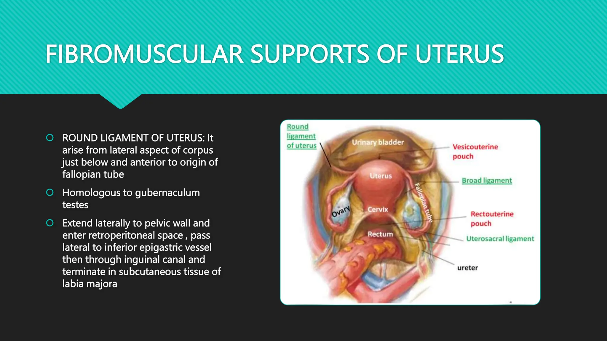
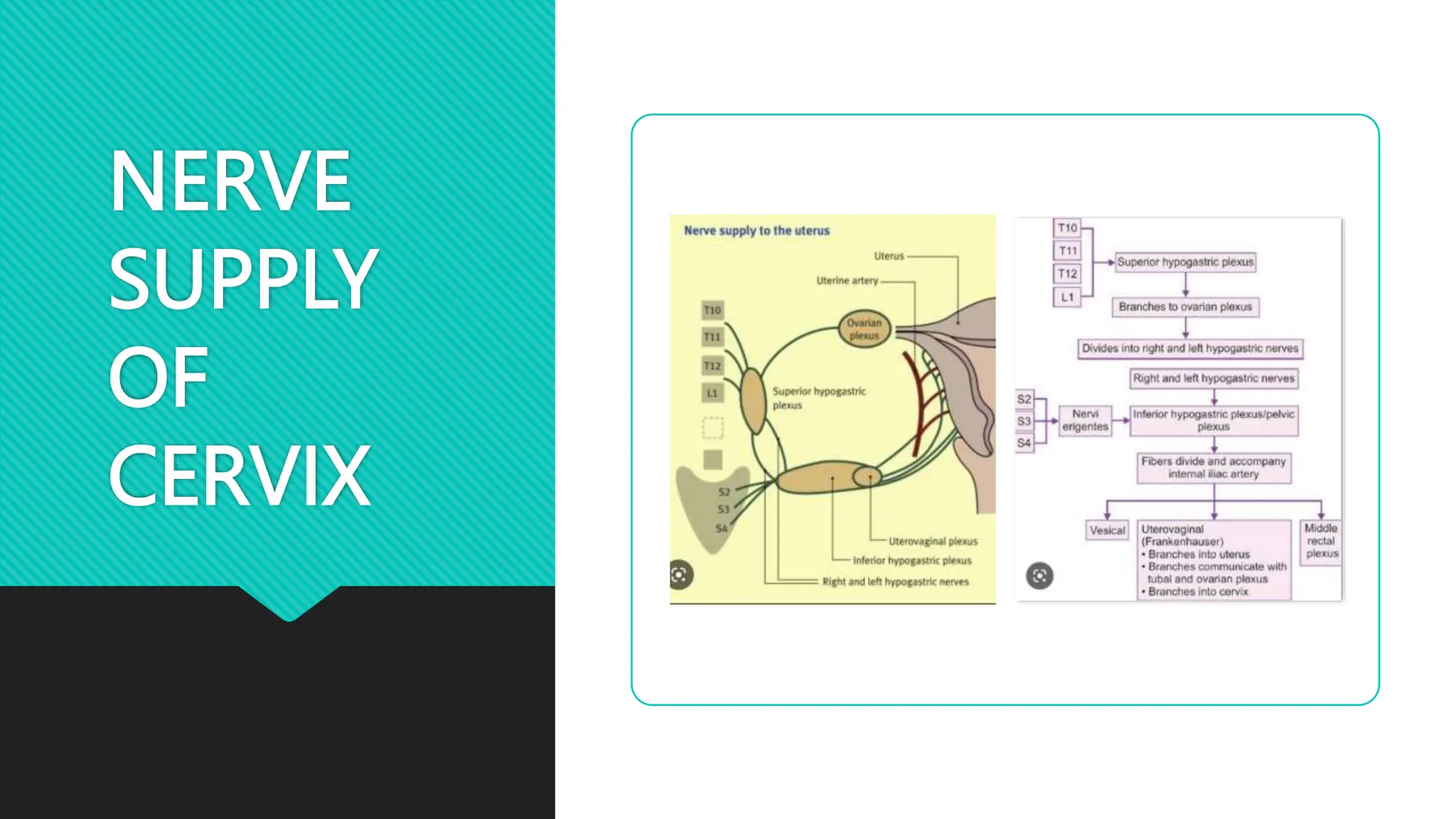
CERVIX
DR.G SAI SUNAYANA
DNB RESIDENT
RADIATION ONCOLOGY
ANATOMY OF CERVIX
cervix is inferior part of uterus which connects to
the vagina.
PARTS OF UTERUS
ANATOMY OF CERVIX
 PARTS OF UTERUS
 FUNDUS : superior part of uterus above the line joining the opening of fallopian tubes
 CORNU : part of uterus where fallopian tubes open into it
 BODY : middle portion of the uterus
 ISTHMUS : narrowest and inferior part of uterus
 CERVIX : lowest part of uterus which opens into vagina
ANATOMY OF CERVIX
 CERVIX : 3 cms in length and 2.5 cms in width
 It is in between uterine corpus and vagina
 The opening of cervix into uterus is INTERNAL OS
 The opening of cervix into vagina is EXTERNAL OS
 The part between the two openings is ENDOCERVICAL CANAL or ENDOCERVIX
 The part outside the external os is ECTOCERVIX
SQUAMOCOLUMNAR
JUNCTION
 Endocervix is lined by single
layer of columnar epithelium
 Ectocervix is lined by stratifies
squamous epithelium
TRANSFORMATION
ZONE CHANGES
RELATIONS OF CERVIX
 Based on the attachment of vaginal wall to
cervix
 It is divided into VAGINAL PART and
SUPRAVAGINAL PART
SUPRAVAGINAL PORTION
VAGINAL
PORTION
 Relations:
 Anteriorly : Anterior fornix of
vagina
 Posteriorly : Posterior fornix
 On each side : Lateral fornices
SUPPORTS OF
UTERUS
MUSCULAR
SUPPORTS
 PELVIC DIAPHRAGM: LEVATOR
ANI (pubococcygeus
puborectalis,iliococcygeus) and
Coccygeus
MUSCULAR
SUPPORTS
 PERINEAL BODY
 Confluence of the following
muscles
 BULBOSPONGIOSUS
 LEVATOR ANI
 EXTERNAL ANAL SPHINCTER
 SPHINCTER URETHRA
 SUPERFICIAL AND DEEP
TRANSVERSE PERINEAL
MUSCLES
MUSCULAR
SUPPORTS
 UROGENITAL DIAPHRAGMs
 Deep transverse perineal
muscle and sphincter urethra
along with inferior fascia
FIBROMUSCULAR SUPPORTS OF UTERUS
 CARDINAL LIGAMENT : Cervix to
posterolateral pelvic wall near the
origin of INTERNAL ILIAC ARTERY
and it contains UTERINE ARTERY
 UTEROSACRAL LIGAMENT: Cervix to
posterior pelvic walls and sacrum
and contain pelvic autonomic nerves
 PUBOCERVICAL LIGAMENT : Pubis
to anterior surface of cervix
FIBROMUSCULAR SUPPORTS OF UTERUS
 ROUND LIGAMENT OF UTERUS: It
arise from lateral aspect of corpus
just below and anterior to origin of
fallopian tube
 Homologous to gubernaculum
testes
 Extend laterally to pelvic wall and
enter retroperitoneal space , pass
lateral to inferior epigastric vessel
then through inguinal canal and
terminate in subcutaneous tissue of
labia majora
SECONDARY
SUPPORTS
 BROAD LIGAMENT:
 Double layers of peritoneum
extend from lateral wall of uterus
to pelvic walls
 Divided as mesosalphinx ,
mesoovarium mesometrium
infundibulopelvic ligament
 Cardinal and uterosacral
ligaments lie in lower portion of
broad ligament
 CONTENTS OF BROAD LIGAMENT:
 Fallopian tube
 Round ligament of uterus
 Ovarian ligament
 Uterovaginal plexus and ovarian plexus
 Lymphatics
 Fibroareolar tissue or parametrium
BLOOD SUPPLY OF
CERVIX
 Descending branch of uterine
artery
 ascending branch of vaginal
artery
NERVE
SUPPLY
OF
CERVIX
LYMPHATIC
DRAINAGE OF
CERVIX
 Drain principally into
PARACERVICAL and
PARAMETRIAL nodes
 Then into OBTURATOR ,
INTERNAL ILIAC,EXTERNAL ILIAC
and COMMON ILIAC NODES
 Finally into PARA AORTIC LYMPH
NODES
THANK YOU

Anatomy of cervix

  • 1.
    CERVIX DR.G SAI SUNAYANA DNBRESIDENT RADIATION ONCOLOGY
  • 2.
    ANATOMY OF CERVIX cervixis inferior part of uterus which connects to the vagina.
  • 3.
  • 4.
    ANATOMY OF CERVIX PARTS OF UTERUS  FUNDUS : superior part of uterus above the line joining the opening of fallopian tubes  CORNU : part of uterus where fallopian tubes open into it  BODY : middle portion of the uterus  ISTHMUS : narrowest and inferior part of uterus  CERVIX : lowest part of uterus which opens into vagina
  • 5.
    ANATOMY OF CERVIX CERVIX : 3 cms in length and 2.5 cms in width  It is in between uterine corpus and vagina  The opening of cervix into uterus is INTERNAL OS  The opening of cervix into vagina is EXTERNAL OS  The part between the two openings is ENDOCERVICAL CANAL or ENDOCERVIX  The part outside the external os is ECTOCERVIX
  • 7.
    SQUAMOCOLUMNAR JUNCTION  Endocervix islined by single layer of columnar epithelium  Ectocervix is lined by stratifies squamous epithelium
  • 8.
  • 9.
    RELATIONS OF CERVIX Based on the attachment of vaginal wall to cervix  It is divided into VAGINAL PART and SUPRAVAGINAL PART
  • 10.
  • 11.
    VAGINAL PORTION  Relations:  Anteriorly: Anterior fornix of vagina  Posteriorly : Posterior fornix  On each side : Lateral fornices
  • 12.
  • 13.
    MUSCULAR SUPPORTS  PELVIC DIAPHRAGM:LEVATOR ANI (pubococcygeus puborectalis,iliococcygeus) and Coccygeus
  • 14.
    MUSCULAR SUPPORTS  PERINEAL BODY Confluence of the following muscles  BULBOSPONGIOSUS  LEVATOR ANI  EXTERNAL ANAL SPHINCTER  SPHINCTER URETHRA  SUPERFICIAL AND DEEP TRANSVERSE PERINEAL MUSCLES
  • 15.
    MUSCULAR SUPPORTS  UROGENITAL DIAPHRAGMs Deep transverse perineal muscle and sphincter urethra along with inferior fascia
  • 16.
    FIBROMUSCULAR SUPPORTS OFUTERUS  CARDINAL LIGAMENT : Cervix to posterolateral pelvic wall near the origin of INTERNAL ILIAC ARTERY and it contains UTERINE ARTERY  UTEROSACRAL LIGAMENT: Cervix to posterior pelvic walls and sacrum and contain pelvic autonomic nerves  PUBOCERVICAL LIGAMENT : Pubis to anterior surface of cervix
  • 17.
    FIBROMUSCULAR SUPPORTS OFUTERUS  ROUND LIGAMENT OF UTERUS: It arise from lateral aspect of corpus just below and anterior to origin of fallopian tube  Homologous to gubernaculum testes  Extend laterally to pelvic wall and enter retroperitoneal space , pass lateral to inferior epigastric vessel then through inguinal canal and terminate in subcutaneous tissue of labia majora
  • 18.
    SECONDARY SUPPORTS  BROAD LIGAMENT: Double layers of peritoneum extend from lateral wall of uterus to pelvic walls  Divided as mesosalphinx , mesoovarium mesometrium infundibulopelvic ligament  Cardinal and uterosacral ligaments lie in lower portion of broad ligament
  • 19.
     CONTENTS OFBROAD LIGAMENT:  Fallopian tube  Round ligament of uterus  Ovarian ligament  Uterovaginal plexus and ovarian plexus  Lymphatics  Fibroareolar tissue or parametrium
  • 20.
    BLOOD SUPPLY OF CERVIX Descending branch of uterine artery  ascending branch of vaginal artery
  • 21.
  • 22.
    LYMPHATIC DRAINAGE OF CERVIX  Drainprincipally into PARACERVICAL and PARAMETRIAL nodes  Then into OBTURATOR , INTERNAL ILIAC,EXTERNAL ILIAC and COMMON ILIAC NODES  Finally into PARA AORTIC LYMPH NODES
  • 23.