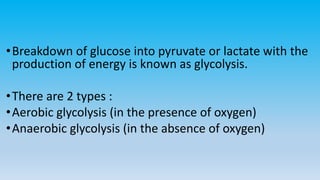
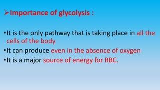
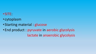
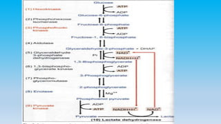
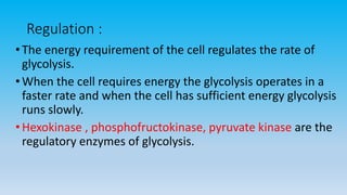
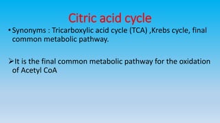
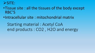
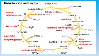
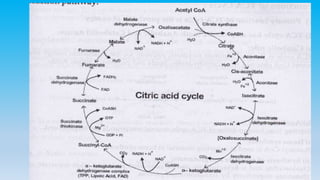
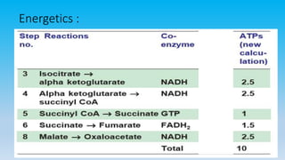
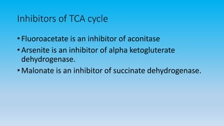
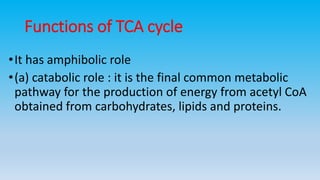
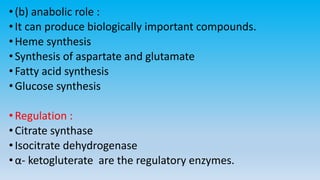
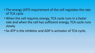
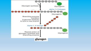
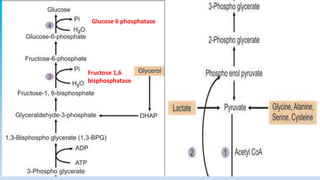
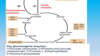
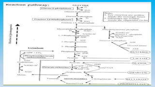
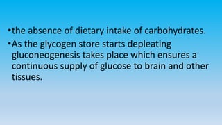
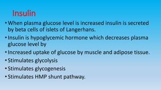
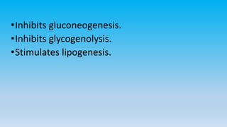
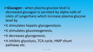
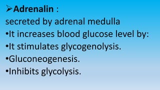
Carbohydrate
metabolism
Adarsh benny
Overview of metabolism
• The term metabolism can be defined as the entire biochemical
reactions that are taking place in the body.
• The compounds that take part in the reactions are called
metabolites.
• Metabolism consists of 2 categories :
(i) Anabolism : the chemical reaction pathways leading to the
synthesis of a compound is termed as anabolism. It requires
energy.
(ii) Catabolism: the chemical reaction pathway leading to the
degradation of compound is called catabolism. It releases
energy.
Glycolysis (Embden - Meyerhoff
pathway)
•Breakdown of glucose into pyruvate or lactate with the
production of energy is known as glycolysis.
•There are 2 types :
•Aerobic glycolysis (in the presence of oxygen)
•Anaerobic glycolysis (in the absence of oxygen)
Importance of glycolysis :
•It is the only pathway that is taking place in all the
cells of the body
•It can produce even in the absence of oxygen
•It is a major source of energy for RBC.
•SITE:
•cytoplasm
•Starting material : glucose
•End product : pyruvate in aerobic glycolysis
lactate in anaerobic glycolysis
• ENERGETICS:
• Net ATP produced = total no. of ATP’S produced – total no. of ATP’S
utilized
• Aerobic glycolysis : 2+2+5=9 ATP’S
• Total 9 ATP’S
• 2 ATP’S are used during the initial reaction so net ATP produced is
(9-2= 7 ATP’S)
• Anaerobic glycolysis:
• Total no. of ATP’S produced = 4
• Total no. of ATP’S utilised during the reaction = 2
• (4-2=2 ATP’S)
Regulation :
•The energy requirement of the cell regulates the rate of
glycolysis.
•When the cell requires energy the glycolysis operates in a
faster rate and when the cell has sufficient energy glycolysis
runs slowly.
•Hexokinase , phosphofructokinase, pyruvate kinase are the
regulatory enzymes of glycolysis.
Inhibitors of glycolysis
• Arsenate inhibit the enzyme glyceraldehyde-3-phosphate
dehydrogenase
• Fluoride inhibits the enzyme enolase. It is commonly used to
prevent glycolysis during the blood glucose estimation in the
laboratory.
Rapaport leubering cycle (2,3 BPG)
Rapaport Leubering Cycle
•This takes place in the RBC’S .
•2,3 BPG is a regulator of oxygen transport in
erythrocytes.
•In the presence of 2,3 BPG, oxy Hb will unload O2
more easily in tissues.
Citric acid cycle
•Synonyms : Tricarboxylic acid cycle (TCA) ,Krebs cycle, final
common metabolic pathway.
It is the final common metabolic pathway for the oxidation
of Acetyl CoA
Starting material : Acetyl CoA
end products : CO2 , H2O and energy
SITE:
•Tissue site : all the tissues of the body except
RBC’S
•Intracellular site : mitochondrial matrix
Citrate synthase
aconitase
Isocitrate dehydrogenase
Alpha
ketoglutarate
dehydrogenase
succinate thiokinase
succinate
dehydrogenase
fumarase
malate
dehydrogenase
Energetics :
• In the entire reactions 3 molecules of NADH + H is
formed so it wil produce  3x2.5= 7.5 ATP’S
• 1 FADH2 wil produce  1.5 ATP’S
• 1 ATP
• So total 10 ATP’S are formed from 1 molecule of
Acetyl CoA.
Inhibitors of TCA cycle
• Fluoroacetate is an inhibitor of aconitase
• Arsenite is an inhibitor of alpha ketogluterate
dehydrogenase.
• Malonate is an inhibitor of succinate dehydrogenase.
Functions of TCA cycle
•It has amphibolic role
•(a) catabolic role : it is the final common metabolic
pathway for the production of energy from acetyl CoA
obtained from carbohydrates, lipids and proteins.
•(b) anabolic role :
•It can produce biologically important compounds.
•Heme synthesis
•Synthesis of aspartate and glutamate
•Fatty acid synthesis
•Glucose synthesis
•Regulation :
•Citrate synthase
•Isocitrate dehydrogenase
•α- ketogluterate are the regulatory enzymes.
•The energy (ATP) requirement of the cell regulates the rate
of TCA cycle
•When the cell requires energy, TCA cycle runs in a faster
rate and when the cell has sufficient energy, TCA cycle runs
slowly.
•So ATP is the inhibitor and ADP is activator of TCA cycle.
Anaplerotic reactions
•In its anabolic role several intermediates of TCA
cycle are used up for biosynthesis of many
compounds.
•These intermediates should be replenished
•The reactions which replenish these
intermediates are called anaplerotic reactions or
anaplerosis.
Substrate level phosphorylation
•Production of ATP where the energy is trapped
directly from the substrate without the involvment
of electron transport chain.
Glycogen metabolism
•Glycogen is the major storage form of glucose mainly in the
liver and muscle
•It is a homopolysaccharide made up of many glucose
residues joined together by α (1,4) glycosidic bonds
•and at the branching points by α (1,6) glycosidic bonds.
Functions of glycogen :
•The major function of liver glycogen is to provide glucose
during fasting.
•When blood glucose level lowers liver glycogen is broken
down and helps to maintain blood glucose level.
•GLYCOGENESIS (GLYCOGEN SYNTHESIS):
•Formation of glycogen from glucose is called glycogenesis
•SITE :
•Cytosol of liver and skeletal muscle
•Starting compound : glucose
•End product : glycogen
1. Activation of Glucose
•UDP glucose is formed from glucose-1-phosphate and UTP
(uridine triphosphate) by the enzyme UDPglucose
pyrophosphorylase
Glucose-1-phosphate +UTP ———————→ UDP-glucose
UDP-glucosepyrophosphorylase
2) initiation of glycogen synthesis
a special protein glycogenin (glycogen primer) acts as
an initiator Glycogenin takes up glucose residue from
UDP glucose
It then extends the glucose chain by accepting seven
more glucose residues from UDP glucose
3. Branching Enzyme
• The glycogen synthase can add glucose units only in
alpha-1,4 linkage. A branching enzyme is needed to
create the alpha-1,6 linkages
• When the chain is lengthened to 11 - 12 glucose
residues, the branching enzyme will transfer 6 to 8
glucose residues from this chain and forms this alpha-
1,6 linkage
glycogen
Glycogenolysis (glycogen breakdown)
•It is the breakdown of glycogen to form glucose,
•SITE:
•Cytosol of liver and skeletal muscle
.
•It is broken down by the combined action of 3
enzymes:
•(i) glycogen phosphorylase : it acts on glycogen and
breaks the terminal α (1,4) glycosidic bonds of
glycogen and releases the terminal glucose molecule
•(ii) transferase : it transfers 3 glucose residues from
one chain to another chain and thus exposing the
(1,6) glycosidic bonds.
•(iii) debranching enzyme : it splits alpha (1,6)
bond at the branch point and releases free
glucose .
Gluconeogenesis
•Synthesis of new glucose molecules from non carbohydrate
sources is termed as gluconeogenesis or neoglucogenesis.
•SITES :
•(a) tissue site : liver (90%) and renal cortex(10%)
•(b) intracellular site : partly in cytosol and partly in
mitochondria.
•Starting compounds
•(a) lactate.
•(b) glycerol : from fats
•(c) glucogenic amino acids like glutamate, aspartate and
alanine
•(d) propionyl CoA : from β- oxidation
•Reaction pathway : it involves
•Reactions of TCA cycle.
•Reversible reactions of glycolysis.
Glucose 6 phosphatase
Fructose 1,6
bisphosphatase
Phosphoenolpyruvate
carboxykinase
Pyruvate
carboxylase
4 key enzymes; on irreversible steps
of glycolysis
• Pyruvate carboxylase
• Phosphoenolpyruvate carboxykinase
• Fructose 1,6 bisphosphatase.
• Glucose 6 phosphatase
Significance of gluconeogenesis :
•Gluconeogenesis meets the needs of the body for glucose
and maintains blood glucose homeostasis when
carbohydrate is not available in sufficient amounts from the
diet.
•Some tissues such as brain, erythrocytes require continuous
supply of glucose as a source of energy.
•the absence of dietary intake of carbohydrates.
•As the glycogen store starts depleating
gluconeogenesis takes place which ensures a
continuous supply of glucose to brain and other
tissues.
Assignment
Cori cycle
Glycosuria
•It refers to excretion of sugars (mainly glucose) in
urine.
•Glucose is reabsorbed almost completely in renal
tubules and hence urine contains almost no glucose.
•When blood glucose level exceeds 180mg/dl, glucose
will be excreted in urine.
•This level is called the renal threshold for glucose.
Types
• Hyperglycemic glycosuria: when blood glucose level
exceeds the renal threshold glucose is excreted in urine.
• Renal glycosuria : glucose is excreted in urine even though
the blood glucose level is within normal limits.
•Alimentary glycosuria : it is following a rich carbohydrate
meal
• Glycosuria of pregnancy.
Hormonal regulation of glucose
• Normal fasting plasma glucose level is 70-110mg/dl
•Levels above this range are called hyperglycemia.
• Below are called hypoglycaemia.
• Blood glucose level is under the influence of various
hormones.
• These hormones are of 2 types:
(i) Hypoglycemic hormone : insulin
(ii) Hyperglycemic hormones : glucagon,
adrenalin,glucocorticoids, GH, thyroid hormone.
Insulin
•When plasma glucose level is increased insulin is secreted
by beta cells of islets of Langerhans.
•Insulin is hypoglycemic hormone which decreases plasma
glucose level by
•Increased uptake of glucose by muscle and adipose tissue.
•Stimulates glycolysis
•Stimulates glycogenesis
•Stimulates HMP shunt pathway.
•Inhibits gluconeogenesis.
•Inhibits glycogenolysis.
•Stimulates lipogenesis.
Glucagon : when plasma glucose level is
decreased glucagon is secreted by alpha-cells of
islets of Langerhans which increase plasma glucose
level by
•It stimulates hepatic glycogenolysis.
•It stimulates gluconeogenesis.
•It decreases glycogenesis.
•It inhibits glycolysis, TCA cycle, HMP shunt
pathway etc.
CLAUDIA JOHNNY
Adrenalin :
secreted by adrenal medulla
•It increases blood glucose level by:
•It stimulates glycogenolysis.
•Gluconeogenesis.
•Inhibits glycolysis.
Cortisol :secreted by adrenal cortex
•It increases blood glucose level by
•It stimulates gluconeogenesis.
•It stimulates glycogenolysis
•It decreases glycolysis.
•It inhibits insulin secretion.
CLAUDIA JOHNNY
Growth hormone : it is secreted by anterior pituitary
•It increases the blood glucose level by :
•It decreases the uptake of glucose by muscle tissues.
•It decreases glycolysis.
Thyroid hormone : it is secreted by thyroid glands
•Thyroid hormones increases the blood glucose level by
•It increases the absorption of glucose from intestine
•It stimulates glycogenolysis.
Renal control mechanism
•When blood glucose rises to relatively high levels the
kidney regulates the absorption.
•Glucose is continuously filtered by the glomeruli but is
normally reabsorbed completely in renal tubules.
•The capacity of the tubular system to reabsorb glucose is
limited.
•If the blood glucose level is raised above 180mg/dl
complete reabsorption of glucose does not occur and extra
amount appear in the urine causing glycosuria.
•180mg/dl is the limiting level of glucose in the
blood above which tubular reabsorption does
not occur which is known as renal threshold
value for glucose.
Glycogen Storage Disease
•GLYCOGENOSIS (GLYCOGEN STORAGE DISEASE)
TYPE NAME ENZYME DEFECT
I Von-Gierkes disease Glucose-6-phosphatase in
liver
II Pompes disease Lysosomal acid maltase
III Coris disease Debranching enzyme
IV Andersons disease Branching enzyme
V McArdles disease Muscle glycogen
phosphorylase
VI Hers disease Liver glycogen
phosphorylase
• Glycogen Storage Disease
• Type- 1
• It is also called Von Gierke's Disease.
• Most common type of glycogen storage disease is type I.
• Glucose-6-phosphatase is deficient.
• Glucose-6-phosphate is accumulated, so it is channeled to
HMP shunt pathway, producing more ribose and more
nucleotides
• Purines are then catabolized to uric acid, leading to
hyperuricemia
• Glycogen gets deposited in liver. Massive liver enlargement
may lead to cirrhosis
•Children usually die in early childhood.
•Treatment is to give small quantity of food at
frequent intervals.
• ketosis is also seen
Diabetes mellitus
•It is a metabolic disease caused either due to insulin
deficiency or failure in insulin action.
•CLASSIFICATION :
It is classified into 2 types
(a) Type I diabetes mellitus (insulin dependent diabetes
mellitus IDDM )
(b) Type II diabetes mellitus (Non-insulin dependent
diabetes mellitus,NIDDM)
Type I diabetes mellitus (insulin dependent
diabetes mellitus IDDM )
• It is due to decreased insulin production.
• Circulating insulin level is very low.
• These patients are dependent on insulin injections.
Onset is usually below 30 years of age, most
commonly during adolescence.
• They are more prone to develop ketosis.
•It may be due to
•Autoimmune disorder; Circulating antibodies against
insulin is seen
•Type II diabetes mellitus (Non-insulin dependent
diabetes mellitus,NIDDM) :
•95% of the patients belong to this type.
•The disease is due to the decreased biological response to
insulin, otherwise called insulin resistance
•Type 2 disease is commonly seen in individuals above 40
years.
•These patients are less prone to develop ketosis
•About 60% patients are obese
Metabolic changes in Diabetes
• in Carbohydrate Metabolism
• Net effect is an inhibition of glycolysis and stimulation of
gluconeogenesis leading to hyperglycemia.
• in Lipid Metabolism
• Fatty acid breakdown leads to high FFA levels
• The excess of acetyl CoA therefore, is diverted to ketone bodies,
leading to ketogenesis
• in Protein Metabolism
• Increased breakdown of proteins and amino acids
Clinical Presentations in Diabetes Mellitus
• When the blood glucose level exceeds the renal threshold glucose is
excreted in urine (glucosuria)
• Polyuria
• more water is taken (polydypsia)
• patient will take more food (polyphagia).
Acute Metabolic Complications
• Diabetic Keto Acidosis
• ketonemia, excretion in urine (ketonuria) and smell of acetone in
breath. All these three together constitute the condition known as
ketosis
Chronic Complications of Diabetes Mellitus
• cataract of lens
• Pregnancy: Diabetic mothers tend to have big babie, Chances of
abortion, premature birth
• Neuropathy
• Atherosclerosis
Diagnosis of DM
Blood glucose estimation :
• Normal fasting plasma glucose level is 70-110mg/dl.
• Post prandial plasma glucose : < 140mg/dl (after 2hrs of food
intake)
• Random plasma glucose : 70-150mg/dl
Urine testing:
• Urine is tested for the presence of glucose by benedict's test
• Urinary glucose is poor marker of diabetes mellitus because the
renal threshold for glucose is 180mg/dl blood glucose level
should exceed this value.
Glucose tolerance test (GTT) /OGTT (oral GTT)
: it is the test to measure the capacity of the body to utilize
an additional load of glucose entering into the body.
•It is performed in diagnosis of doubtful cases of diabetes
mellitus.
•PROCEDURE :
•In the morning a sample of blood and urine is collected in
the fasting condition (8-12hrs) this is denoted as the ‘0’ hour
sample.
•Glucose load dose : the dose is 75gm anhydrous glucose in
250-300ml of water.
Glycated haemoglobin (HbA1)
•Glucose is attached to Hb
•the rate of addition is directly proportional to the blood
glucose level.
•Normal level : 4 to 7%
•Diabetics : 8 to 15%
•Significance :
•Glycated Hb measurement is an index of long term control
of blood glucose once attached cannot be removed from hb
•Once formed remains inside the RBC throughout the life
span of RBCs (120days)
•Therefore it reflects the blood glucose level over a period of
2-3 months.
Management of diabetes mellitus
•Diet , exercise , drugs and insulin are the options for
management of diabetes mellitus.
•Insulin injections : it is used in type I diabetes, also used in
type II where oral drugs are not sufficient.
•Diet and exercise : it can control about 50% of type II
diabetes.
•A diabetic patient is advised to take a diet low in
carbohydrates and fat and rich in protein and fibers.

carbohydrate metabolism -bpt.pptx

  • 1.
  • 2.
    Overview of metabolism •The term metabolism can be defined as the entire biochemical reactions that are taking place in the body. • The compounds that take part in the reactions are called metabolites. • Metabolism consists of 2 categories : (i) Anabolism : the chemical reaction pathways leading to the synthesis of a compound is termed as anabolism. It requires energy. (ii) Catabolism: the chemical reaction pathway leading to the degradation of compound is called catabolism. It releases energy.
  • 3.
    Glycolysis (Embden -Meyerhoff pathway)
  • 4.
    •Breakdown of glucoseinto pyruvate or lactate with the production of energy is known as glycolysis. •There are 2 types : •Aerobic glycolysis (in the presence of oxygen) •Anaerobic glycolysis (in the absence of oxygen)
  • 5.
    Importance of glycolysis: •It is the only pathway that is taking place in all the cells of the body •It can produce even in the absence of oxygen •It is a major source of energy for RBC.
  • 6.
    •SITE: •cytoplasm •Starting material :glucose •End product : pyruvate in aerobic glycolysis lactate in anaerobic glycolysis
  • 8.
    • ENERGETICS: • NetATP produced = total no. of ATP’S produced – total no. of ATP’S utilized • Aerobic glycolysis : 2+2+5=9 ATP’S • Total 9 ATP’S • 2 ATP’S are used during the initial reaction so net ATP produced is (9-2= 7 ATP’S) • Anaerobic glycolysis: • Total no. of ATP’S produced = 4 • Total no. of ATP’S utilised during the reaction = 2 • (4-2=2 ATP’S)
  • 10.
    Regulation : •The energyrequirement of the cell regulates the rate of glycolysis. •When the cell requires energy the glycolysis operates in a faster rate and when the cell has sufficient energy glycolysis runs slowly. •Hexokinase , phosphofructokinase, pyruvate kinase are the regulatory enzymes of glycolysis.
  • 11.
    Inhibitors of glycolysis •Arsenate inhibit the enzyme glyceraldehyde-3-phosphate dehydrogenase • Fluoride inhibits the enzyme enolase. It is commonly used to prevent glycolysis during the blood glucose estimation in the laboratory.
  • 12.
    Rapaport leubering cycle(2,3 BPG) Rapaport Leubering Cycle
  • 13.
    •This takes placein the RBC’S . •2,3 BPG is a regulator of oxygen transport in erythrocytes. •In the presence of 2,3 BPG, oxy Hb will unload O2 more easily in tissues.
  • 14.
    Citric acid cycle •Synonyms: Tricarboxylic acid cycle (TCA) ,Krebs cycle, final common metabolic pathway. It is the final common metabolic pathway for the oxidation of Acetyl CoA
  • 15.
    Starting material :Acetyl CoA end products : CO2 , H2O and energy SITE: •Tissue site : all the tissues of the body except RBC’S •Intracellular site : mitochondrial matrix
  • 16.
    Citrate synthase aconitase Isocitrate dehydrogenase Alpha ketoglutarate dehydrogenase succinatethiokinase succinate dehydrogenase fumarase malate dehydrogenase
  • 18.
    Energetics : • Inthe entire reactions 3 molecules of NADH + H is formed so it wil produce  3x2.5= 7.5 ATP’S • 1 FADH2 wil produce  1.5 ATP’S • 1 ATP • So total 10 ATP’S are formed from 1 molecule of Acetyl CoA.
  • 19.
    Inhibitors of TCAcycle • Fluoroacetate is an inhibitor of aconitase • Arsenite is an inhibitor of alpha ketogluterate dehydrogenase. • Malonate is an inhibitor of succinate dehydrogenase.
  • 20.
    Functions of TCAcycle •It has amphibolic role •(a) catabolic role : it is the final common metabolic pathway for the production of energy from acetyl CoA obtained from carbohydrates, lipids and proteins.
  • 21.
    •(b) anabolic role: •It can produce biologically important compounds. •Heme synthesis •Synthesis of aspartate and glutamate •Fatty acid synthesis •Glucose synthesis •Regulation : •Citrate synthase •Isocitrate dehydrogenase •α- ketogluterate are the regulatory enzymes.
  • 22.
    •The energy (ATP)requirement of the cell regulates the rate of TCA cycle •When the cell requires energy, TCA cycle runs in a faster rate and when the cell has sufficient energy, TCA cycle runs slowly. •So ATP is the inhibitor and ADP is activator of TCA cycle.
  • 23.
    Anaplerotic reactions •In itsanabolic role several intermediates of TCA cycle are used up for biosynthesis of many compounds. •These intermediates should be replenished •The reactions which replenish these intermediates are called anaplerotic reactions or anaplerosis.
  • 25.
    Substrate level phosphorylation •Productionof ATP where the energy is trapped directly from the substrate without the involvment of electron transport chain.
  • 27.
    Glycogen metabolism •Glycogen isthe major storage form of glucose mainly in the liver and muscle •It is a homopolysaccharide made up of many glucose residues joined together by α (1,4) glycosidic bonds •and at the branching points by α (1,6) glycosidic bonds.
  • 28.
    Functions of glycogen: •The major function of liver glycogen is to provide glucose during fasting. •When blood glucose level lowers liver glycogen is broken down and helps to maintain blood glucose level.
  • 29.
    •GLYCOGENESIS (GLYCOGEN SYNTHESIS): •Formationof glycogen from glucose is called glycogenesis •SITE : •Cytosol of liver and skeletal muscle •Starting compound : glucose •End product : glycogen
  • 30.
    1. Activation ofGlucose •UDP glucose is formed from glucose-1-phosphate and UTP (uridine triphosphate) by the enzyme UDPglucose pyrophosphorylase Glucose-1-phosphate +UTP ———————→ UDP-glucose UDP-glucosepyrophosphorylase
  • 31.
    2) initiation ofglycogen synthesis a special protein glycogenin (glycogen primer) acts as an initiator Glycogenin takes up glucose residue from UDP glucose It then extends the glucose chain by accepting seven more glucose residues from UDP glucose
  • 32.
    3. Branching Enzyme •The glycogen synthase can add glucose units only in alpha-1,4 linkage. A branching enzyme is needed to create the alpha-1,6 linkages • When the chain is lengthened to 11 - 12 glucose residues, the branching enzyme will transfer 6 to 8 glucose residues from this chain and forms this alpha- 1,6 linkage
  • 33.
  • 34.
    Glycogenolysis (glycogen breakdown) •Itis the breakdown of glycogen to form glucose, •SITE: •Cytosol of liver and skeletal muscle .
  • 36.
    •It is brokendown by the combined action of 3 enzymes: •(i) glycogen phosphorylase : it acts on glycogen and breaks the terminal α (1,4) glycosidic bonds of glycogen and releases the terminal glucose molecule •(ii) transferase : it transfers 3 glucose residues from one chain to another chain and thus exposing the (1,6) glycosidic bonds.
  • 37.
    •(iii) debranching enzyme: it splits alpha (1,6) bond at the branch point and releases free glucose .
  • 38.
    Gluconeogenesis •Synthesis of newglucose molecules from non carbohydrate sources is termed as gluconeogenesis or neoglucogenesis. •SITES : •(a) tissue site : liver (90%) and renal cortex(10%) •(b) intracellular site : partly in cytosol and partly in mitochondria. •Starting compounds •(a) lactate.
  • 39.
    •(b) glycerol :from fats •(c) glucogenic amino acids like glutamate, aspartate and alanine •(d) propionyl CoA : from β- oxidation •Reaction pathway : it involves •Reactions of TCA cycle. •Reversible reactions of glycolysis.
  • 40.
  • 41.
  • 43.
    4 key enzymes;on irreversible steps of glycolysis • Pyruvate carboxylase • Phosphoenolpyruvate carboxykinase • Fructose 1,6 bisphosphatase. • Glucose 6 phosphatase
  • 44.
    Significance of gluconeogenesis: •Gluconeogenesis meets the needs of the body for glucose and maintains blood glucose homeostasis when carbohydrate is not available in sufficient amounts from the diet. •Some tissues such as brain, erythrocytes require continuous supply of glucose as a source of energy.
  • 45.
    •the absence ofdietary intake of carbohydrates. •As the glycogen store starts depleating gluconeogenesis takes place which ensures a continuous supply of glucose to brain and other tissues.
  • 46.
  • 47.
    Glycosuria •It refers toexcretion of sugars (mainly glucose) in urine. •Glucose is reabsorbed almost completely in renal tubules and hence urine contains almost no glucose. •When blood glucose level exceeds 180mg/dl, glucose will be excreted in urine. •This level is called the renal threshold for glucose.
  • 48.
    Types • Hyperglycemic glycosuria:when blood glucose level exceeds the renal threshold glucose is excreted in urine. • Renal glycosuria : glucose is excreted in urine even though the blood glucose level is within normal limits. •Alimentary glycosuria : it is following a rich carbohydrate meal • Glycosuria of pregnancy.
  • 49.
    Hormonal regulation ofglucose • Normal fasting plasma glucose level is 70-110mg/dl •Levels above this range are called hyperglycemia. • Below are called hypoglycaemia. • Blood glucose level is under the influence of various hormones. • These hormones are of 2 types: (i) Hypoglycemic hormone : insulin (ii) Hyperglycemic hormones : glucagon, adrenalin,glucocorticoids, GH, thyroid hormone.
  • 50.
    Insulin •When plasma glucoselevel is increased insulin is secreted by beta cells of islets of Langerhans. •Insulin is hypoglycemic hormone which decreases plasma glucose level by •Increased uptake of glucose by muscle and adipose tissue. •Stimulates glycolysis •Stimulates glycogenesis •Stimulates HMP shunt pathway.
  • 51.
  • 52.
    Glucagon : whenplasma glucose level is decreased glucagon is secreted by alpha-cells of islets of Langerhans which increase plasma glucose level by •It stimulates hepatic glycogenolysis. •It stimulates gluconeogenesis. •It decreases glycogenesis. •It inhibits glycolysis, TCA cycle, HMP shunt pathway etc. CLAUDIA JOHNNY
  • 53.
    Adrenalin : secreted byadrenal medulla •It increases blood glucose level by: •It stimulates glycogenolysis. •Gluconeogenesis. •Inhibits glycolysis.
  • 54.
    Cortisol :secreted byadrenal cortex •It increases blood glucose level by •It stimulates gluconeogenesis. •It stimulates glycogenolysis •It decreases glycolysis. •It inhibits insulin secretion. CLAUDIA JOHNNY
  • 55.
    Growth hormone :it is secreted by anterior pituitary •It increases the blood glucose level by : •It decreases the uptake of glucose by muscle tissues. •It decreases glycolysis. Thyroid hormone : it is secreted by thyroid glands •Thyroid hormones increases the blood glucose level by •It increases the absorption of glucose from intestine •It stimulates glycogenolysis.
  • 56.
    Renal control mechanism •Whenblood glucose rises to relatively high levels the kidney regulates the absorption. •Glucose is continuously filtered by the glomeruli but is normally reabsorbed completely in renal tubules. •The capacity of the tubular system to reabsorb glucose is limited. •If the blood glucose level is raised above 180mg/dl complete reabsorption of glucose does not occur and extra amount appear in the urine causing glycosuria.
  • 57.
    •180mg/dl is thelimiting level of glucose in the blood above which tubular reabsorption does not occur which is known as renal threshold value for glucose.
  • 58.
  • 59.
    •GLYCOGENOSIS (GLYCOGEN STORAGEDISEASE) TYPE NAME ENZYME DEFECT I Von-Gierkes disease Glucose-6-phosphatase in liver II Pompes disease Lysosomal acid maltase III Coris disease Debranching enzyme IV Andersons disease Branching enzyme V McArdles disease Muscle glycogen phosphorylase VI Hers disease Liver glycogen phosphorylase
  • 60.
    • Glycogen StorageDisease • Type- 1 • It is also called Von Gierke's Disease. • Most common type of glycogen storage disease is type I. • Glucose-6-phosphatase is deficient. • Glucose-6-phosphate is accumulated, so it is channeled to HMP shunt pathway, producing more ribose and more nucleotides • Purines are then catabolized to uric acid, leading to hyperuricemia • Glycogen gets deposited in liver. Massive liver enlargement may lead to cirrhosis
  • 61.
    •Children usually diein early childhood. •Treatment is to give small quantity of food at frequent intervals. • ketosis is also seen
  • 62.
    Diabetes mellitus •It isa metabolic disease caused either due to insulin deficiency or failure in insulin action. •CLASSIFICATION : It is classified into 2 types (a) Type I diabetes mellitus (insulin dependent diabetes mellitus IDDM ) (b) Type II diabetes mellitus (Non-insulin dependent diabetes mellitus,NIDDM)
  • 63.
    Type I diabetesmellitus (insulin dependent diabetes mellitus IDDM ) • It is due to decreased insulin production. • Circulating insulin level is very low. • These patients are dependent on insulin injections. Onset is usually below 30 years of age, most commonly during adolescence. • They are more prone to develop ketosis.
  • 64.
    •It may bedue to •Autoimmune disorder; Circulating antibodies against insulin is seen
  • 65.
    •Type II diabetesmellitus (Non-insulin dependent diabetes mellitus,NIDDM) : •95% of the patients belong to this type. •The disease is due to the decreased biological response to insulin, otherwise called insulin resistance •Type 2 disease is commonly seen in individuals above 40 years. •These patients are less prone to develop ketosis •About 60% patients are obese
  • 66.
    Metabolic changes inDiabetes • in Carbohydrate Metabolism • Net effect is an inhibition of glycolysis and stimulation of gluconeogenesis leading to hyperglycemia. • in Lipid Metabolism • Fatty acid breakdown leads to high FFA levels • The excess of acetyl CoA therefore, is diverted to ketone bodies, leading to ketogenesis
  • 67.
    • in ProteinMetabolism • Increased breakdown of proteins and amino acids
  • 68.
    Clinical Presentations inDiabetes Mellitus • When the blood glucose level exceeds the renal threshold glucose is excreted in urine (glucosuria) • Polyuria • more water is taken (polydypsia) • patient will take more food (polyphagia).
  • 69.
    Acute Metabolic Complications •Diabetic Keto Acidosis • ketonemia, excretion in urine (ketonuria) and smell of acetone in breath. All these three together constitute the condition known as ketosis
  • 70.
    Chronic Complications ofDiabetes Mellitus • cataract of lens • Pregnancy: Diabetic mothers tend to have big babie, Chances of abortion, premature birth • Neuropathy • Atherosclerosis
  • 71.
    Diagnosis of DM Bloodglucose estimation : • Normal fasting plasma glucose level is 70-110mg/dl. • Post prandial plasma glucose : < 140mg/dl (after 2hrs of food intake) • Random plasma glucose : 70-150mg/dl Urine testing: • Urine is tested for the presence of glucose by benedict's test • Urinary glucose is poor marker of diabetes mellitus because the renal threshold for glucose is 180mg/dl blood glucose level should exceed this value.
  • 72.
    Glucose tolerance test(GTT) /OGTT (oral GTT) : it is the test to measure the capacity of the body to utilize an additional load of glucose entering into the body. •It is performed in diagnosis of doubtful cases of diabetes mellitus. •PROCEDURE : •In the morning a sample of blood and urine is collected in the fasting condition (8-12hrs) this is denoted as the ‘0’ hour sample. •Glucose load dose : the dose is 75gm anhydrous glucose in 250-300ml of water.
  • 73.
    Glycated haemoglobin (HbA1) •Glucoseis attached to Hb •the rate of addition is directly proportional to the blood glucose level. •Normal level : 4 to 7% •Diabetics : 8 to 15%
  • 74.
    •Significance : •Glycated Hbmeasurement is an index of long term control of blood glucose once attached cannot be removed from hb •Once formed remains inside the RBC throughout the life span of RBCs (120days) •Therefore it reflects the blood glucose level over a period of 2-3 months.
  • 75.
    Management of diabetesmellitus •Diet , exercise , drugs and insulin are the options for management of diabetes mellitus. •Insulin injections : it is used in type I diabetes, also used in type II where oral drugs are not sufficient. •Diet and exercise : it can control about 50% of type II diabetes. •A diabetic patient is advised to take a diet low in carbohydrates and fat and rich in protein and fibers.