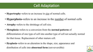
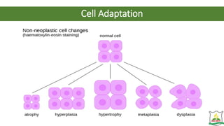
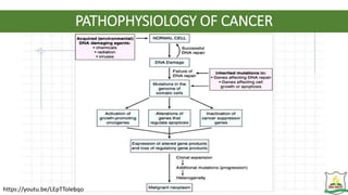
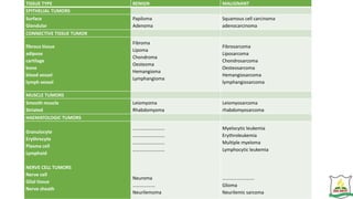
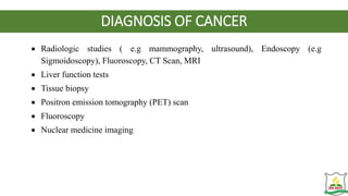
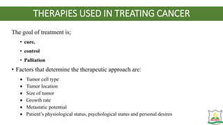
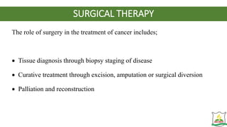
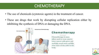
SURGERY AND SURGICAL NURSING
TOPIC:
ONCOLOGY
BY
SAMUEL TWUMASI (Mr.)
S.D.A NURSING AND MIDWIFERY TRAINING COLLEGE
KWADASO-BAREKESE, KUMASI
2023
LEARNING OBJECTIVES
By the end of the lesson, the student will be able to:
• Define Oncology
• Identify the characteristics of a normal cell
• Describe cell adaptation
• Define Cancer & Describe the pathophysiology of cancer
• Differentiate between benign and malignant tumours
• Describe carcinogenesis and Identify agents or factors considered as
carcinogenic
LEARNING OBJECTIVES CONT’D
• Describe the classification of cancer
• Identify the diagnosis and risk factors of cancer
• Identify the signs and symptoms as well as the warning signs of cancer
• Identify the treatment modalities for cancer
• Describe the nurses’ role in the various treatment modalities.
OVERVIEW
• Oncology (from the Ancient Greek ‘onkos’- meaning bulk, mass, or tumor, and the
suffix-logy- meaning "study of") is a branch of medicine that deals with tumors
(cancer).
• A medical professional who practices oncology is an Oncologist.
• Oncology is concerned with:
• The diagnosis of any cancer in a person
• Therapy (e.g., surgery, chemotherapy, radiotherapy and other modalities)
• Follow-up of cancer patients after successful treatment
• Palliative care of patients with terminal malignancies
• Ethical questions surrounding cancer care
Characteristics of a Normal Cell
• Show specific morphology (shape)
• Have limited cell division
• Grow in orderly and well regulated manner
• Undergo Apoptosis (programmed cell death)
• Adhere tightly together
• Non migratory
• Are euploid (equal number of required chromosomes)
• Perform specific differentiated functions
Cell Differentiation
Cell Adaptation
• Hypertrophy–refers to an increase in size of normal cells.
• Hyperplasia–refers to an increase in the number of normal cells
• Atrophy–refers to the shrinkage of cell size.
• Metaplasia–refers to a conversion from the normal patterns of
differentiation of one type of cell into another type of cell not actually normal
for that tissue. Replacement of other mature cell.
• Dysplasia–refers to an alteration in the shape, size, appearance and
distribution of cells into abnormal form (not-reversible)
Cell Adaptation
Definition of Cancer
• Neoplasia–refers to an abnormal cell growth or tumor- a mass of new
tissue functioning independently and serving no useful purpose
• Cancer is a group of diseases characterized by uncontrolled and
unregulated cellular growth.
• It is basically a disorder of gene expression beginning with the
transformation of a single normally functioning cell into an abnormal or
cancer cell.
• It appears that most cancers result from the interaction of genetic and
internal factors with environmental factors or carcinogens.
• Tumors, abnormal growth of tissue, are clusters of cells that are
capable of growing and dividing uncontrollably; their growth is not
regulated.
• The term "cancer" is used when a tumor is malignant, which
is to say it has the potential to cause harm, including death.
INCIDENCE
• Cancer is major health problem that affect all ethnic groups.
• Though considered a disease of the aged (people over 65 years), cancer
affect people of all ages.
• Overall incidence is higher in men than in women.
• More men die of cancer than women.
CARCINOGENS
• There are some factors which are responsible for change of normal cell into
cancer cell.
• Those factors or agents are known as carcinogens.
• It is believed that all cells carry certain cancer producing oncogenes.
• Oncogenes are the genes that are responsible for induction of tumors.
of tumors.
• Under certain conditions these genes are triggered to multiply rapidly into
malignant neoplasm.
Environmental factors:
• Tobacco,
• smokes, Heavy smoking cause lung, oral cavity and oesophagus cancer.
• Certain diets,
• environmental pollutants,
• Excessive intake of alcohol cause liver cancer.
Chemical carcinogen:
• styrene and bisphenol-A (in polyethene)
• Nickel compounds, (in coins, wires, ceramics, batteries)
• Cadmium, (in jewellery)
• Arsenic, nitrosamines, (in textiles, papers, pharmaceutical
purposes)
• Trichloroethylene, (in paint or polish removers, correction
fluids)
• arylamines, (cosmetics, pesticides, etc)
• benzopyrene, (found in crude oil, coal tar, tobacco, and many
foods, especially grilled meats).
• aflatoxins, (produced by certain fungi in crops like maize)
• reactive oxygen radicals etc
• Physical carcinogen:
• UV rays (ultraviolet),
• ionizing radiation (x-rays and gamma rays)
• Endogenous factors:
• Mutations,
• change in DNA replication,
• Immune system defects,
• Ageing
Biological carcinogen:
• Virus:
• Virus has also been associated with various types of cancers. These viruses are called
oncoviruses .
• Rous sarcoma virus (RSV) is the first discovered retro-virus causing cancer.
causing cancer.
• (Oncovirus); Human papilloma virus (HPV), Epstein-BarrVirus, (EBV), Hepatitis B
virus, Herpes virus
• Hepatitis B and C virus is casually related with hepato-cellular carcinoma.
• Cytomegalovirus (CMV) is associated with kaposi’s sarcoma.
• Human papilloma virus (HPV) is a chief suspect of cervix cancer.
• Bacteria; Helicobacter pylori,
PATHOPHYSIOLOGY OF CANCER
https://youtu.be/LEpTTolebqo
Pathophysiology of Cancer Cont’d.
• First step: Mutation and tumor initiation
• Genetic alteration leads to mutation in a single cell which results into
abnormal proliferation of that cell known as tumor cell.
• Second step: Cell proliferation and Tumor progression
• Tumor progression continues as additional mutations occur within cells of
• The mutated cells have some selective advantage over normal cell as such
and division. The descendants of a cell bearing such additional mutation
become dominant within the tumor population
• Third step: Clonal selection and malignancy
• Cell proliferation of tumor then leads to new clone of tumor cells with
properties (such as survival, invasion, or metastasis) that confer a selective
called clonal selection.
• Clonal selection continues throughout tumor development, so tumors
rapid-growing and increasingly malignant.
Pathophysiology of Cancer Cont’d.
• Fourth step: Metastasis
• Metastasis is a complex process in which cancer cells break
tumor and circulate through the bloodstream or lymphatic
body.
• At new sites, the cells continue to multiply and eventually form
comprised of cells that reflect the tissue of origin.
• The ability of tumors, such as pancreatic cancer and uveal (iris,
of eye) cancers, to metastasize contributes greatly to their
• Many fundamental questions remain about the clonal structures
phylogenetic relationships among metastases, the scale of
in metastatic and primary sites, how the tumor disseminates,
tumor micro-environment plays in the determination of the
WARNING SIGNS OF CANCER
• The acronym CAUTION UP is used
COMMON MANIFESTATIONS OF CANCER
 Pain: one of the most serious concerns of clients with cancer. It may be acute or chronic
 Hemorrhage: due to tumor angiogenesis and erosion through blood vessels
 Bone marrow suppression: a common effect of cancer and its treatment, leading to anaemia
,leukopenia and thrombocytopenia.
 Infection: this is a common manifestation due to impaired immune defenses and direct effect
of the tumor cells.
 Anorexia-cachexia syndrome: tumor cells secrete substances that alter taste and smell and
produce early satiety.
 Unexplained rapid weight loss: cancer cells support their growth process by breaking down
(catabolizing) body tissues and muscle proteins.
 Disruption of function: this may result from obstruction or pressure e.g urine retention from
prostatic tumors obstructing the urethra)
TYPES OF CANCER
• Abnormal proliferation of any of the different kinds of cells in the body can result in
Cancer.
• So there are more than a hundred different types of cancer varying on their behavior,
pathophysiology, site of origin and response to treatment or therapy.
• A tumor can be either benign or malignant.
• Benign tumor: A tumor that remains confined to its original location, neither
invading surrounding normal tissue nor spreading to distant body sites is known as
benign tumor. For examples; Skin wart
• Malignant tumor: A tumor which is capable of both invading surrounding normal
tissue and spreading (metastasis) throughout the body via the circulatory or lymphatic
systems is known as
• malignant tumor.
• Only malignant tumors are properly referred to as CANCER.
CHARACTERISTICS BENIGN TUMOR MALIGNANT TUMOR
Cell characteristics Cells are well differentiated and resemble
normal cells of the tissue from which the tumor
originate
Cells are undifferentiated and often bear little or no
resemblance to the normal cells of the tissue of
origin
Mode of growth Grows by expansion and does not infiltrate the
surrounding tissue; usually encapsulated
Grows at the periphery and sends out processes that
infiltrate and destroy other surrounding tissue( grows
by infiltration)
Rate of growth Usually slow Rate of growth is usually variable and depends on the
level of differentiation; the more anaplastic the
tumor, the faster its growth
Metastasis Does not spread by metastasis Gain access to the blood and lymph channels and
metastasize to other areas of the body
General effects Does not cause generalized effect unless its
location interferes with vital functions
Often cause generalized effects such as anemia,
weakness and weight loss
Tissue destruction Does not usually cause tissue damage unless its
location interferes with blood flow
Often causes extensive tissue damage as the tumor
outgrows its blood supply or encroaches on blood
flow to the area
Ability to cause death Does not usually cause death unless its location Usually cause death unless growth is controlled.
CLASSIFICATION OF CANCER
Cancer can be classified in the following manner:
• Classification by Grade
• Classification by Stage
• Classification by anatomic site
CLASSIFICATION OF CANCER
CLASSIFICATION BY GRADE
• Cells that are well differentiated closely resemble normal
specialized cells and belong to low grade tumors.
• Cells that are undifferentiated are highly abnormal with respect
to surrounding tissues. These are high grade tumors.
Grade 1 – well differentiated cells with slight abnormality.
Grade 2 – cells are moderately differentiated and slightly more
abnormal
Grade 3 – cells are poorly differentiated and very abnormal
Grade 4 – cells are immature and primitive and undifferentiated
CLASSIFICATION OF CANCER CONT’D
CLASSIFICATION BY STAGE
• Classifying the extent of spread of disease is termed staging.
• Clinical staging : this classification system determines the anatomic extent of
the malignant disease process by stages.
Stage 0 : cancer being in situ.
Stage I : limited to the tissue of origin.
Stage II : limited local spread.
Stage III : extensive local and regional spread.
Stage IV is advanced cancer with distant spread and metastasis.
CLASSIFICATION OF CANCER CONT’D
CLASSIFICATION BY STAGE
• There are several types of staging methods.
• The most commonly used method uses classification in terms of
• tumor size (T),
• the degree of regional spread or node involvement (N), and
• distant metastasis (M).
• This is called the TNM staging.
CLASSIFICATION OF CANCER CONT’D
CLASSIFICATION BY STAGE (TNM STAGING)
Primary Tumor (T)
T0 -No sign of primary tumor
Tx -Tumor cannot be found or assessed
Tis -Carcinoma in situ
T1, T2, T3, T4 -Increasing size and/or extension of primary tumor
Regional Lymph Nodes (N)
N0 -No evidence of tumor cells in regional lymph nodes
Nx -regional lymph nodes cannot be assessed
N1, N2, N3, -Increasing involvement of regional lymph nodes
Distant Metastasis (M)
M0 -No distant metastasis
Mx -Distant metastasis cannot be assessed
M1 -Distant metastasis
CLASSIFICATION OF CANCER CONT’D.
CLASSIFICATION BY ANATOMIC SITE
• Pathologically, cancers (malignant) are classified into three
Sarcomas, Leukemia:
• 1. Carcinomas:
• This type of cancer arises from epithelial cells or tissues lining
the various organs.
• 2. Sarcomas:
• These cancers arise from connective and muscular tissue
• 3. Lymphomas or Leukemia:
• It is the malignant growth of leucocytes (WBC).
• Persons affected with this cancer show the excessive production
cancer) and cancer of bone marrow.
TISSUE TYPE BENIGN MALIGNANT
EPITHELIAL TUMORS
Surface
Glandular
Papiloma
Adenoma
Squamous cell carcinoma
adenocarcinoma
CONNECTIVE TISSUE TUMOR
fibrous tissue
adipose
cartilage
bone
blood vessel
lymph vessel
Fibroma
Lipoma
Chondroma
Oesteoma
Hemangioma
Lymphangioma
Fibrosarcoma
Liposarcoma
Chondrosarcoma
Oesteosarcoma
Hemangiosarcoma
lymphangiosarcoma
MUSCLE TUMORS
Smooth muscle
Striated
Leiomyoma
Rhabdomyoma
Leiomyosarcoma
rhabdomyosarcoma
HAEMATOLOGIC TUMORS
Granulocyte
Erythrocyte
Plasma cell
Lymphoid
NERVE CELL TUMORS
Nerve cell
Glial tissue
Nerve sheath
………………………
………………………
………………………
………………………
Neuroma
……………….
Neurilemoma
Myelocytic leukemia
Erythroleukemia
Multiple myeloma
Lymphocytic leukemia
………………………
Glioma
Neurilemic sarcoma
DIAGNOSIS OF CANCER
• The diagnostic plan for the person in whom cancer is suspected include:
I. Health history
II.Physical examination
III.Identification of risk factors: include controllable/modifiable risk
factors as well as uncontrollable risk factors
IV.Specific diagnostic studies include:
 Cytology study (e.g Papanicolaou (Pap) test), Chest X-rays, Complete blood
count, chemistry profile, Tumor marker identification
DIAGNOSIS OF CANCER
 Radiologic studies ( e.g mammography, ultrasound), Endoscopy (e.g
Sigmoidoscopy), Fluoroscopy, CT Scan, MRI
 Liver function tests
 Tissue biopsy
 Positron emission tomography (PET) scan
 Fluoroscopy
 Nuclear medicine imaging
THERAPIES USED IN TREATING CANCER
The goal of treatment is;
• cure,
• control
• Palliation
• Factors that determine the therapeutic approach are:
 Tumor cell type
 Tumor location
 Size of tumor
 Growth rate
 Metastatic potential
 Patient’s physiological status, psychological status and personal desires
SURGICAL THERAPY
The role of surgery in the treatment of cancer includes;
 Tissue diagnosis through biopsy staging of disease
 Curative treatment through excision, amputation or surgical diversion
 Palliation and reconstruction
SURGICAL THERAPY
The role of surgery in the treatment of cancer includes;
 Tissue diagnosis through biopsy staging of disease
 Curative treatment through excision, amputation or surgical diversion
 Palliation and reconstruction
SURGICAL THERAPY CONT’D.
• TYPES OF SURGICAL PROCEDURES
• Diagnostic Surgery
• Needle biopsies use a needle to aspirate fluid or tissues and are easily
done under local anaesthesia.
• Establishing a tissue diagnosis through biopsy can be done by
incisional biopsy where a small portion of tissue is removed and
examined or by excision biopsy, whereby the whole tumor is removed.
• Endoscopy allows visualization of potentially cancerous lesions and
biopsy without the need for invasive surgery.
• Curative surgery
• This is the definitive surgery for many cancers especially benign
cancers.
• Curative surgery encompasses both removal of the tumor mass and
a safe margin of healthy tissue around it.
• Cancer in situ can be treated with CRYOSURGERY (the use of
extreme cold to destroy cancer cells or tumors), Electrosurgery,
Chemosurgery, Laser surgery.
• Adjuvant surgery
• Used to remove residual mass in radio or chemo-sensitive tumors like
osteosarcomas and testicular teratomas.
• Prophylactic surgery
• This is usually done to prevent the development of cancer in people
who have high risk of developing cancer. E.g removal of the colon in a
patient with familial polyposis.
• Ablative surgery
 May be done to remove hormonal influence on tumor growth through procedures
such as oophorectomy, colectomy, mastectomy, orchidectomy etc.
• Palliative and supportive surgery
 Performed to improve the quality of life of the patient rather than as curative procedure.
E.g creation of a colostomy to allow healing of rectal abscess, suprapubic cystostomy
for the patient with advanced prostatic cancer.
• Reconstructive surgery
 This is performed following curative or radical surgery in an attempt to improve
function or obtain a more desirable cosmetic effect.
CHEMOTHERAPY
• The use of chemicals (cytotoxic agents) in the treatment of cancer.
• These are drugs that work by disrupting cellular replication either by
inhibiting the synthesis of DNA or damaging the DNA.
SAFETY PRECAUTIONS IN ADMINISTERING CHEMOTHERAPIES
 Use a biological safety cabinet for the preparation of all chemotherapeutic
agents
 Wear surgical gloves when handling these drugs and excretions of patients
who are receiving chemotherapy
 Wear disposable long sleeved gown when preparing and administering
chemotherapy
 Use tight fittings on all IV tubings used to deliver chemotherapy
 Dispose off all equipment used in chemotherapy preparation appropriately
 Dispose off all chemotherapy waste as hazardous material
RADIATION THERAPY
• Radiation therapy is the use of ionizing radiation to interrupt cellular growth.
• It is also used to control malignant disease when a tumour cannot be removed
surgically or when local nodal metastasis is present or it can be used
prophylactically to prevent leukemic infiltration to the brain or spinal cord.
• Radiation therapy may be used to cure the cancer, as in Hodgkin disease,
testicular seminomas, thyroid carcinomas, localized cancers of the head and
neck.
• There are two types of ionizing radiation:
• electromagnetic rays (x-rays and gamma rays) and
• particles (electrons [beta particles], protons, neutrons and alpha particles).
Precautionary Measures for the Nurse in Radiation Safety
Nurses caring for the patients should be given instructions about safe
times and distances in patient’s care.
 Minimize the amount of time near a radioactive source.
 Maximize the distance from the radioactive source.
 Use required shielding to minimize exposure.
 Pregnant nurses should not care for patients receiving radiation therapy.
Precautionary Measures for the Nurse in Radiation Safety
 The nurse should plan the visits to the patient to minimize the amount of
time the nurse is in contact with the patient.
 The nurse should remain as far as possible from the radiation site (i.e. at
the entrance to the room).
 The nurse should wear film badges or pocket ion chambers to monitor
radiation exposure.
 The nurse should wear rubber gloves to dispose of any matter that may
be contaminated.
The nurse should follow specific laundry and housekeeping instructions.
Nursing Care for a Patient with Internal Radiation
 Place a notice on the door of the room of the patient receiving radiation therapy.
 Explain the procedure to the patient concerning the radiation therapy.
 Give patient emotional support.
 Ensure that the patient has absolute bed rest.
 Move patient from side to side with her back supported by a pillow and the head of
the bed raised to 15 degrees.
 Encourage the patient to do deep-breathing and coughing exercise.
 Encourage the patient to flex and extend the feet to stretch the calf muscles.
 Give patient elastic compression stockings to prevent venous pooling.
 Give patient a low-residue diet to prevent frequent bowel movements.
SIDE EFFECTS OF CANCER TREATMENT
Some of the common side effects
are:
 Bone marrow suppression.
This may cause anemia, leucopenia,
thrombocytopenia
 Stomatitis /mucositis
 Diarrhea
 Fatigue
 Alopecia
 Constipation
 Fatigue
 Weight loss
 Anorexia
 Nausea and vomiting
 Malaise
 Ototoxicity
 Teratogenic effect
NURSING CARE OF PATIENT WITH CANCER
• Most patients with cancer experience pain which may be
caused by:
 Nerve compression
 Ischemia
 Organ obstruction / infiltration
 Bone metastasis
 Skin inflammation or ulceration
• Pain may also be caused by some cancer treatment.
PAIN MANAGEMENT
 The nurse assesses the patient’s pain and note the site, source, quality, duration and
frequency of pain. Note also the aggravating factors such as fear, apprehension,
patient’s pain perception and fatigue.
 Reassure patient that pain is real and will offer assistance in reducing it.
 Both pharmacology and non pharmacology measures should be used in pain
management. None of these measures should be over looked due to poor prognosis.
 The patient and family are involved in the management of patient pain.
 Administer prescribed analgesic to relieve pain.
 Encourage the use of previously successful pain relieving strategies that the patient
has used.
 Teach patient how to use new strategies such as relaxation, imagery, distraction etc
to relieve pain and discomfort.
INFECTION PREVENTION
 Assess patient for evidence of infection. i.e check vital signs 4 hourly and monitor WBC
count daily
 Inspect sites such as skin folds, wounds, perineum, IV sites, oral cavity etc which may
serve as entry ports for pathogens
 Report fever, chills, swelling, pain as well as exudates on any body surface
 Report changes in respiratory or mental status, malaise, urinary frequency or burning
 Nurse patient in a private room(or side ward ) and avoid patient contact with people who
have known or recent infection to reduce risk for infection
 All health personnel should practice proper hand washing before and after entering
patient’s room
 Avoid rectal or vaginal procedures such as suppositories or taking rectal temperature and
vaginal tampons. This is to prevent trauma to tissues
 Use stool softeners to prevent constipation or straining
INFECTION PREVENTION CONT’D.
 Assist patient to practice meticulous personal hygiene
 Encourage patient to ambulate in his room (unless contraindicated) to prevent blood stasis,
prevent stasis of pulmonary secretions, minimize chances of skin breakdown and enhance
his wellbeing.
 Instruct patient to use electric razor to minimize skin trauma
 Avoid fresh fruits, raw meats, fish and vegetables. Remove fresh flowers and potted plants
from patient’s room. (fresh fruits and vegetables harbor bacteria not removed by ordinary
washing, flowers and potted plants are sources of organisms)
 Avoid IM injections
 Avoid insertion of urinary catheter. (if absolutely necessary, use strict aseptic technique )
 Advise patient to avoid exposure to animal excreta, vaginal douches and rectal or vaginal
manipulation during sexual contact
HAIR LOSS (ALOPECIA)
• Hair loss is a potential adverse effect of radiation therapy and various chemotherapeutic agents. This
may present as a major problem to patients some of whom may refuse treatment for fear of hair loss.
• INTERVENTIONS
 Explain to the patient potential for hair loss.
 Reassure him/her about regrowth which usually begins once therapy is completed.
 Assess the potential impact of hair loss on self image, interpersonal relationship and sexuality.
 Prevent hair loss by cutting long hair before treatment, use of mild shampoo (avoid excessive
shampooing) avoid excessive combing or use wide-toothed comb. These helps to minimize hair loss
due to weight and manipulation of the hair
 Prevent trauma to scalp by lubricating the scalp with vitamin A and D ointment to decrease itching.
 Suggest the use of wigs or hair piece before hair loss
 Advise patient to wear hat or scarf to conceal hair loss
NAUSEA AND VOMITING INTERVENTION
 Assess patient’s previous experience of nausea and vomiting including causes, and interventions that
have proved useful to patient
 Consult with dietician as needed
 Adjust diet before and after drug administration according to patient’s preference and tolerance.
Treatment should be begin when the stomach is relatively empty and daily intake of a full meal 3-
4hrs after treatment (note: nausea and vomiting occurs 1-3 hours after treatment)
 Prevent unpleasant odor, sight or sounds in the environment
 Use distraction, relaxation techniques before, during and after chemotherapy
 Ensure adequate hydration before and after drug administration
 Encourage frequent oral hygiene
 Administer prescribed anti-emetics (such as Odansetron or prokinetic drugs like Metochlopramide.
Dexamethasone and Cyclizine for raised intracranial pressure, Haloperidol or a Phenothiazine for
morphine or hyperkalaemia-inuced vomiting)
PROMOTING NUTRITION
 Plan diet with patient according to his preference
 Meals should contain high protein and calories
 Avoid unpleasant odor or sight during meal time
 Serve food in bits and at regular intervals
 Encourage adequate fluid intake to prevent dehydration
 Promote relaxed, quite environment during meal time with increased social interaction
 Give mouth care to stimulate appetite and promote salivation
 Serve nutritional supplements and appetite stimulants as prescribed
 Provide parenteral nutrition when necessary (e.g in non-functioning GIT)
 Provide enteral tube feeding of liquid diet or blended food as prescribed
 Involve family and friends and advice them not to pressurize the patient to eat
ASSISTING PATIENT IN THE GRIEVING PROCESS
 Reassure patient
 Encourage patient to verbalize his fears and concerns about the disease
 Explore previous coping mechanisms have been successful
 Involve family and patient in patient’s care
 Allow patient to vent negative feelings such as anger
 Involve spiritual leader if desired
 Refer for professional counseling of patient and family
 Allow patient and family to go through the grieving process at their
own pace
PREVENTION OF CANCER
Prevention of cancer involves primary and secondary prevention.
1. Primary Prevention
 Educate individuals on the avoidance of known cancer risk, that is, the
carcinogens.
 Advice and encourage individuals on diet and lifestyle changes.
 Encourage individuals to participate in cancer prevention programmes.
PREVENTION OF CANCER CONT’D.
• Secondary Prevention
 Individuals should be screened for cancer, such as mammography and
breast examination for breast cancer and testicular examination for
cancer of the testes.
 Certain vaccinations have been associated with the prevention of some
cancers. For example, many women receive a vaccination for the
human papillomavirus because of the virus's relationship with cervical
cancer.
 Hepatitis B vaccines prevent the hepatitis B virus, which can cause liver
cancer.
MYTH ABOUT CANCER
• Getting a biopsy makes cancer spread
• Eating sugar causes cancer to grow
• You won’t need surgery if the tumor is solid
• A lump in your breast is always breast cancer
• Chemotherapy is painful
• Pregnant women can’t get cancer treatment
• Hair will never grow back after chemotherapy
• Cancer will always come back
• Every 4th February is considered World Cancer Day
• World Cancer Day 2022-2024 theme:
Close the Care Gap
• The Month of October is considered:
Breast Cancer Awareness Month
SAMPLE MCQS
1. A male client has an abnormal result on a Papanicolaou test.
After admitting, he read his chart while the nurse was out of
the room, the client asks what dysplasia means. Which definition
should the nurse provide?
a. Presence of completely undifferentiated tumor cells that don't
resemble cells of the tissues of their origin
b. Increase in the number of normal cells in a normal arrangement in
a tissue or an organ
c. Replacement of one type of fully differentiated cell by another in
tissues where the second type normally isn't found
d. Alteration in the size, shape, and organization of differentiated
cells
• 2. For a female client with newly diagnosed
cancer, the nurse formulates a nursing diagnosis
of Anxiety related to the threat of death
secondary to cancer diagnosis. Which expected
outcome would be appropriate for this client?
a. "Client verbalizes feelings of anxiety."
b. "Client doesn't guess at prognosis."
c. "Client uses any effective method to reduce
tension."
d. "Client stops seeking information."
• 3. A male client with a cerebellar brain tumor is
admitted to an acute care facility. The nurse
formulates a nursing diagnosis of Risk for injury.
Which "related-to" phrase should the nurse add
to complete the nursing diagnosis statement?
a. Related to visual field deficits
b. Related to difficulty swallowing
c. Related to impaired balance
d. Related to psychomotor seizures
4. A client, age 41, visits the gynecologist. After
examining her, the physician suspects cervical
cancer. The nurse reviews the client's history for
risk factors for this disease. Which history finding
is a risk factor for cervical cancer?
a. Onset of sporadic sexual activity at age 17
b. Spontaneous abortion at age 19
c. Pregnancy complicated with eclampsia at age 27
d. Human papillomavirus infection at age 32
• 5. A male client with a nagging cough makes an
appointment to see the physician after reading that
this symptom is one of the seven warning signs of
cancer. What is another warning sign of cancer?
a. Persistent nausea
b. Rash
c. Indigestion
d. Chronic ache or pain
6. Which of the following is NOT a cancer of
connective tissue origin?
a. Osteoma
b. Adenocarcinoma
c. Hemangiosarcoma
d. Chondrosarcoma
QUIZ 2
•1. Define ONCOLOGY
•2. State any 3 characteristics of a normal cell
•3. Differentiate between Hyperplasia and
Hypertrophy

ONCOLOGY.pptx

  • 1.
    SURGERY AND SURGICALNURSING TOPIC: ONCOLOGY BY SAMUEL TWUMASI (Mr.) S.D.A NURSING AND MIDWIFERY TRAINING COLLEGE KWADASO-BAREKESE, KUMASI 2023
  • 2.
    LEARNING OBJECTIVES By theend of the lesson, the student will be able to: • Define Oncology • Identify the characteristics of a normal cell • Describe cell adaptation • Define Cancer & Describe the pathophysiology of cancer • Differentiate between benign and malignant tumours • Describe carcinogenesis and Identify agents or factors considered as carcinogenic
  • 3.
    LEARNING OBJECTIVES CONT’D •Describe the classification of cancer • Identify the diagnosis and risk factors of cancer • Identify the signs and symptoms as well as the warning signs of cancer • Identify the treatment modalities for cancer • Describe the nurses’ role in the various treatment modalities.
  • 4.
    OVERVIEW • Oncology (fromthe Ancient Greek ‘onkos’- meaning bulk, mass, or tumor, and the suffix-logy- meaning "study of") is a branch of medicine that deals with tumors (cancer). • A medical professional who practices oncology is an Oncologist. • Oncology is concerned with: • The diagnosis of any cancer in a person • Therapy (e.g., surgery, chemotherapy, radiotherapy and other modalities) • Follow-up of cancer patients after successful treatment • Palliative care of patients with terminal malignancies • Ethical questions surrounding cancer care
  • 5.
    Characteristics of aNormal Cell • Show specific morphology (shape) • Have limited cell division • Grow in orderly and well regulated manner • Undergo Apoptosis (programmed cell death) • Adhere tightly together • Non migratory • Are euploid (equal number of required chromosomes) • Perform specific differentiated functions
  • 6.
  • 7.
    Cell Adaptation • Hypertrophy–refersto an increase in size of normal cells. • Hyperplasia–refers to an increase in the number of normal cells • Atrophy–refers to the shrinkage of cell size. • Metaplasia–refers to a conversion from the normal patterns of differentiation of one type of cell into another type of cell not actually normal for that tissue. Replacement of other mature cell. • Dysplasia–refers to an alteration in the shape, size, appearance and distribution of cells into abnormal form (not-reversible)
  • 8.
  • 9.
    Definition of Cancer •Neoplasia–refers to an abnormal cell growth or tumor- a mass of new tissue functioning independently and serving no useful purpose • Cancer is a group of diseases characterized by uncontrolled and unregulated cellular growth. • It is basically a disorder of gene expression beginning with the transformation of a single normally functioning cell into an abnormal or cancer cell. • It appears that most cancers result from the interaction of genetic and internal factors with environmental factors or carcinogens.
  • 10.
    • Tumors, abnormalgrowth of tissue, are clusters of cells that are capable of growing and dividing uncontrollably; their growth is not regulated. • The term "cancer" is used when a tumor is malignant, which is to say it has the potential to cause harm, including death.
  • 12.
    INCIDENCE • Cancer ismajor health problem that affect all ethnic groups. • Though considered a disease of the aged (people over 65 years), cancer affect people of all ages. • Overall incidence is higher in men than in women. • More men die of cancer than women.
  • 13.
    CARCINOGENS • There aresome factors which are responsible for change of normal cell into cancer cell. • Those factors or agents are known as carcinogens. • It is believed that all cells carry certain cancer producing oncogenes. • Oncogenes are the genes that are responsible for induction of tumors. of tumors. • Under certain conditions these genes are triggered to multiply rapidly into malignant neoplasm.
  • 14.
    Environmental factors: • Tobacco, •smokes, Heavy smoking cause lung, oral cavity and oesophagus cancer. • Certain diets, • environmental pollutants, • Excessive intake of alcohol cause liver cancer.
  • 15.
    Chemical carcinogen: • styreneand bisphenol-A (in polyethene) • Nickel compounds, (in coins, wires, ceramics, batteries) • Cadmium, (in jewellery) • Arsenic, nitrosamines, (in textiles, papers, pharmaceutical purposes) • Trichloroethylene, (in paint or polish removers, correction fluids) • arylamines, (cosmetics, pesticides, etc) • benzopyrene, (found in crude oil, coal tar, tobacco, and many foods, especially grilled meats). • aflatoxins, (produced by certain fungi in crops like maize) • reactive oxygen radicals etc
  • 16.
    • Physical carcinogen: •UV rays (ultraviolet), • ionizing radiation (x-rays and gamma rays) • Endogenous factors: • Mutations, • change in DNA replication, • Immune system defects, • Ageing
  • 17.
    Biological carcinogen: • Virus: •Virus has also been associated with various types of cancers. These viruses are called oncoviruses . • Rous sarcoma virus (RSV) is the first discovered retro-virus causing cancer. causing cancer. • (Oncovirus); Human papilloma virus (HPV), Epstein-BarrVirus, (EBV), Hepatitis B virus, Herpes virus • Hepatitis B and C virus is casually related with hepato-cellular carcinoma. • Cytomegalovirus (CMV) is associated with kaposi’s sarcoma. • Human papilloma virus (HPV) is a chief suspect of cervix cancer. • Bacteria; Helicobacter pylori,
  • 18.
  • 19.
    Pathophysiology of CancerCont’d. • First step: Mutation and tumor initiation • Genetic alteration leads to mutation in a single cell which results into abnormal proliferation of that cell known as tumor cell. • Second step: Cell proliferation and Tumor progression • Tumor progression continues as additional mutations occur within cells of • The mutated cells have some selective advantage over normal cell as such and division. The descendants of a cell bearing such additional mutation become dominant within the tumor population • Third step: Clonal selection and malignancy • Cell proliferation of tumor then leads to new clone of tumor cells with properties (such as survival, invasion, or metastasis) that confer a selective called clonal selection. • Clonal selection continues throughout tumor development, so tumors rapid-growing and increasingly malignant.
  • 20.
    Pathophysiology of CancerCont’d. • Fourth step: Metastasis • Metastasis is a complex process in which cancer cells break tumor and circulate through the bloodstream or lymphatic body. • At new sites, the cells continue to multiply and eventually form comprised of cells that reflect the tissue of origin. • The ability of tumors, such as pancreatic cancer and uveal (iris, of eye) cancers, to metastasize contributes greatly to their • Many fundamental questions remain about the clonal structures phylogenetic relationships among metastases, the scale of in metastatic and primary sites, how the tumor disseminates, tumor micro-environment plays in the determination of the
  • 21.
    WARNING SIGNS OFCANCER • The acronym CAUTION UP is used
  • 22.
    COMMON MANIFESTATIONS OFCANCER  Pain: one of the most serious concerns of clients with cancer. It may be acute or chronic  Hemorrhage: due to tumor angiogenesis and erosion through blood vessels  Bone marrow suppression: a common effect of cancer and its treatment, leading to anaemia ,leukopenia and thrombocytopenia.  Infection: this is a common manifestation due to impaired immune defenses and direct effect of the tumor cells.  Anorexia-cachexia syndrome: tumor cells secrete substances that alter taste and smell and produce early satiety.  Unexplained rapid weight loss: cancer cells support their growth process by breaking down (catabolizing) body tissues and muscle proteins.  Disruption of function: this may result from obstruction or pressure e.g urine retention from prostatic tumors obstructing the urethra)
  • 23.
    TYPES OF CANCER •Abnormal proliferation of any of the different kinds of cells in the body can result in Cancer. • So there are more than a hundred different types of cancer varying on their behavior, pathophysiology, site of origin and response to treatment or therapy. • A tumor can be either benign or malignant. • Benign tumor: A tumor that remains confined to its original location, neither invading surrounding normal tissue nor spreading to distant body sites is known as benign tumor. For examples; Skin wart • Malignant tumor: A tumor which is capable of both invading surrounding normal tissue and spreading (metastasis) throughout the body via the circulatory or lymphatic systems is known as • malignant tumor. • Only malignant tumors are properly referred to as CANCER.
  • 24.
    CHARACTERISTICS BENIGN TUMORMALIGNANT TUMOR Cell characteristics Cells are well differentiated and resemble normal cells of the tissue from which the tumor originate Cells are undifferentiated and often bear little or no resemblance to the normal cells of the tissue of origin Mode of growth Grows by expansion and does not infiltrate the surrounding tissue; usually encapsulated Grows at the periphery and sends out processes that infiltrate and destroy other surrounding tissue( grows by infiltration) Rate of growth Usually slow Rate of growth is usually variable and depends on the level of differentiation; the more anaplastic the tumor, the faster its growth Metastasis Does not spread by metastasis Gain access to the blood and lymph channels and metastasize to other areas of the body General effects Does not cause generalized effect unless its location interferes with vital functions Often cause generalized effects such as anemia, weakness and weight loss Tissue destruction Does not usually cause tissue damage unless its location interferes with blood flow Often causes extensive tissue damage as the tumor outgrows its blood supply or encroaches on blood flow to the area Ability to cause death Does not usually cause death unless its location Usually cause death unless growth is controlled.
  • 25.
    CLASSIFICATION OF CANCER Cancercan be classified in the following manner: • Classification by Grade • Classification by Stage • Classification by anatomic site
  • 26.
    CLASSIFICATION OF CANCER CLASSIFICATIONBY GRADE • Cells that are well differentiated closely resemble normal specialized cells and belong to low grade tumors. • Cells that are undifferentiated are highly abnormal with respect to surrounding tissues. These are high grade tumors. Grade 1 – well differentiated cells with slight abnormality. Grade 2 – cells are moderately differentiated and slightly more abnormal Grade 3 – cells are poorly differentiated and very abnormal Grade 4 – cells are immature and primitive and undifferentiated
  • 27.
    CLASSIFICATION OF CANCERCONT’D CLASSIFICATION BY STAGE • Classifying the extent of spread of disease is termed staging. • Clinical staging : this classification system determines the anatomic extent of the malignant disease process by stages. Stage 0 : cancer being in situ. Stage I : limited to the tissue of origin. Stage II : limited local spread. Stage III : extensive local and regional spread. Stage IV is advanced cancer with distant spread and metastasis.
  • 28.
    CLASSIFICATION OF CANCERCONT’D CLASSIFICATION BY STAGE • There are several types of staging methods. • The most commonly used method uses classification in terms of • tumor size (T), • the degree of regional spread or node involvement (N), and • distant metastasis (M). • This is called the TNM staging.
  • 29.
    CLASSIFICATION OF CANCERCONT’D CLASSIFICATION BY STAGE (TNM STAGING) Primary Tumor (T) T0 -No sign of primary tumor Tx -Tumor cannot be found or assessed Tis -Carcinoma in situ T1, T2, T3, T4 -Increasing size and/or extension of primary tumor Regional Lymph Nodes (N) N0 -No evidence of tumor cells in regional lymph nodes Nx -regional lymph nodes cannot be assessed N1, N2, N3, -Increasing involvement of regional lymph nodes Distant Metastasis (M) M0 -No distant metastasis Mx -Distant metastasis cannot be assessed M1 -Distant metastasis
  • 30.
    CLASSIFICATION OF CANCERCONT’D. CLASSIFICATION BY ANATOMIC SITE • Pathologically, cancers (malignant) are classified into three Sarcomas, Leukemia: • 1. Carcinomas: • This type of cancer arises from epithelial cells or tissues lining the various organs. • 2. Sarcomas: • These cancers arise from connective and muscular tissue • 3. Lymphomas or Leukemia: • It is the malignant growth of leucocytes (WBC). • Persons affected with this cancer show the excessive production cancer) and cancer of bone marrow.
  • 31.
    TISSUE TYPE BENIGNMALIGNANT EPITHELIAL TUMORS Surface Glandular Papiloma Adenoma Squamous cell carcinoma adenocarcinoma CONNECTIVE TISSUE TUMOR fibrous tissue adipose cartilage bone blood vessel lymph vessel Fibroma Lipoma Chondroma Oesteoma Hemangioma Lymphangioma Fibrosarcoma Liposarcoma Chondrosarcoma Oesteosarcoma Hemangiosarcoma lymphangiosarcoma MUSCLE TUMORS Smooth muscle Striated Leiomyoma Rhabdomyoma Leiomyosarcoma rhabdomyosarcoma HAEMATOLOGIC TUMORS Granulocyte Erythrocyte Plasma cell Lymphoid NERVE CELL TUMORS Nerve cell Glial tissue Nerve sheath ……………………… ……………………… ……………………… ……………………… Neuroma ………………. Neurilemoma Myelocytic leukemia Erythroleukemia Multiple myeloma Lymphocytic leukemia ……………………… Glioma Neurilemic sarcoma
  • 32.
    DIAGNOSIS OF CANCER •The diagnostic plan for the person in whom cancer is suspected include: I. Health history II.Physical examination III.Identification of risk factors: include controllable/modifiable risk factors as well as uncontrollable risk factors IV.Specific diagnostic studies include:  Cytology study (e.g Papanicolaou (Pap) test), Chest X-rays, Complete blood count, chemistry profile, Tumor marker identification
  • 33.
    DIAGNOSIS OF CANCER Radiologic studies ( e.g mammography, ultrasound), Endoscopy (e.g Sigmoidoscopy), Fluoroscopy, CT Scan, MRI  Liver function tests  Tissue biopsy  Positron emission tomography (PET) scan  Fluoroscopy  Nuclear medicine imaging
  • 34.
    THERAPIES USED INTREATING CANCER The goal of treatment is; • cure, • control • Palliation • Factors that determine the therapeutic approach are:  Tumor cell type  Tumor location  Size of tumor  Growth rate  Metastatic potential  Patient’s physiological status, psychological status and personal desires
  • 35.
    SURGICAL THERAPY The roleof surgery in the treatment of cancer includes;  Tissue diagnosis through biopsy staging of disease  Curative treatment through excision, amputation or surgical diversion  Palliation and reconstruction
  • 36.
    SURGICAL THERAPY The roleof surgery in the treatment of cancer includes;  Tissue diagnosis through biopsy staging of disease  Curative treatment through excision, amputation or surgical diversion  Palliation and reconstruction
  • 37.
    SURGICAL THERAPY CONT’D. •TYPES OF SURGICAL PROCEDURES • Diagnostic Surgery • Needle biopsies use a needle to aspirate fluid or tissues and are easily done under local anaesthesia. • Establishing a tissue diagnosis through biopsy can be done by incisional biopsy where a small portion of tissue is removed and examined or by excision biopsy, whereby the whole tumor is removed. • Endoscopy allows visualization of potentially cancerous lesions and biopsy without the need for invasive surgery.
  • 38.
    • Curative surgery •This is the definitive surgery for many cancers especially benign cancers. • Curative surgery encompasses both removal of the tumor mass and a safe margin of healthy tissue around it. • Cancer in situ can be treated with CRYOSURGERY (the use of extreme cold to destroy cancer cells or tumors), Electrosurgery, Chemosurgery, Laser surgery.
  • 39.
    • Adjuvant surgery •Used to remove residual mass in radio or chemo-sensitive tumors like osteosarcomas and testicular teratomas. • Prophylactic surgery • This is usually done to prevent the development of cancer in people who have high risk of developing cancer. E.g removal of the colon in a patient with familial polyposis.
  • 40.
    • Ablative surgery May be done to remove hormonal influence on tumor growth through procedures such as oophorectomy, colectomy, mastectomy, orchidectomy etc. • Palliative and supportive surgery  Performed to improve the quality of life of the patient rather than as curative procedure. E.g creation of a colostomy to allow healing of rectal abscess, suprapubic cystostomy for the patient with advanced prostatic cancer. • Reconstructive surgery  This is performed following curative or radical surgery in an attempt to improve function or obtain a more desirable cosmetic effect.
  • 41.
    CHEMOTHERAPY • The useof chemicals (cytotoxic agents) in the treatment of cancer. • These are drugs that work by disrupting cellular replication either by inhibiting the synthesis of DNA or damaging the DNA.
  • 42.
    SAFETY PRECAUTIONS INADMINISTERING CHEMOTHERAPIES  Use a biological safety cabinet for the preparation of all chemotherapeutic agents  Wear surgical gloves when handling these drugs and excretions of patients who are receiving chemotherapy  Wear disposable long sleeved gown when preparing and administering chemotherapy  Use tight fittings on all IV tubings used to deliver chemotherapy  Dispose off all equipment used in chemotherapy preparation appropriately  Dispose off all chemotherapy waste as hazardous material
  • 43.
    RADIATION THERAPY • Radiationtherapy is the use of ionizing radiation to interrupt cellular growth. • It is also used to control malignant disease when a tumour cannot be removed surgically or when local nodal metastasis is present or it can be used prophylactically to prevent leukemic infiltration to the brain or spinal cord. • Radiation therapy may be used to cure the cancer, as in Hodgkin disease, testicular seminomas, thyroid carcinomas, localized cancers of the head and neck. • There are two types of ionizing radiation: • electromagnetic rays (x-rays and gamma rays) and • particles (electrons [beta particles], protons, neutrons and alpha particles).
  • 44.
    Precautionary Measures forthe Nurse in Radiation Safety Nurses caring for the patients should be given instructions about safe times and distances in patient’s care.  Minimize the amount of time near a radioactive source.  Maximize the distance from the radioactive source.  Use required shielding to minimize exposure.  Pregnant nurses should not care for patients receiving radiation therapy.
  • 45.
    Precautionary Measures forthe Nurse in Radiation Safety  The nurse should plan the visits to the patient to minimize the amount of time the nurse is in contact with the patient.  The nurse should remain as far as possible from the radiation site (i.e. at the entrance to the room).  The nurse should wear film badges or pocket ion chambers to monitor radiation exposure.  The nurse should wear rubber gloves to dispose of any matter that may be contaminated. The nurse should follow specific laundry and housekeeping instructions.
  • 46.
    Nursing Care fora Patient with Internal Radiation  Place a notice on the door of the room of the patient receiving radiation therapy.  Explain the procedure to the patient concerning the radiation therapy.  Give patient emotional support.  Ensure that the patient has absolute bed rest.  Move patient from side to side with her back supported by a pillow and the head of the bed raised to 15 degrees.  Encourage the patient to do deep-breathing and coughing exercise.  Encourage the patient to flex and extend the feet to stretch the calf muscles.  Give patient elastic compression stockings to prevent venous pooling.  Give patient a low-residue diet to prevent frequent bowel movements.
  • 47.
    SIDE EFFECTS OFCANCER TREATMENT Some of the common side effects are:  Bone marrow suppression. This may cause anemia, leucopenia, thrombocytopenia  Stomatitis /mucositis  Diarrhea  Fatigue  Alopecia  Constipation  Fatigue  Weight loss  Anorexia  Nausea and vomiting  Malaise  Ototoxicity  Teratogenic effect
  • 48.
    NURSING CARE OFPATIENT WITH CANCER • Most patients with cancer experience pain which may be caused by:  Nerve compression  Ischemia  Organ obstruction / infiltration  Bone metastasis  Skin inflammation or ulceration • Pain may also be caused by some cancer treatment.
  • 49.
    PAIN MANAGEMENT  Thenurse assesses the patient’s pain and note the site, source, quality, duration and frequency of pain. Note also the aggravating factors such as fear, apprehension, patient’s pain perception and fatigue.  Reassure patient that pain is real and will offer assistance in reducing it.  Both pharmacology and non pharmacology measures should be used in pain management. None of these measures should be over looked due to poor prognosis.  The patient and family are involved in the management of patient pain.  Administer prescribed analgesic to relieve pain.  Encourage the use of previously successful pain relieving strategies that the patient has used.  Teach patient how to use new strategies such as relaxation, imagery, distraction etc to relieve pain and discomfort.
  • 50.
    INFECTION PREVENTION  Assesspatient for evidence of infection. i.e check vital signs 4 hourly and monitor WBC count daily  Inspect sites such as skin folds, wounds, perineum, IV sites, oral cavity etc which may serve as entry ports for pathogens  Report fever, chills, swelling, pain as well as exudates on any body surface  Report changes in respiratory or mental status, malaise, urinary frequency or burning  Nurse patient in a private room(or side ward ) and avoid patient contact with people who have known or recent infection to reduce risk for infection  All health personnel should practice proper hand washing before and after entering patient’s room  Avoid rectal or vaginal procedures such as suppositories or taking rectal temperature and vaginal tampons. This is to prevent trauma to tissues  Use stool softeners to prevent constipation or straining
  • 51.
    INFECTION PREVENTION CONT’D. Assist patient to practice meticulous personal hygiene  Encourage patient to ambulate in his room (unless contraindicated) to prevent blood stasis, prevent stasis of pulmonary secretions, minimize chances of skin breakdown and enhance his wellbeing.  Instruct patient to use electric razor to minimize skin trauma  Avoid fresh fruits, raw meats, fish and vegetables. Remove fresh flowers and potted plants from patient’s room. (fresh fruits and vegetables harbor bacteria not removed by ordinary washing, flowers and potted plants are sources of organisms)  Avoid IM injections  Avoid insertion of urinary catheter. (if absolutely necessary, use strict aseptic technique )  Advise patient to avoid exposure to animal excreta, vaginal douches and rectal or vaginal manipulation during sexual contact
  • 52.
    HAIR LOSS (ALOPECIA) •Hair loss is a potential adverse effect of radiation therapy and various chemotherapeutic agents. This may present as a major problem to patients some of whom may refuse treatment for fear of hair loss. • INTERVENTIONS  Explain to the patient potential for hair loss.  Reassure him/her about regrowth which usually begins once therapy is completed.  Assess the potential impact of hair loss on self image, interpersonal relationship and sexuality.  Prevent hair loss by cutting long hair before treatment, use of mild shampoo (avoid excessive shampooing) avoid excessive combing or use wide-toothed comb. These helps to minimize hair loss due to weight and manipulation of the hair  Prevent trauma to scalp by lubricating the scalp with vitamin A and D ointment to decrease itching.  Suggest the use of wigs or hair piece before hair loss  Advise patient to wear hat or scarf to conceal hair loss
  • 53.
    NAUSEA AND VOMITINGINTERVENTION  Assess patient’s previous experience of nausea and vomiting including causes, and interventions that have proved useful to patient  Consult with dietician as needed  Adjust diet before and after drug administration according to patient’s preference and tolerance. Treatment should be begin when the stomach is relatively empty and daily intake of a full meal 3- 4hrs after treatment (note: nausea and vomiting occurs 1-3 hours after treatment)  Prevent unpleasant odor, sight or sounds in the environment  Use distraction, relaxation techniques before, during and after chemotherapy  Ensure adequate hydration before and after drug administration  Encourage frequent oral hygiene  Administer prescribed anti-emetics (such as Odansetron or prokinetic drugs like Metochlopramide. Dexamethasone and Cyclizine for raised intracranial pressure, Haloperidol or a Phenothiazine for morphine or hyperkalaemia-inuced vomiting)
  • 54.
    PROMOTING NUTRITION  Plandiet with patient according to his preference  Meals should contain high protein and calories  Avoid unpleasant odor or sight during meal time  Serve food in bits and at regular intervals  Encourage adequate fluid intake to prevent dehydration  Promote relaxed, quite environment during meal time with increased social interaction  Give mouth care to stimulate appetite and promote salivation  Serve nutritional supplements and appetite stimulants as prescribed  Provide parenteral nutrition when necessary (e.g in non-functioning GIT)  Provide enteral tube feeding of liquid diet or blended food as prescribed  Involve family and friends and advice them not to pressurize the patient to eat
  • 55.
    ASSISTING PATIENT INTHE GRIEVING PROCESS  Reassure patient  Encourage patient to verbalize his fears and concerns about the disease  Explore previous coping mechanisms have been successful  Involve family and patient in patient’s care  Allow patient to vent negative feelings such as anger  Involve spiritual leader if desired  Refer for professional counseling of patient and family  Allow patient and family to go through the grieving process at their own pace
  • 56.
    PREVENTION OF CANCER Preventionof cancer involves primary and secondary prevention. 1. Primary Prevention  Educate individuals on the avoidance of known cancer risk, that is, the carcinogens.  Advice and encourage individuals on diet and lifestyle changes.  Encourage individuals to participate in cancer prevention programmes.
  • 57.
    PREVENTION OF CANCERCONT’D. • Secondary Prevention  Individuals should be screened for cancer, such as mammography and breast examination for breast cancer and testicular examination for cancer of the testes.  Certain vaccinations have been associated with the prevention of some cancers. For example, many women receive a vaccination for the human papillomavirus because of the virus's relationship with cervical cancer.  Hepatitis B vaccines prevent the hepatitis B virus, which can cause liver cancer.
  • 59.
    MYTH ABOUT CANCER •Getting a biopsy makes cancer spread • Eating sugar causes cancer to grow • You won’t need surgery if the tumor is solid • A lump in your breast is always breast cancer • Chemotherapy is painful • Pregnant women can’t get cancer treatment • Hair will never grow back after chemotherapy • Cancer will always come back
  • 60.
    • Every 4thFebruary is considered World Cancer Day • World Cancer Day 2022-2024 theme: Close the Care Gap • The Month of October is considered: Breast Cancer Awareness Month
  • 61.
    SAMPLE MCQS 1. Amale client has an abnormal result on a Papanicolaou test. After admitting, he read his chart while the nurse was out of the room, the client asks what dysplasia means. Which definition should the nurse provide? a. Presence of completely undifferentiated tumor cells that don't resemble cells of the tissues of their origin b. Increase in the number of normal cells in a normal arrangement in a tissue or an organ c. Replacement of one type of fully differentiated cell by another in tissues where the second type normally isn't found d. Alteration in the size, shape, and organization of differentiated cells
  • 62.
    • 2. Fora female client with newly diagnosed cancer, the nurse formulates a nursing diagnosis of Anxiety related to the threat of death secondary to cancer diagnosis. Which expected outcome would be appropriate for this client? a. "Client verbalizes feelings of anxiety." b. "Client doesn't guess at prognosis." c. "Client uses any effective method to reduce tension." d. "Client stops seeking information."
  • 63.
    • 3. Amale client with a cerebellar brain tumor is admitted to an acute care facility. The nurse formulates a nursing diagnosis of Risk for injury. Which "related-to" phrase should the nurse add to complete the nursing diagnosis statement? a. Related to visual field deficits b. Related to difficulty swallowing c. Related to impaired balance d. Related to psychomotor seizures
  • 64.
    4. A client,age 41, visits the gynecologist. After examining her, the physician suspects cervical cancer. The nurse reviews the client's history for risk factors for this disease. Which history finding is a risk factor for cervical cancer? a. Onset of sporadic sexual activity at age 17 b. Spontaneous abortion at age 19 c. Pregnancy complicated with eclampsia at age 27 d. Human papillomavirus infection at age 32
  • 65.
    • 5. Amale client with a nagging cough makes an appointment to see the physician after reading that this symptom is one of the seven warning signs of cancer. What is another warning sign of cancer? a. Persistent nausea b. Rash c. Indigestion d. Chronic ache or pain
  • 66.
    6. Which ofthe following is NOT a cancer of connective tissue origin? a. Osteoma b. Adenocarcinoma c. Hemangiosarcoma d. Chondrosarcoma
  • 67.
    QUIZ 2 •1. DefineONCOLOGY •2. State any 3 characteristics of a normal cell •3. Differentiate between Hyperplasia and Hypertrophy

Editor's Notes

  • #6 Differentiation–refers to the process whereby cells develop specific structures and functions in order to specialize in certain tasks
  • #19 Alteration of a gene is called mutation
  • #31 For benign tumours, add the suffix ”oma” to the organ or anatomic site involved. E.g.: Osteo=bone; benign bone cancer= OSTEOMA
  • #35 Once a diagnosis has been made, a combination of treatment approaches is used to maximize the likelihood of a cure. The concept of cure in cancer is expressed in terms of 5 year survival from initial diagnosis and treatment.
  • #62 D.
  • #63 A
  • #64 C.
  • #65 D.
  • #66 C.
  • #67 B