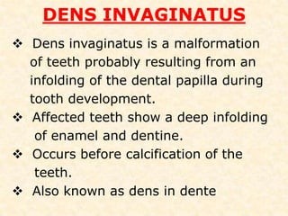
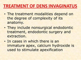
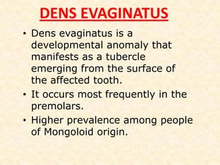
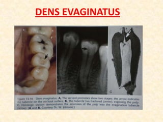
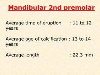
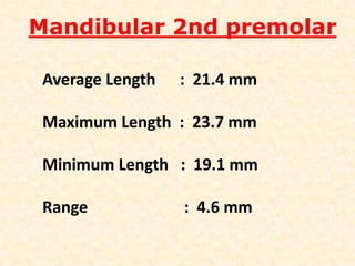
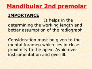
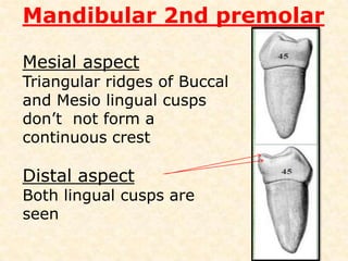
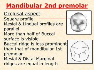
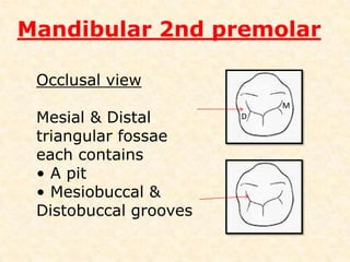
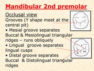
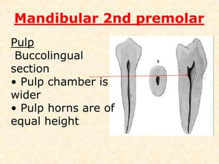
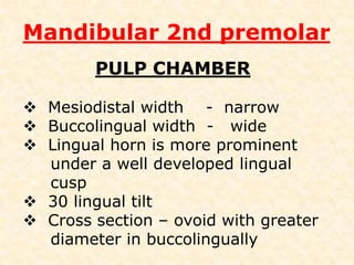
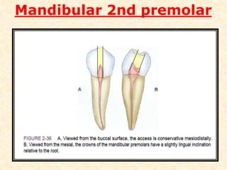
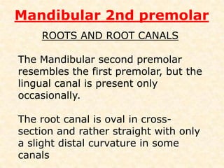
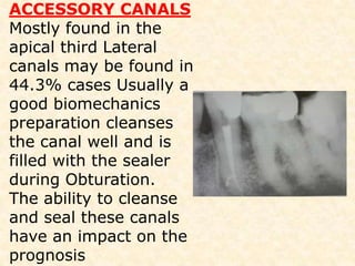
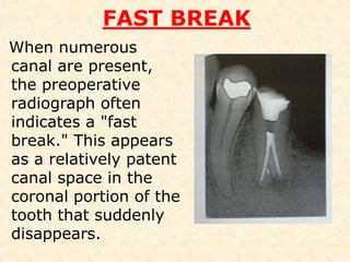
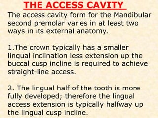
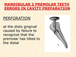
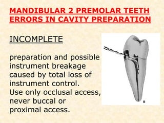
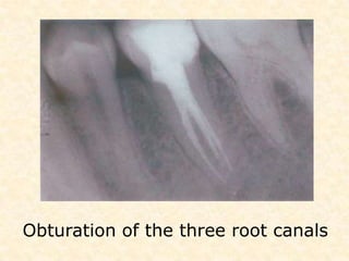
This document provides information about the anatomy and root canal treatment of the mandibular second premolar tooth. It describes the external and internal anatomy of the tooth, including average measurements, root canal morphology, and anatomical variations that may be present. It also discusses errors that can occur during root canal treatment of this tooth and provides a case example of successfully treating a mandibular second premolar with three root canals.
The success of endodontic treatment is directly related to the clinician’s ability to remove maximum number of bacteria from the root canal system.Some teeth are more difficult than others due to access limitations or anatomy, but the problem that faces all of us, in every tooth, is our inability to measure the absence of bacteria within the root canal and the potential for microanatomy to exist at any level of the root—whether it is a five-canal molar or a single-rooted anterior.
For more information, you can book an appointment at
Dr Sachdeva's Dental Aesthetic And Implant Institute,
I 101, Ashok Vihar Phase 1, Delhi- 110052
Contact us at
• Phone : +919818894041,01142464041
• Our Websites:
• www.sachdevadentalcare.com
• www.dentalclinicindelhi.com
• www.dentalimplantindia.co.in
• www.dentalcoursesdelhi.com
• www.facialaestheticsdelhi.com
• Google+ link: https://goo.gl/vqAmvr
• Facebook link: https://goo.gl/tui98A
• Youtube link: https://goo.gl/mk7jfm
• Linkedin link: https://goo.gl/PrPgpB
• Slideshare link : http://goo.gl/0HY6ep
• Twitter Page : https://goo.gl/tohkcI
• Instagram page : https://goo.gl/OOGVig
Indian Dental Academy: will be one of the most relevant and exciting training center with best faculty and flexible training programs for dental professionals who wish to advance in their dental practice,Offers certified courses in Dental implants,Orthodontics,Endodontics,Cosmetic Dentistry, Prosthetic Dentistry, Periodontics and General Dentistry.
Internal anatomy of permanent/ orthodontic course by indian dental academyIndian dental academy
Indian Dental Academy: will be one of the most relevant and exciting training center with best faculty and flexible training programs for dental professionals who wish to advance in their dental practice,Offers certified courses in Dental implants,Orthodontics,Endodontics,Cosmetic Dentistry, Prosthetic Dentistry, Periodontics and General Dentistry.
To make your Root canal easy contact Dental Implant India. We are specialized in root canal and other dental problem. We are the best dentist in North Delhi. To book an appointment visit - http://www.dentalimplantindia.co.in/dental-clinic-in-north-delhi.html
Developing Health Sciences students’ information skills through online self-p...Sarah Gallagher
Initial feedback on a cross cohort evaluation of an online self-paced information skills programme in three second year health sciences programmes at the Unviersity of Otago: Medicine, Pharmacy and Physiotherapy. Presented at Spotlight on Teaching 2013, University of Otago.
Indian Dental Academy: will be one of the most relevant and exciting training center with best faculty and flexible training programs for dental professionals who wish to advance in their dental practice,Offers certified courses in Dental implants,Orthodontics,Endodontics,Cosmetic Dentistry, Prosthetic Dentistry, Periodontics and General Dentistry.
Internal anatomy of permanent/ orthodontic course by indian dental academyIndian dental academy
Indian Dental Academy: will be one of the most relevant and exciting training center with best faculty and flexible training programs for dental professionals who wish to advance in their dental practice,Offers certified courses in Dental implants,Orthodontics,Endodontics,Cosmetic Dentistry, Prosthetic Dentistry, Periodontics and General Dentistry.
To make your Root canal easy contact Dental Implant India. We are specialized in root canal and other dental problem. We are the best dentist in North Delhi. To book an appointment visit - http://www.dentalimplantindia.co.in/dental-clinic-in-north-delhi.html
Developing Health Sciences students’ information skills through online self-p...Sarah Gallagher
Initial feedback on a cross cohort evaluation of an online self-paced information skills programme in three second year health sciences programmes at the Unviersity of Otago: Medicine, Pharmacy and Physiotherapy. Presented at Spotlight on Teaching 2013, University of Otago.
Books on Prescription evaluation & panel discussionSarah Gallagher
Slides for a public panel held on 11 March 2015 at Dunedin Public Libraries and hosted by LIANZA Otago Southland. Panelists included representatives from: WellSouth, Waitaki District Libraries & Archive, University of Otago Health Sciences Library & University of Otago Student Health Services. The panelists talked about their involvement in the Books on Prescription programme that is run through WellSouth. WellSouth also reported on a recent evaluation of the programme across the Otago Southland region. Speakers were: Katie Jahnke, Sophie Carty, Philip Van Zijl, Richard German, Jodie Black & Sarah Gallagher.
A presentation for the school community about what we have achieved over the past year on our library redevelopment journey. http://sacredheartdn.school.nz/library
Fui o mais sucinto possível, como falei, amo café, portanto o convite fica lançado para que possamos interagir e construir algo juntxs.
Com essa apresentação, minha expectativa é alcançar coletivos e ONGs para realizar trabalhos de impacto social. Analisando a melhor forma de otimizar para que esse auxílio seja constante e consistente.
Essa vontade já é antiga e recebeu impulso graças ao grupo dots, portanto agradecer à Kuki Baily por inspirar essa onda de bondade.
The pulp cavity is the central cavity within a tooth and is entirely enclosed by dentin except at apical foramen.
It is divided into:
1. Coronal portion pulp chamber
2. Radicular portion root canal
PULP CHAMBER
ROOF OF PULP CAVITY: consists of dentin covering the pulp chamber occlussaly or incisally.
PULP HORN : Accentuation of the roof of pulp chamber directly under a cusp or developmental lobe.
FLOOR OF PULP CHAMBER: runs parallel to the roof and consists of dentin bounding the pulp chamber near cervical area of tooth, particularly dentin forming the furcation area.
CANAL ORIFICES: openings in the floor of pulp chamber leading to the root canals.
ROOT CANALS
Portion of the pulp cavity from the canal orifice to the apical foramen
Divided into 3 section( for convenience)
Coronal third
Middle third
Apical third
The root canal curvature
Straight canal extending with minimal apical curvature
Gradual curvature of canal with straight apical ending
Gradual curvature of entire canal
Sharp curvature of canal near the apex
Success of negotiating narrow curved canal depends on
Degree of curvature
Size and constriction of root canal
Size and flexibility of endodontic instrument blade
Skill of operator
Classification based on canal cross-section
Round/circular
Oval
Long oval
Flattened(flat/ribbon)
Irregular
Vertucci’s Classification
Weine’s Classification
ISTHMUS
A narrow passage or anatomic part connecting two larger structures (root canals)
APICAL FORAMEN
In young incompletely developed teeth the apical foramen is funnel shaped with wider portion extending outward
As root develops the apical foramen becomes narrower
Apical foramen is not the most constricted part of root apex\apical foramen is not always located at the centre of the root apex
LATERAL CANALS AND ACCESSARY FORAMINA
Lateral canals frequently occur in apical third of root
May occur in areas of bifurcation and trifurcation of multirooted teeth
With increasing age, number of accessory foramina reduce due to calcification of contained soft tissue
INFLUENCE OF AGING
METHODS OF DETERMINING PULP ANATOMY
CLINICAL METHODS
Anatomy studies
Radiographs
Explorations
High resolution compound tomography
Visualisation endogram
Fiberoptic endoscope
Magnetic resonance imaging
IN VITRO METHODS
sectioning of teeth
use of dyes
Contrasting media
Scanning electron microscope analysis
VARIATIONS IN INTERNAL ANATOMY
Variations in development
Gemination
Fusion
Concrescence
Taurodontism
Talon’s cusp
Dilaceration
Extra root canal
Dens invaginatus
Dens evaginatus
Maxillary Central Incisor
Maxillary Lateral Incisor
Maxillary Canine
Mandibular Central and Lateral Incisors
Mandibular Canine
Maxillary First Premolar
Maxillary Second Premolar
The typical second premolar has one
root and one canal and sometimes
has an apical distal curvature.
The Type I canal form is p
9. Mandibular 2nd premolar Average time of eruption : 11 to 12 years Average age of calcification : 13 to 14 years Average length : 22.3 mm
10. Significance of average time of eruption,ageof calcification,toothlength & root curvature: IT HELPS IN DIAGNOSIS AND TREATMENT PLAN TREATMENT IS DIFFERENT IN ADULT AND YOUNG NECROTIC PULP RCT ADULT IRREVERSIBLE PULPITIS
11. YOUNG Irreversible Pulpit's Necrotic Pulp Reversible Pulpit's Pulp Capping or Pulpotomy Closed Apex Open Apex Apexification Obturation Apexogenesis RCT
12. Mandibular 2nd premolar Average Length : 21.4 mm Maximum Length : 23.7 mm Minimum Length : 19.1 mm Range : 4.6 mm
13. Mandibular 2nd premolar IMPORTANCE It helps in the determining the working length and better assumption of the radiograph Consideration must be given to the mental foramen which lies in close proximity to the apex. Avoid over instrumentation and overfill.
14. Mandibular 2nd premolar Buccal aspect Long pointed buccal cusp in the occlusal profile Mesial cusp ridge is shorter than distal Cusp tip is a little mesial to the tooth midline
15. Mandibular 2nd premolar Buccal aspect Mesial & Distal outlines are markedly converging Cervical line is flat mesiodistal compared to that of canine Root is conical with pointed apex
16. Mandibular 2nd premolar Lingual aspect mesiodistal diameter = that from Buccal aspect Occlusal surface cannot be seen fully Occlusal plane is perpendicular to tooth Axis
17. Mandibular 2nd premolar 2 lingual cusps (most commonly) • Mesiolingual – major, 2/3 MD diameter, same height as Buccal • Distolingual – minor Lingual groove 2/3
18. Mandibular 2nd premolar Mesial aspect Triangular ridges of Buccal and Mesio lingual cusps don’t not form a continuous crest Distal aspect Both lingual cusps are seen
19. Mandibular 2nd premolar Occlusal aspect Square profile Mesial & Lingual profiles are parallel More than half of Buccal surface is visible Buccal ridge is less prominent than that of mandibular 1st premolar Mesial & Distal Marginal ridges are equal in length
20. Mandibular 2nd premolar Occlusal view Mesial & Distal triangular fossae each contains • A pit • Mesiobuccal & Distobuccal grooves M D
21. Mandibular 2nd premolar Occlusal view Grooves (Y shape meet at the central pit) • Mesial groove separates Buccal & Mesiolingual triangular ridges – runs obliquely • Lingual groove separates lingual cusps • Distal groove separates Buccal & Distolingual triangular ridges B ML DL
22. Mandibular 2nd premolar Pulp Buccolingual section • Pulp chamber is wider • Pulp horns are of equal height
28. Mandibular 2nd premolar ROOTS AND ROOT CANALS The Mandibular second premolar resembles the first premolar, but the lingual canal is present only occasionally. The root canal is oval in cross-section and rather straight with only a slight distal curvature in some canals
31. ROOTS AND ROOT CANALS One root canal dividing in to two at apex Single canal that has divided and cross over at the apex
32. ROOT CANAL ORIFICES 1 CANAL SEPARATE IN TO 2 CANALS DIVISION IS BUCCAL AND LINGUAL LINGUAL CANAL SPLITS FROM THE MAIN CANAL AT SHARP ANGLE IT IS VISUAL CONFIGURATION AS LOWER CASE LETTER h BUCCAL CANAL IS STRAIGHT PORTION OF THE h
33. ACCESSORY CANALS Mostly found in the apical third Lateral canals may be found in 44.3% cases Usually a good biomechanics preparation cleanses the canal well and is filled with the sealer during Obturation. The ability to cleanse and seal these canals have an impact on the prognosis
34.
35. Mandibular 2nd premolar Anatomic relationships in situ The mental canal and foramen are close to the root apex Radiograph appearance may shows peiapical pathosis
36. Anatomic relationships in situ Avoid over instrumentation and overfill When viewing an x-ray of this area, the mental foramen is sometimes misdiagnosed as a premolar abscess. Therefore, before performing root canal therapy, make sure all diagnostic tests confirm your finding.
37. FAST BREAK When numerous canalare present, the preoperative radiograph often indicates a "fast break." This appears as a relatively patent canal space in the coronal portion of the tooth that suddenly disappears.
38. FAST BREAK Note: If a straight-on preoperative radiograph of a Mandibular 2 premolar shows the pulp canal disappearing in mid-root, this is an important indication that two canals are present.
39.
40. THE ACCESS CAVITY The access cavity form for the Mandibular second premolar varies in at least two ways in its external anatomy. 1.The crown typically has a smaller lingual inclination less extension up the buccal cusp incline is required to achieve straight-line access. 2. The lingual half of the tooth is more fully developed; therefore the lingual access extension is typically halfway up the lingual cusp incline.
41. THE ACCESS CAVITY The Mandibular second premolar can have two lingual cusps, sometimes of equal size. When this occurs, the access preparation is centered mesiodistally on a line connecting the buccal cusp and the lingual groove between the lingual cusp tips.
42. THE ACCESS CAVITY Buccolingual ovoid outline form reflects the anatomy of the pulp chamber and position of the centrally located canal.
43.
44. CROSS SECTIONAL IN MIDROOT LEVEL AND APICAL Midroot level: the canal continues to be long ovoid and requires perimeter filing Apical third level: the canals, generally round, are shaped into round, tapered preparations. Preparation terminates at the cementodentinal junction, 0.5 to 1.0 mm from the radiographic apex.
45. MANDIBULAR 2 PREMOLAR TEETH ERRORS IN CAVITY PREPARATION PERFORATION at the disto gingival caused by failure to recognize that the premolar has tilted to the distal
46. MANDIBULAR 2 PREMOLAR TEETH ERRORS IN CAVITY PREPARATION INCOMPLETE preparation and possible instrument breakage caused by total loss of instrument control. Use only occlusal access, never buccal or proximal access.
47. MANDIBULAR 2 PREMOLAR TEETH ERRORS IN CAVITY PREPARATION BIFURCATION Of a canal completely missed, caused by failure to adequately explore the canal with a curved instrument
48. MANDIBULAR 2 PREMOLAR TEETH ERRORS IN CAVITY PREPARATION APICAL PERFORATION Of an invitingly straight conical canal. Failure to establish the exact length of the tooth leads to trephination of the foramen
49. MANDIBULAR 2 PREMOLAR TEETH ERRORS IN CAVITY PREPARATION PERFORATION at the apical curvature caused by failure to recognize, by exploration, buccal curvature. A standard bucco lingual radiograph will not show buccal or lingual curvature
59. Various prophylactic treatments like selective grinding, application of resin, restorations and partial Pulpotomy can be done.
60.
61. A 20- year-old male with non contributory medical history was referred to the clinics of the SaudiBoard in Advanced Restorative at the Faculty of Dentistry, for evaluation of root canal therapy of a mandibular 2 premolar. Clinical examination revealed that the tooth responded positively to percussion but not to palpation. Radiographic examination revealed short and inadequate root canal filling
63. The tooth was isolated with rubber dam, the old amalgam filling was removed and the access cavity preparation was established. Three canals were located, buccally, lingually and an extra canal in the middle. The working length was checked radiographically
64. Working length radiograph showing files in the three root canals.
65. The canals were conventionally instrumented to a # 35K file using crown-down pressureless technique, irrigated with 5.25 percent sodium hypochlorite, dried with sterile paper points and sealed with calcium hydroxide paste The access opening was closed with Cavit. The patient returned asymptomatic after 1 week, the tooth was isolated with rubber dam; the canals were instrumented with file #35 and irrigated with sodium hypochlorite to remove all the remnants of the calcium hydroxide, and then dried with paper points
66. Master cone was selected and the canals were filled with gutta-percha and AH26 sealer cement using lateral condensation. Access opening was sealed with amalgam restoration. Post-operative radiograph was taken to confirm the quality of the filling .The patient was referred to the prosthetic clinic for crown construction.
68. DISCUSSION Location and thorough instrumentation of all the canals in the root of a diseased tooth normally ensure success of the endodontic therapy. Presented is a case of mandibular second premolar which was referred for endodontic therapy. Clinical and radiographicexamination revealed inadequate root canal filling. Three canals were located. Endodontic therapy was performed under aseptic conditions