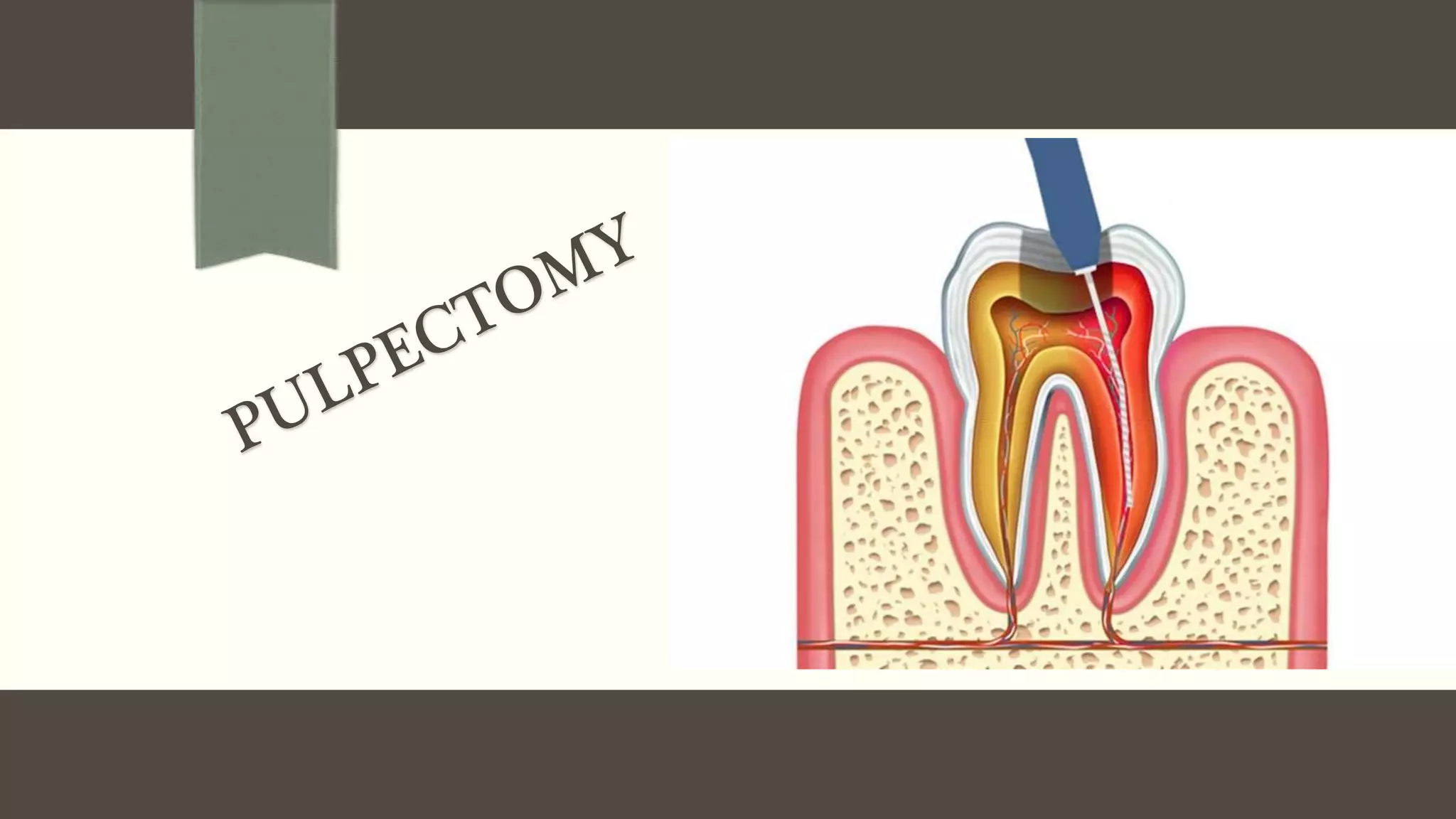
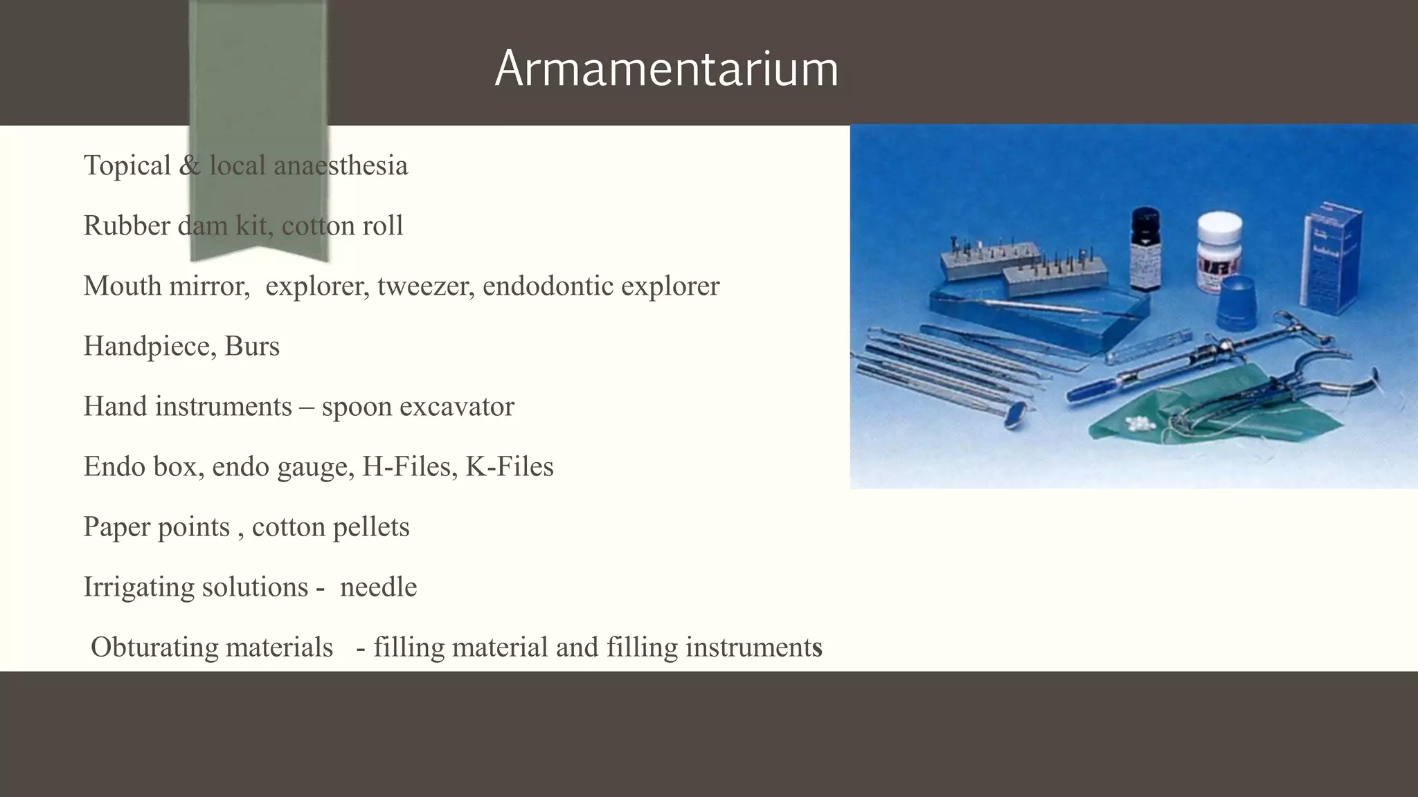
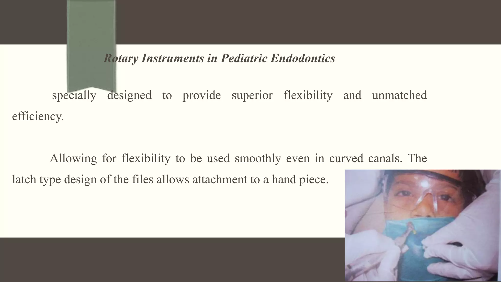
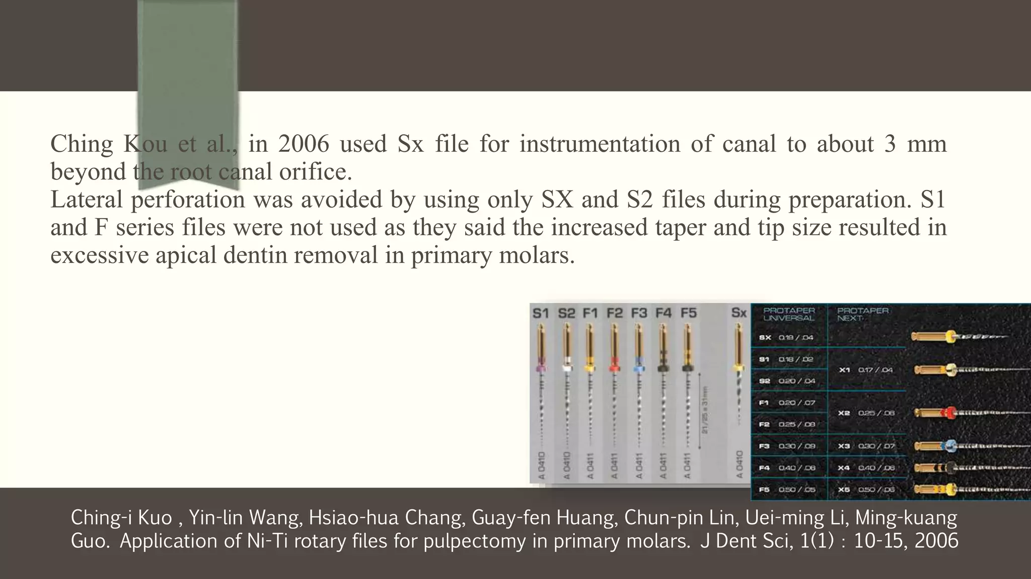
Primary Pulp Organs- function for short period of time (Avg – 8.3 years)
Pulp organ growth – 1 year
Pulp maturation - 3 years
Pulp regression - 3-6 years
Diagnostic considerations
The length of time the tooth in question are to be retained
The health of the patient
The status of the remaining dentition
The restorability of the tooth
Patient and parent co operation
Mathewson (1995): Complete removal of necrotic pulpal tissue from the root canals and
coronal portion of the primary teeth to maintain a tooth in the dental arch.
Finn: removal of all pulpal tissue from the tooth, including both coronal and root portions.
AAPD 2008- Root canal procedure for pulp tissue that is irreversibly infected or necrotic due
to caries or trauma. The root canals are debrided, enlarged, disinfected, and filled with a
resorbable material.
Definitions
Objectives – AAPD – clinical guidelines 2008
• Following treatment, the infectious process should resolve in 6 months
• Radiographic evidence of a successful filling without gross overextension or
underfilling.
• Should permit resorption of the primary root structures and filling materials to
permit normal eruption of succedaneous tooth
• No radiographic evidence of further breakdown of the supporting tissues.
• Alleviate and prevent further sensitivity, pain or swelling.
• No internal or external root resorption or other pathology.
Goals of pulp therapy (Mathewson)
Successful treatment of the cariously involved pulp, allowing the tooth to remain
in a non pathologic state
Maintenance of arch length and tooth space
Restoration of comfort with the ability to chew
Prevention of speech abnormalities and abnormal habits
Rationale
(Uk National Clinical Guidelines In Paediatric Dentistry International
Journal Of Paediatric Dentistry 2006)
To remove irreversibly inflamed or necrotic radicular pulp tissue and
gently clean RCS
To obturate the root canals with a filling material…………………..
According to Mc Donald
• The canals are accessible
• evidence of normal supporting bone
• Retention of a second primary molar when the succedaneous second
premolar is congenitally missing.
• Special effort made to retain second primary molar even with
necrotic pulp before the eruption of first permanent molar
Applicable to vital teeth where hemorrhage from the amputated radicular stumps is dark red, a slow
ooze, and is uncontrollable.
Rajesh Singla, Samir Dutta, Nikhil Marwah – single visit vs multi root canal therapy – Jaypee’s
International JL of Clinical Pediatric Dentistry – 2008;1(1):17-24
Isolation and sealing problems
Anterior esthetic problems
Restorative considerations
Vital pulp exposure and symptomatic pulpitis
SINGLE VISIT PULPECTOMY (KENNEDY))
MULTIVISIT PULPECTOMY (KENNEDY)
Paterson and curzon (1991) Indicated where infection, an abscess or
a chronic sinus exist.
Contraindications
Excessive mobility and/or reduced bone support
Non-restorable tooth
Underlying dentigerous or follicular cyst
Less than two-third of root length remaining
Perforation of pulpal floor
Medically compromised children – leukemia, rheumatic and CHD
Internal resorption
Deep caries and a large radiolucency with the possibility of
damaging/ affecting the succedaneous tooth.
Difference between primary and permanent pulp
Pulp chamber in deciduous teeth is larger compared to the crown size
The roots are thin and slender with narrow pulp horns
Mesial pulp horns extend closer towards outer surface
Accessory canals extend from pulp chamber to the inter radicular area at the
furcation.
Deciduous pulp is highly vascularized.
Increased rate of reparative dentin formation in primary tooh
Nerve fibres are few in number thus exhibiting reduced sensitivity to pain
Root canals are ribbon shaped or have a hour glass appearance.
The canals are narrow m-d.
Multiple ramifications are common in deciduous pulp canal making complete
debridement difficult
Topical & local anaesthesia
Rubber dam kit, cotton roll
Mouth mirror, explorer, tweezer, endodontic explorer
Handpiece, Burs
Hand instruments – spoon excavator
Endo box, endo gauge, H-Files, K-Files
Paper points , cotton pellets
Irrigating solutions - needle
Obturating materials - filling material and filling instruments
Armamentarium
Sterilization of instruments
Clean aseptic field
Important role in success
Diagnostic instruments
Local anesthesia
Isolation
Access opening - Burs (No.330 FG high speed, No.8 RA slow speed)
Pulp extirpation & w-l determination
Classification of root canal instruments
Exploring - E.g. smooth broach and endodontic explorer.
Debridement - E.g. barbed broach,
Shaping - E.g. reamers and files.
Obturating - E.g. pluggers, spreaders, and lentulospirals.
Smooth broaches/ Miller needles - smooth, pointed and tapered with either round, pentagonal or square
cross-section.
Barbed broach - short handled instrument - extirpation of pulp , removal of necrotic debris, absorbent
points, cotton pledgets, other foreign material
K-REAMERS
Have ½ to 1 cutting blade per mm
Made from either triangular or square blank
SMALLER SIZES -SQUARE
LARGER SIZES –TRIANGULAR
Triangular cross section is more flexible
Angle of blades to long axis is 20 degree
Have fewer cutting blades than K-files.
Primarily designed to be used in a rotary reaming motion
CUTTING ACTION- REAMERS
Twisted clockwise about ¼ turn to 1/2 turn to engage their blades into dentin and then withdrawn
o In a straight canal – 1/2 turn
o In curved canals – 1/4 turn
The instrument produces a round tapered preparation
Reamers are almost completely out of clinical practice now a days
HEDSTROEM FILES
ANSI No. 58
ISO No. 3630/1
Also known as H-type files
Has a characteristic Christmas tree appearance
The angle between cutting edge and the long axis of the instrument is about 60-65
degree.
Periphery bears spiral grooves (like a screw)
Single helix tear drop appearance in cross section
The spiraling flutes with the cutting blades are produced by machining a single
helix into the shaft of a piece of round tapered stainless steel wire.
Designed primarily for a linear filing motion
Due to the positive rake angle, they cut in only one direction, during retraction (withdrawal stroke)
Unsuitable to be used in rotary or reaming motion, due to its greater fragility
3 times more efficient than K files
K-FILES
ADA/ANSI SPECIFICATION No. 28
K-files have 1 ½ to 2 ½ cutting blades per mm of their working end
About twice the number of spirals on a K- reamer of corresponding size
Angle of the flutes to the long axis is about 25-40 degree
The tighter spirals establish a cutting angle that achieves its principal cutting
action on withdrawal
It can even cut in the push motion
The cutting action of the file can be achieved in either a filing (rasping) or
reaming (drilling) motion
K-Flex File
introduced with rhomboidal shaped blanks, which are twisted.
Increased flexibility and cutting efficiency.
alternating high and low flutes to make the instrument more efficient to remove
debris.
Safety H-files
Kerr manufacturing co introduced these in 1998.
A non-cutting side characterizes the spiral of the working end of these files with
smoothened edges to prevent ledging in curved canals.
These blunt flutes have to be oriented to the region of the canal wall that requires
no cutting.
Elizabeth, S. Bair in 1999-2000. 55% - Ni 45% - Ti. Flexibility and instrument
design - closely follow original root canal path.
Advantages
Tissue and debris are more easily and quickly removed
Faster results.
Allows easy access to all canals.
It possesses a memory effect
Disadvantages
Cost
Learning the technique.
Nickel;-Titanium root canal files
Rotary Instruments in Pediatric Endodontics
specially designed to provide superior flexibility and unmatched
efficiency.
Allowing for flexibility to be used smoothly even in curved canals. The
latch type design of the files allows attachment to a hand piece.
Ching Kou et al., in 2006 used Sx file for instrumentation of canal to about 3 mm
beyond the root canal orifice.
Lateral perforation was avoided by using only SX and S2 files during preparation. S1
and F series files were not used as they said the increased taper and tip size resulted in
excessive apical dentin removal in primary molars.
Ching-i Kuo , Yin-lin Wang, Hsiao-hua Chang, Guay-fen Huang, Chun-pin Lin, Uei-ming Li, Ming-kuang
Guo. Application of Ni-Ti rotary files for pulpectomy in primary molars. J Dent Sci, 1(1):10-15, 2006
Nagaratna PJ et al., in 2006 instrumented root canal with profile 0.04 taper 29 series
rotary instruments starting from size 2 to 7 in reduction gear hand piece.
Bahrololoomi Z et al. in 2007 performed instrumentation with 25-mm-long flexmaster
Ni-Ti rotary files (VDW, Germany)
Kummer TR et al. in 2008 prepared root canal with the Hero 642 system
(MicroMega) and a reducing 50:1 handpiece (MicroMega).
Azar MR, Mokhtare M in 2011 and Azar MR et al.,[9] in 2012 used 21 mm long
Mtwo NiTi rotary files driven with a torque limited rotation with maximum speed
of 280 rpm for preparing root canals.
Mtwo files
Advantages
Provide a more consistently dense fill due to uniform debridement.
Allow for greater apical enlargement.
Prevent apical exposure
Provide better shape than traditional hand-filling.
Significantly reduce instrumentation time.
Disadvantages
Skill is required for practice for a beginner.
Resorption of roots in primary teeth may cause a problem
Repeated use increases risk of fracture, especially in curved canals.
Irrigants
Sodium hypochlorite
Hydrogen peroxide
Saline
Chlorhexidine
Endodontic techniques for primary teeth have limitations in disinfection and
root canal filling. For this reason, sodium hypochlorite is recommended as the
main irrigating medium because of its broad antimicrobial spectrum and its
unique capacity to dissolve necrotic tissue remnants.
Patricia Nivoloni, Maristela Barbosa Portela,Rogerio Gleiserd and Laura Guimarães Primo. Histopathologic
and SEM analysis of primary teeth with pulpectomy failure. OOO - 2009;108:29-33
Seow et al (1991) suggested the use of an ultrasound
method to biomechanically clean primary teeth prior to
filling. A combination of mechanical cleaning and use of
ultrasound removed 95% of the test bacteria.
(RIFTKIN ET AL)
Should resorb at a similar rate as the primary root
Should be harmless to the periapical tissues and to the permanent tooth germ; if pressed
beyond the apex it should resorb readily
It should have a stable disinfecting power
It should be inserted easily into the root canal and be removed easily
Should adhere to the walls of the canal and should not shrink
It should not be soluble in water
Should be radio opaque and not discolor the tooth.
OBTURATING MATERIALS
Zinc Oxide-Eugenol Paste
Most commonly used filling material for primary teeth
77 – 80%
Bonastre (1837) discovered zinc oxide eugenol
Dentistry by Chisholm (1876).
.
Camp in 1984 introduced the endodontic pressure syringe to overcome the
problem of underfilling
Underfilling, however, is frequently clinically acceptable.
Overfilling, may cause a mild past foreign body reaction.
Cox et al ZOE paste has been found to be more effectively bacteriostatic than
formocresol
Advantages
• Resorbable
• Radioopaque
• Anti-inflammatory & analgesic action.
• Cytotoxicity remained lower
• Easily available.
• Cheaper/ cost effective
• Good plasticity
• Insoluble in tissue fluids
Common problems with ZOE as deciduous tooth obturation material
ZOE is harder and resorbs slowly compared to the root.
As the resorption reaches the pulpal floor, the permanent tooth may get deflected
from its normal path of eruption due to the presence and obstruction from bulk of
ZOE
Coll et al found a higher than expected rate of ectopic eruption following RCT in
primary teeth using zinc oxide eugenol as a dressing material
ZnOE has also been used in combination with different fixative agents,
viz, - formaldehyde (Goodman J. 1985; Starkey PE 1973)
- formocresol (Coll JA, Josell S, Casper JS 1985)
- Paraformaldehyde (Berk H, Krakow A 1972) and
- cresol (Goodman J. 1985)
all of which have inherent cytotoxicity apart from other drawbacks
Iodoform Paste
Several authors have reported use of KRI paste. It resorb rapidly and has
no undesirable effects on succedaneous teeth - abscessed primary teeth.
KRI paste that extrudes into the periapical tissue is rapidly replaced with a normal
tissue.
Found to have a long lasting bactericidal potential. Since iodoform paste does not
set into a hard mass, it can be removed if re-treatment is required.
Walkhoff in 1928
Iodoform – 80.8%
Camphor – 4.86%
Parachlorophenol – 2.025%
Menthol – 1.215%
Advantages
Bactericidal in root canal
Resorbs from the apical tissues-1-2weeks
Harmless to permanent tooth germs
Radio opaque
Does’nt set to a hard mass
Easily inserted and removed
KRI PASTE
MAISTO’S PASTE
Maisto in 1967
Zinc-oxide - 14g
Iodoform - 42g
Thymol - 2g
Lanolin – 0.5g
Chloramphenol camphor – 3 cc
Eiyahu Mass ( 1989)
Used Maisto paste for obturation
In the follow up after 3 ½ years , tooth was functional
Overfilling in the distal canal was resorbed upto middle part of the root
Development of underlying tooth bud seem to be normal
No pathologic resorption of root was seen
Mass E, Zilberman UL, Endodontic treatment of infected primary teeth
using Maisto’s paste, J Dent Child ,1989;56:117-120
Calcium Hydroxide
Generally not used in pulp therapy for primary teeth. However, clinical and
histopathologic investigations of calcium hydroxide and iodoform mixture
(Vitapex) have been published by Fuchino and Nishino (1980).
Easy to apply and resorbs at a slightly faster rate than that of the root.
It has no toxic effects on permanent successor and is radio opaque.
VITAPEX/ METAPEX
Iodoform - 40.4%
CH - 30.3%
Silicone oil - 22.4%
Others – 6.9
Advantages
1) When extruded periapically resorbs within 2 weeks, intra and extra
radicularly.
2) Easy delivery system
3) Proven beneficial effect of iodoform
4 ) Radio opaque and does not set to a hard mass
Vitapex when extruded into furcal or apical areas, can either get
diffused or resorbed by macrophages, in as short a time as 1 or 2
weeks up to 2 to 3 months and causes no foreign body reaction, with
success rate of 96% to 100% ( Nurko et al 1999)
It is probable that the rapid elimination of extruded Vitapex and the fact
that it does not set to a hard mass can be considered as one of the
most important advantages of Vitapex over ZOE.
Comparison of zinc oxide and eugenol, and Vitapex for root canal treatment
of
necrotic primary teeth. Int J Paediatr Dent 2004 Nov;14(6):417-24
The overall success rates of Vitapex and ZOE were 100% and 78.5%,
respectively
Both ZOE and Vitapex gave encouraging results. Vitapex, however, can be
used more safely whenever there is a doubt about the patient's return for
follow-up.
Success of pulpectomy with zinc oxide-eugenol vs calcium hydroxide /iodoform paste in
primary molars: a clinical study. Pediatr Dent. 2008 Jul-
Aug;30(4):303-8
At 6 and 12 months, the ZOE success rates were 48% and 85%, respectively, and the
Vitapex success rates were 78% and 89%.
Vitapex appeared to resolve furcation pathology at a faster rate than zinc oxide-eugenol
at 6 months, while at 12 months, both materials yielded similar results.
Walkhoff
paste
KRI paste Maisto paste Vitapex Endoflas Guedes-Pino
paste
Parachloro-
phenol
Camphor
Menthol
Iodoform
80.8% Campor
4.86% PCP
2.025%
Menthol
1.21%
ZnO 14g
iodoform 42 g
Thymol 2g
Chlorphenol
Camphor 3 cc
Lanolin 0.5g
Calcium
hydroxide
iodoform oily
additives
ZnO 56.5%
Barium sulfate
1.63%
iodoform
40.6%
Calcium
hydroxide
1.07% Eugenol
Pentachloroph
enol
0.30g
iodoform 0.25
g calcium
hydroxide 0.1
ml
camphorated
paramonochi-
orophenol
Colla Cote
It is a soft, white, pliable, biocompatible sponge - bovine collagen.
It can be applied to moist or bleeding canals.
absorbable collagen barrier which prevents or diminishes extravasation of root
canal filling material
Also provides a scaffold for bone growth
Marlin.s.Johnson, Leandro.R.Britto, and Marcio Guelmann determined whether
placement of a resorbable collagen barrier at the apical one third of the root canal could
prevent extrusion of the pulpectomy filling material.
The result showed that collacote at the apical 1/3 of the canals did not completely prevent,
but did significantly decrease, the risk for overfilling in primary molars.
Endoflas
Endoflas is a root canal sealer material, which is composed of zinc oxide,
barium sulfate, iodoform, calcium hydroxide, eugenol and pentachlorophenol
One condition for success of Endoflas is the prevention of microleakage.
A permanent restoration should be placed
A retrospective study done on primary teeth using endoflas has shown 70%
success rate.
Gutta-Percha (Not indicated for primary teeth)
Since gutta-percha is not a resorbable material, its use is contraindicated in the primary
teeth.
When the succedaneous , permanent tooth is missing and the retained primary
tooth becomes pulpally involved, the canals are filled with GP after pulpectomy
(Cohen)
Procedure
Local anesthesia
Rate of solution deposition: 1
ml/min or at atleast 1 ml/30 sec
Isolation – Rubber dam
Essential in any endodontic procedure as
it is the best method of isolating the tooth
from the oral cavity.
First introduced by Barnum (1864), it is
useful in providing a clean, dry and
sterilizable field.
Access Opening for pulpectomy in primary teeth
Rules for Proper Access Preparation
Endodontic Dogma: “Careful cavity preparation and root canal obturation are the
keystones to successful RCT.”
The objective of entry is to gain direct access to the apical foramina and not merely
to the canal orifices.
interior anatomy of the tooth under treatment must be determined
prepared always through the occlusal or the lingual surface and never the
proximal or the gingival surface.
unsupported cusps of posterior teeth must be reduced to avoid weakening of the
tooth structure.
To achieve optimal preparation three factors of internal
anatomy must be considered:
Size of the Pulp Chamber
Shape of the Pulp Chamber
Number, position and curvature of the root canals.
Access Opening for Primary Teeth
Have traditionally been through the lingual surface. This remains the
surface of choice except for the primary maxillary incisors(Cohen).
Procedure for pulp extirpation
Healey 1994 pass the barbed broach along a canal wall towards the end of the canal

As it reaches to the apical constriction, move it into the center of the mass of pulp tissue

Rotate the broach several times in a watch winding fashion to entangle the pulp that is withdrawn from the
canal.
Working length determination
Tactile sensation
Radiographic method
Electronic apex locator
The multiple ramifications - make complete debridement impossible
Also, the ribbon shape of the root canals…………………….
Attempts to prepare a circular apical 1/3 mechanically result in lateral perforation of the
canal because of its hour glass shape.
Because of the bizarre anatomy of the root canals the use of barbed broaches as in
conventional endodontics may be unsuccessful - increased danger of instrument fracture.
Kennedy
H files number 15 or 20 are strongly recommended - the maximum size used should be 30.
A preliminary working length is determined by measurement of a radiograph taken with a
paralleling technique.
Cohen
Because of the thin walls sonic and ultrasonic cleaning devices should not be used to
prepare the canals of the primary tooth. Also GG drills or peso drills are contra indicated
because of the danger of perforation or stripping of the roots
Instruments should be gently curved to help negotiate the canals.- lessens the risk
of perforation.
Shaping of the canals proceeds in much the same manner as is done to receive a
gutta-percha filling.
minimum size of 30 to 35.
Copious irrigation during cleaning and shaping must be maintained. Debridement
is more often accomplished by chemical than mechanical means.
After canal debridement, flushed with NaOCl and are then dried with sterile paper pints; a
pellet of cotton is barely moistened with camphorated parachlorophenol and sealed into
the pulp chamber with temporary cement.
At a subsequent appointment the canal is re-entered. As long as the patient is free of all
signs and symptoms of inflammation, the canals are again irrigated with sodium
hypochlorite and dried preparatory to filling.
Obturation Techniques
Using Reamer
The canals are dried with paper points.
Ideal consistency
Then a no. 15 or a no.20 reamer is taken
First the reamer is rotated clockwise inside the canals for 10-15 rotations
Secondly, the reamer is moved vertically up and down 10-15 times, simultaneously tilting the
reamer in all directions horizontally
Thirdly the reamer is withdrawn anticlockwise from the canal
Using wet cotton:
ZOE mixed and taken into the root canal with a file or reamer. Then a small
wet cotton pellet (squeezed thoroughly) is taken and used to condense the
material inside the canal from the chamber. repeated 5-8 times, apply
pressure using the cotton pellet towards each canal separately
Using Lentulo spirals
ZOE is taken inside the canal with either a hand lentulo or an engine driven
lentulo spiral.
Obturation using injectable syringes
After the canal is dried thoroughly, the syringe loaded with the obturating material is
taken inside the canal and the material is extruded slowly, continuously withdrawing
the syringe from the canal. This method is a simpler and faster method for obturating
the root canals of primary teeth.
Metapex
Vitapex
Calcicure
Endodontic pressure syringe
Designed by Greenberg and Katz.
Using this syringe, a very thick mix of ZOE can be forced through an extremely narrow gauge
needle
allows effective placement of filling material into the apical portion first. Accordingly, none of
the sealer is lost in the access cavity, at the orifice of the canals, or along the walls of the
coronal portion of the canal.
canal should be enlarged to size 40 or 50.
Other methods
Jiffy tube: The regular mix of ZOE is backloaded into the tube, placed into the canal
orifice and the material expressed into the canal with a downward squeezing motion
until the orifice appeared visibly filled.
Tuberculin syringe: thin mix of ZOE and the material was expressed into the canal
by slow finger pressure on the plunger until the canal was visibly filled at the orifice.
Yacobi and Kenny (1991) stated that over filling is not preferable to
underfilling, although it has been showed that if small quantities of
paste are extruded through the apex they resorb
Clinical evaluation of root canal obturation methods in primary teeth. Pediatric
Dentistry 2006; 28(1):39- 47
Omar Bawazir, Fouad Salama.- evaluate in vivo two different obturation
techniques in primary teeth – lentulo spirals mounted in a slow speed hand piece
and hand held on 24 children.
Evaluation was done immediately after pulpectomy and 6 months following
treatment. 96% success was found when – lentulo spirals mounted in a slow speed
hand piece was used and 92% success was found when – lentulo spirals hand held.
Pulpectomy Technique – Mathewson – Single Visit
.
Local anaesthesia and Rubber Dam isolation
Prepare a cavity preparation
Use large round bur to remove remaining caries and debris in pulp chamber
Evaluate haemorrhage or purulent exudates.
With endodontic file, remove diseased pulp tissue from all canals. As file is withdrawn, it carries pulpal material with
it.
.
Start with No. 15 and finish with No.35
If a point of resistance is encountered do not attempt to go beyond it
Irrigate canals repeatedly with dilute NaOCl, dry with cotton pellets and paper points. Never put
air directly into canal.
When haemorrhage is controlled and canals are dry, fill with ZOE cement. Mix on a pad, lift with
amalgam carrier and insert into pulp chamber
Use an amalgam plugger to constantly apply pressure and pack the cement into the canals. This
condensing pressure forces the cement mass into the canals.
Alternative method: use a thin mix of ZOE cement on a file or paper point and place it in the pulp
canals. Then shape a thick mix of ZOE in a cone and pack it in the canals using a moist cotton pellet as
condensor.
Obtain a periapical radiograph to be certain the canals are filled.
Completed procedure includes ZOE filled pulp chamber and root canals, and stainless steel crown.
Place the patient on periodic recall program to evaluate the success. Teeth that are symptom free
clinically and radiograpically with exfoliation within normal limits are considered successful.
Technique for anterior teeth (Mathewson)
Isolate- rubber dam
Open into pulp chamber – 330 high speed bur
With no: 15 endodontic file , remove the diseased pulp tissue, use a large file . A rubber stopper is used as a
marker
Irrigate the canals gently with a soln of NaOCl
Dry canals – paper points and cotton pellets
Use a thick mix of ZOE in the canal. A large endodontic condenser or amalgam plugger is applied to pack
the cement into the canal
To check success obtain an IOPA.
Restore with SSC or composite resin crown. ZOE is condensed to the apices
Teeth should be evaluated periodically for normal exfoliation
Starkey's Complete Pulpectomy Technique for Primary Molars
(Mc Donald)
Rubber dam applied - following anesthesia
Roof of pulp chamber removed to gain access to root canals
The contents of the pulp chamber and all debris from the occlusal 1/3 of the canals should
be removed, with care taken to avoid forcing any of the infected contents through the
apical foramen.
A moistened pellet of CMCP or 1:5 concentration of Buckley's formocresol with excess
moisture blotted, should be placed in the pulp chamber. It may be sealed with ZOE.
Several days later, at the second appointment: If the tooth has remained asymptomatic during the interval, remaining
contents of canals should be removed - fine barbed broach, H file
The apex of each root should be penetrated slightly with smallest file
A treatment pellet should again be placed and seal with ZOE.
After another interval of few days, seal removed. If asymptomatic, canals may be prepared and filled
If tooth painful / moisture in canals - treatment repeated.
Currently, primary teeth pulpectomies are commonly completed in a
single appointment. However, if the tooth has painful necrosis with
purulence in canals, 2 or 3 visits, should improve prognosis.
Partial pulpectomy (Mc Donald)
Completed in one appointment, removal of the coronal pulp and the pulp filaments - fine barbed broach -
hemorrhage
A H file will be helpful in the removal of remnants of the pulp
After pulp extirpation , 3% H2O2 followed by NaOCl
dried with sterile paper points.
a thin mix of unreinforced ZOE paste may be prepared and paper points covered with the material are used to
coat the root canal walls. Small K files may be used to file the paste into the walls.
Excess thin paste may be removed with paper points and H files. A thick mix should be prepared rolled into a
point and carried into the canal. Root canal pluggers may be used
It can be evaluated radio-graphically
The tooth should be restored with full coverage
Problems encountered in deciduous teeth pulpectomy
Multiple ramification – makes complete debridement impossible
Ribbon shaped or hour glass shaped canals- discourages gross enlargement of
the canal
In primary teeth attempt to prepare a circular apical 1/3 mechanically may result
in lateral perforation of the canal, due to its hour glass shape.
Evaluation of success of pulpectomy
No purulent discharge from the gingival margin
No abnormal mobility
No post operative pain
No further resorption of root (except physiological)
Resolution of sinus tract by 6 months
Follow-up after Primary Pulpectomy
The rate of success following primary pulpectomy is high. While resorbing
normally without interference with eruption of the permanent tooth, the primary
tooth should remain asymptomatic, firm in the alveolus and free from pathosis.
may occasionally present a problem of over retention.
Post operative follow up at 6 monthly intervals should include an evaluation of signs and symptoms,
periapical radiographs should be taken between 12 and 18 months post operatively. Pathological
mobility, presence of a fistula and in rare instances pain (on percussion) are clinical evidence of
failure.
Radiographic evidence of failure is judged by the appearance , or increased size of a
radiolucency and by external or internal root resorption. Any bone loss is likely to occur at the
furcation of region and not at the apices.
Pulpectomy

Pulpectomy

  • 3.
    Primary Pulp Organs-function for short period of time (Avg – 8.3 years) Pulp organ growth – 1 year Pulp maturation - 3 years Pulp regression - 3-6 years
  • 4.
    Diagnostic considerations The lengthof time the tooth in question are to be retained The health of the patient The status of the remaining dentition The restorability of the tooth Patient and parent co operation
  • 5.
    Mathewson (1995): Completeremoval of necrotic pulpal tissue from the root canals and coronal portion of the primary teeth to maintain a tooth in the dental arch. Finn: removal of all pulpal tissue from the tooth, including both coronal and root portions. AAPD 2008- Root canal procedure for pulp tissue that is irreversibly infected or necrotic due to caries or trauma. The root canals are debrided, enlarged, disinfected, and filled with a resorbable material. Definitions
  • 6.
    Objectives – AAPD– clinical guidelines 2008 • Following treatment, the infectious process should resolve in 6 months • Radiographic evidence of a successful filling without gross overextension or underfilling. • Should permit resorption of the primary root structures and filling materials to permit normal eruption of succedaneous tooth
  • 7.
    • No radiographicevidence of further breakdown of the supporting tissues. • Alleviate and prevent further sensitivity, pain or swelling. • No internal or external root resorption or other pathology.
  • 8.
    Goals of pulptherapy (Mathewson) Successful treatment of the cariously involved pulp, allowing the tooth to remain in a non pathologic state Maintenance of arch length and tooth space Restoration of comfort with the ability to chew Prevention of speech abnormalities and abnormal habits
  • 9.
    Rationale (Uk National ClinicalGuidelines In Paediatric Dentistry International Journal Of Paediatric Dentistry 2006) To remove irreversibly inflamed or necrotic radicular pulp tissue and gently clean RCS To obturate the root canals with a filling material…………………..
  • 10.
    According to McDonald • The canals are accessible • evidence of normal supporting bone • Retention of a second primary molar when the succedaneous second premolar is congenitally missing. • Special effort made to retain second primary molar even with necrotic pulp before the eruption of first permanent molar
  • 11.
    Applicable to vitalteeth where hemorrhage from the amputated radicular stumps is dark red, a slow ooze, and is uncontrollable. Rajesh Singla, Samir Dutta, Nikhil Marwah – single visit vs multi root canal therapy – Jaypee’s International JL of Clinical Pediatric Dentistry – 2008;1(1):17-24 Isolation and sealing problems Anterior esthetic problems Restorative considerations Vital pulp exposure and symptomatic pulpitis SINGLE VISIT PULPECTOMY (KENNEDY))
  • 12.
    MULTIVISIT PULPECTOMY (KENNEDY) Patersonand curzon (1991) Indicated where infection, an abscess or a chronic sinus exist.
  • 13.
    Contraindications Excessive mobility and/orreduced bone support Non-restorable tooth Underlying dentigerous or follicular cyst Less than two-third of root length remaining Perforation of pulpal floor
  • 14.
    Medically compromised children– leukemia, rheumatic and CHD Internal resorption Deep caries and a large radiolucency with the possibility of damaging/ affecting the succedaneous tooth.
  • 15.
    Difference between primaryand permanent pulp Pulp chamber in deciduous teeth is larger compared to the crown size The roots are thin and slender with narrow pulp horns Mesial pulp horns extend closer towards outer surface Accessory canals extend from pulp chamber to the inter radicular area at the furcation.
  • 16.
    Deciduous pulp ishighly vascularized. Increased rate of reparative dentin formation in primary tooh Nerve fibres are few in number thus exhibiting reduced sensitivity to pain Root canals are ribbon shaped or have a hour glass appearance. The canals are narrow m-d. Multiple ramifications are common in deciduous pulp canal making complete debridement difficult
  • 17.
    Topical & localanaesthesia Rubber dam kit, cotton roll Mouth mirror, explorer, tweezer, endodontic explorer Handpiece, Burs Hand instruments – spoon excavator Endo box, endo gauge, H-Files, K-Files Paper points , cotton pellets Irrigating solutions - needle Obturating materials - filling material and filling instruments Armamentarium
  • 18.
    Sterilization of instruments Cleanaseptic field Important role in success
  • 19.
  • 20.
    Access opening -Burs (No.330 FG high speed, No.8 RA slow speed)
  • 21.
    Pulp extirpation &w-l determination
  • 22.
    Classification of rootcanal instruments Exploring - E.g. smooth broach and endodontic explorer. Debridement - E.g. barbed broach, Shaping - E.g. reamers and files. Obturating - E.g. pluggers, spreaders, and lentulospirals.
  • 23.
    Smooth broaches/ Millerneedles - smooth, pointed and tapered with either round, pentagonal or square cross-section. Barbed broach - short handled instrument - extirpation of pulp , removal of necrotic debris, absorbent points, cotton pledgets, other foreign material
  • 24.
    K-REAMERS Have ½ to1 cutting blade per mm Made from either triangular or square blank SMALLER SIZES -SQUARE LARGER SIZES –TRIANGULAR Triangular cross section is more flexible Angle of blades to long axis is 20 degree Have fewer cutting blades than K-files. Primarily designed to be used in a rotary reaming motion
  • 26.
    CUTTING ACTION- REAMERS Twistedclockwise about ¼ turn to 1/2 turn to engage their blades into dentin and then withdrawn o In a straight canal – 1/2 turn o In curved canals – 1/4 turn The instrument produces a round tapered preparation Reamers are almost completely out of clinical practice now a days
  • 27.
    HEDSTROEM FILES ANSI No.58 ISO No. 3630/1 Also known as H-type files Has a characteristic Christmas tree appearance The angle between cutting edge and the long axis of the instrument is about 60-65 degree. Periphery bears spiral grooves (like a screw) Single helix tear drop appearance in cross section
  • 28.
    The spiraling fluteswith the cutting blades are produced by machining a single helix into the shaft of a piece of round tapered stainless steel wire.
  • 29.
    Designed primarily fora linear filing motion Due to the positive rake angle, they cut in only one direction, during retraction (withdrawal stroke) Unsuitable to be used in rotary or reaming motion, due to its greater fragility 3 times more efficient than K files
  • 30.
    K-FILES ADA/ANSI SPECIFICATION No.28 K-files have 1 ½ to 2 ½ cutting blades per mm of their working end About twice the number of spirals on a K- reamer of corresponding size Angle of the flutes to the long axis is about 25-40 degree
  • 31.
    The tighter spiralsestablish a cutting angle that achieves its principal cutting action on withdrawal It can even cut in the push motion The cutting action of the file can be achieved in either a filing (rasping) or reaming (drilling) motion
  • 32.
    K-Flex File introduced withrhomboidal shaped blanks, which are twisted. Increased flexibility and cutting efficiency. alternating high and low flutes to make the instrument more efficient to remove debris.
  • 33.
    Safety H-files Kerr manufacturingco introduced these in 1998. A non-cutting side characterizes the spiral of the working end of these files with smoothened edges to prevent ledging in curved canals. These blunt flutes have to be oriented to the region of the canal wall that requires no cutting.
  • 34.
    Elizabeth, S. Bairin 1999-2000. 55% - Ni 45% - Ti. Flexibility and instrument design - closely follow original root canal path. Advantages Tissue and debris are more easily and quickly removed Faster results. Allows easy access to all canals. It possesses a memory effect Disadvantages Cost Learning the technique. Nickel;-Titanium root canal files
  • 35.
    Rotary Instruments inPediatric Endodontics specially designed to provide superior flexibility and unmatched efficiency. Allowing for flexibility to be used smoothly even in curved canals. The latch type design of the files allows attachment to a hand piece.
  • 36.
    Ching Kou etal., in 2006 used Sx file for instrumentation of canal to about 3 mm beyond the root canal orifice. Lateral perforation was avoided by using only SX and S2 files during preparation. S1 and F series files were not used as they said the increased taper and tip size resulted in excessive apical dentin removal in primary molars. Ching-i Kuo , Yin-lin Wang, Hsiao-hua Chang, Guay-fen Huang, Chun-pin Lin, Uei-ming Li, Ming-kuang Guo. Application of Ni-Ti rotary files for pulpectomy in primary molars. J Dent Sci, 1(1):10-15, 2006
  • 37.
    Nagaratna PJ etal., in 2006 instrumented root canal with profile 0.04 taper 29 series rotary instruments starting from size 2 to 7 in reduction gear hand piece. Bahrololoomi Z et al. in 2007 performed instrumentation with 25-mm-long flexmaster Ni-Ti rotary files (VDW, Germany)
  • 38.
    Kummer TR etal. in 2008 prepared root canal with the Hero 642 system (MicroMega) and a reducing 50:1 handpiece (MicroMega). Azar MR, Mokhtare M in 2011 and Azar MR et al.,[9] in 2012 used 21 mm long Mtwo NiTi rotary files driven with a torque limited rotation with maximum speed of 280 rpm for preparing root canals. Mtwo files
  • 39.
    Advantages Provide a moreconsistently dense fill due to uniform debridement. Allow for greater apical enlargement. Prevent apical exposure Provide better shape than traditional hand-filling. Significantly reduce instrumentation time. Disadvantages Skill is required for practice for a beginner. Resorption of roots in primary teeth may cause a problem Repeated use increases risk of fracture, especially in curved canals.
  • 40.
  • 41.
    Endodontic techniques forprimary teeth have limitations in disinfection and root canal filling. For this reason, sodium hypochlorite is recommended as the main irrigating medium because of its broad antimicrobial spectrum and its unique capacity to dissolve necrotic tissue remnants. Patricia Nivoloni, Maristela Barbosa Portela,Rogerio Gleiserd and Laura Guimarães Primo. Histopathologic and SEM analysis of primary teeth with pulpectomy failure. OOO - 2009;108:29-33
  • 42.
    Seow et al(1991) suggested the use of an ultrasound method to biomechanically clean primary teeth prior to filling. A combination of mechanical cleaning and use of ultrasound removed 95% of the test bacteria.
  • 43.
    (RIFTKIN ET AL) Shouldresorb at a similar rate as the primary root Should be harmless to the periapical tissues and to the permanent tooth germ; if pressed beyond the apex it should resorb readily It should have a stable disinfecting power It should be inserted easily into the root canal and be removed easily Should adhere to the walls of the canal and should not shrink It should not be soluble in water Should be radio opaque and not discolor the tooth. OBTURATING MATERIALS
  • 44.
    Zinc Oxide-Eugenol Paste Mostcommonly used filling material for primary teeth 77 – 80% Bonastre (1837) discovered zinc oxide eugenol Dentistry by Chisholm (1876). .
  • 45.
    Camp in 1984introduced the endodontic pressure syringe to overcome the problem of underfilling Underfilling, however, is frequently clinically acceptable. Overfilling, may cause a mild past foreign body reaction. Cox et al ZOE paste has been found to be more effectively bacteriostatic than formocresol
  • 46.
    Advantages • Resorbable • Radioopaque •Anti-inflammatory & analgesic action. • Cytotoxicity remained lower • Easily available. • Cheaper/ cost effective • Good plasticity • Insoluble in tissue fluids
  • 47.
    Common problems withZOE as deciduous tooth obturation material ZOE is harder and resorbs slowly compared to the root. As the resorption reaches the pulpal floor, the permanent tooth may get deflected from its normal path of eruption due to the presence and obstruction from bulk of ZOE Coll et al found a higher than expected rate of ectopic eruption following RCT in primary teeth using zinc oxide eugenol as a dressing material
  • 48.
    ZnOE has alsobeen used in combination with different fixative agents, viz, - formaldehyde (Goodman J. 1985; Starkey PE 1973) - formocresol (Coll JA, Josell S, Casper JS 1985) - Paraformaldehyde (Berk H, Krakow A 1972) and - cresol (Goodman J. 1985) all of which have inherent cytotoxicity apart from other drawbacks
  • 49.
    Iodoform Paste Several authorshave reported use of KRI paste. It resorb rapidly and has no undesirable effects on succedaneous teeth - abscessed primary teeth. KRI paste that extrudes into the periapical tissue is rapidly replaced with a normal tissue. Found to have a long lasting bactericidal potential. Since iodoform paste does not set into a hard mass, it can be removed if re-treatment is required.
  • 50.
    Walkhoff in 1928 Iodoform– 80.8% Camphor – 4.86% Parachlorophenol – 2.025% Menthol – 1.215% Advantages Bactericidal in root canal Resorbs from the apical tissues-1-2weeks Harmless to permanent tooth germs Radio opaque Does’nt set to a hard mass Easily inserted and removed KRI PASTE
  • 51.
    MAISTO’S PASTE Maisto in1967 Zinc-oxide - 14g Iodoform - 42g Thymol - 2g Lanolin – 0.5g Chloramphenol camphor – 3 cc
  • 52.
    Eiyahu Mass (1989) Used Maisto paste for obturation In the follow up after 3 ½ years , tooth was functional Overfilling in the distal canal was resorbed upto middle part of the root Development of underlying tooth bud seem to be normal No pathologic resorption of root was seen Mass E, Zilberman UL, Endodontic treatment of infected primary teeth using Maisto’s paste, J Dent Child ,1989;56:117-120
  • 53.
    Calcium Hydroxide Generally notused in pulp therapy for primary teeth. However, clinical and histopathologic investigations of calcium hydroxide and iodoform mixture (Vitapex) have been published by Fuchino and Nishino (1980).
  • 54.
    Easy to applyand resorbs at a slightly faster rate than that of the root. It has no toxic effects on permanent successor and is radio opaque.
  • 55.
    VITAPEX/ METAPEX Iodoform -40.4% CH - 30.3% Silicone oil - 22.4% Others – 6.9
  • 56.
    Advantages 1) When extrudedperiapically resorbs within 2 weeks, intra and extra radicularly. 2) Easy delivery system 3) Proven beneficial effect of iodoform 4 ) Radio opaque and does not set to a hard mass
  • 57.
    Vitapex when extrudedinto furcal or apical areas, can either get diffused or resorbed by macrophages, in as short a time as 1 or 2 weeks up to 2 to 3 months and causes no foreign body reaction, with success rate of 96% to 100% ( Nurko et al 1999) It is probable that the rapid elimination of extruded Vitapex and the fact that it does not set to a hard mass can be considered as one of the most important advantages of Vitapex over ZOE.
  • 58.
    Comparison of zincoxide and eugenol, and Vitapex for root canal treatment of necrotic primary teeth. Int J Paediatr Dent 2004 Nov;14(6):417-24 The overall success rates of Vitapex and ZOE were 100% and 78.5%, respectively Both ZOE and Vitapex gave encouraging results. Vitapex, however, can be used more safely whenever there is a doubt about the patient's return for follow-up.
  • 59.
    Success of pulpectomywith zinc oxide-eugenol vs calcium hydroxide /iodoform paste in primary molars: a clinical study. Pediatr Dent. 2008 Jul- Aug;30(4):303-8 At 6 and 12 months, the ZOE success rates were 48% and 85%, respectively, and the Vitapex success rates were 78% and 89%. Vitapex appeared to resolve furcation pathology at a faster rate than zinc oxide-eugenol at 6 months, while at 12 months, both materials yielded similar results.
  • 60.
    Walkhoff paste KRI paste Maistopaste Vitapex Endoflas Guedes-Pino paste Parachloro- phenol Camphor Menthol Iodoform 80.8% Campor 4.86% PCP 2.025% Menthol 1.21% ZnO 14g iodoform 42 g Thymol 2g Chlorphenol Camphor 3 cc Lanolin 0.5g Calcium hydroxide iodoform oily additives ZnO 56.5% Barium sulfate 1.63% iodoform 40.6% Calcium hydroxide 1.07% Eugenol Pentachloroph enol 0.30g iodoform 0.25 g calcium hydroxide 0.1 ml camphorated paramonochi- orophenol
  • 61.
    Colla Cote It isa soft, white, pliable, biocompatible sponge - bovine collagen. It can be applied to moist or bleeding canals. absorbable collagen barrier which prevents or diminishes extravasation of root canal filling material Also provides a scaffold for bone growth
  • 62.
    Marlin.s.Johnson, Leandro.R.Britto, andMarcio Guelmann determined whether placement of a resorbable collagen barrier at the apical one third of the root canal could prevent extrusion of the pulpectomy filling material. The result showed that collacote at the apical 1/3 of the canals did not completely prevent, but did significantly decrease, the risk for overfilling in primary molars.
  • 63.
    Endoflas Endoflas is aroot canal sealer material, which is composed of zinc oxide, barium sulfate, iodoform, calcium hydroxide, eugenol and pentachlorophenol One condition for success of Endoflas is the prevention of microleakage. A permanent restoration should be placed A retrospective study done on primary teeth using endoflas has shown 70% success rate.
  • 64.
    Gutta-Percha (Not indicatedfor primary teeth) Since gutta-percha is not a resorbable material, its use is contraindicated in the primary teeth. When the succedaneous , permanent tooth is missing and the retained primary tooth becomes pulpally involved, the canals are filled with GP after pulpectomy (Cohen)
  • 65.
  • 66.
    Local anesthesia Rate ofsolution deposition: 1 ml/min or at atleast 1 ml/30 sec
  • 67.
    Isolation – Rubberdam Essential in any endodontic procedure as it is the best method of isolating the tooth from the oral cavity. First introduced by Barnum (1864), it is useful in providing a clean, dry and sterilizable field.
  • 68.
    Access Opening forpulpectomy in primary teeth Rules for Proper Access Preparation Endodontic Dogma: “Careful cavity preparation and root canal obturation are the keystones to successful RCT.” The objective of entry is to gain direct access to the apical foramina and not merely to the canal orifices.
  • 69.
    interior anatomy ofthe tooth under treatment must be determined prepared always through the occlusal or the lingual surface and never the proximal or the gingival surface. unsupported cusps of posterior teeth must be reduced to avoid weakening of the tooth structure.
  • 70.
    To achieve optimalpreparation three factors of internal anatomy must be considered: Size of the Pulp Chamber Shape of the Pulp Chamber Number, position and curvature of the root canals.
  • 71.
    Access Opening forPrimary Teeth Have traditionally been through the lingual surface. This remains the surface of choice except for the primary maxillary incisors(Cohen).
  • 72.
    Procedure for pulpextirpation Healey 1994 pass the barbed broach along a canal wall towards the end of the canal  As it reaches to the apical constriction, move it into the center of the mass of pulp tissue  Rotate the broach several times in a watch winding fashion to entangle the pulp that is withdrawn from the canal.
  • 73.
    Working length determination Tactilesensation Radiographic method Electronic apex locator
  • 74.
    The multiple ramifications- make complete debridement impossible Also, the ribbon shape of the root canals……………………. Attempts to prepare a circular apical 1/3 mechanically result in lateral perforation of the canal because of its hour glass shape. Because of the bizarre anatomy of the root canals the use of barbed broaches as in conventional endodontics may be unsuccessful - increased danger of instrument fracture.
  • 75.
    Kennedy H files number15 or 20 are strongly recommended - the maximum size used should be 30. A preliminary working length is determined by measurement of a radiograph taken with a paralleling technique. Cohen Because of the thin walls sonic and ultrasonic cleaning devices should not be used to prepare the canals of the primary tooth. Also GG drills or peso drills are contra indicated because of the danger of perforation or stripping of the roots
  • 76.
    Instruments should begently curved to help negotiate the canals.- lessens the risk of perforation. Shaping of the canals proceeds in much the same manner as is done to receive a gutta-percha filling. minimum size of 30 to 35. Copious irrigation during cleaning and shaping must be maintained. Debridement is more often accomplished by chemical than mechanical means.
  • 77.
    After canal debridement,flushed with NaOCl and are then dried with sterile paper pints; a pellet of cotton is barely moistened with camphorated parachlorophenol and sealed into the pulp chamber with temporary cement. At a subsequent appointment the canal is re-entered. As long as the patient is free of all signs and symptoms of inflammation, the canals are again irrigated with sodium hypochlorite and dried preparatory to filling.
  • 78.
  • 79.
    Using Reamer The canalsare dried with paper points. Ideal consistency Then a no. 15 or a no.20 reamer is taken First the reamer is rotated clockwise inside the canals for 10-15 rotations Secondly, the reamer is moved vertically up and down 10-15 times, simultaneously tilting the reamer in all directions horizontally Thirdly the reamer is withdrawn anticlockwise from the canal
  • 80.
    Using wet cotton: ZOEmixed and taken into the root canal with a file or reamer. Then a small wet cotton pellet (squeezed thoroughly) is taken and used to condense the material inside the canal from the chamber. repeated 5-8 times, apply pressure using the cotton pellet towards each canal separately
  • 81.
    Using Lentulo spirals ZOEis taken inside the canal with either a hand lentulo or an engine driven lentulo spiral.
  • 82.
    Obturation using injectablesyringes After the canal is dried thoroughly, the syringe loaded with the obturating material is taken inside the canal and the material is extruded slowly, continuously withdrawing the syringe from the canal. This method is a simpler and faster method for obturating the root canals of primary teeth. Metapex Vitapex Calcicure
  • 83.
    Endodontic pressure syringe Designedby Greenberg and Katz. Using this syringe, a very thick mix of ZOE can be forced through an extremely narrow gauge needle allows effective placement of filling material into the apical portion first. Accordingly, none of the sealer is lost in the access cavity, at the orifice of the canals, or along the walls of the coronal portion of the canal. canal should be enlarged to size 40 or 50.
  • 84.
    Other methods Jiffy tube:The regular mix of ZOE is backloaded into the tube, placed into the canal orifice and the material expressed into the canal with a downward squeezing motion until the orifice appeared visibly filled. Tuberculin syringe: thin mix of ZOE and the material was expressed into the canal by slow finger pressure on the plunger until the canal was visibly filled at the orifice.
  • 85.
    Yacobi and Kenny(1991) stated that over filling is not preferable to underfilling, although it has been showed that if small quantities of paste are extruded through the apex they resorb
  • 86.
    Clinical evaluation ofroot canal obturation methods in primary teeth. Pediatric Dentistry 2006; 28(1):39- 47 Omar Bawazir, Fouad Salama.- evaluate in vivo two different obturation techniques in primary teeth – lentulo spirals mounted in a slow speed hand piece and hand held on 24 children. Evaluation was done immediately after pulpectomy and 6 months following treatment. 96% success was found when – lentulo spirals mounted in a slow speed hand piece was used and 92% success was found when – lentulo spirals hand held.
  • 87.
    Pulpectomy Technique –Mathewson – Single Visit . Local anaesthesia and Rubber Dam isolation Prepare a cavity preparation Use large round bur to remove remaining caries and debris in pulp chamber Evaluate haemorrhage or purulent exudates. With endodontic file, remove diseased pulp tissue from all canals. As file is withdrawn, it carries pulpal material with it.
  • 88.
    . Start with No.15 and finish with No.35 If a point of resistance is encountered do not attempt to go beyond it Irrigate canals repeatedly with dilute NaOCl, dry with cotton pellets and paper points. Never put air directly into canal. When haemorrhage is controlled and canals are dry, fill with ZOE cement. Mix on a pad, lift with amalgam carrier and insert into pulp chamber Use an amalgam plugger to constantly apply pressure and pack the cement into the canals. This condensing pressure forces the cement mass into the canals.
  • 89.
    Alternative method: usea thin mix of ZOE cement on a file or paper point and place it in the pulp canals. Then shape a thick mix of ZOE in a cone and pack it in the canals using a moist cotton pellet as condensor. Obtain a periapical radiograph to be certain the canals are filled. Completed procedure includes ZOE filled pulp chamber and root canals, and stainless steel crown. Place the patient on periodic recall program to evaluate the success. Teeth that are symptom free clinically and radiograpically with exfoliation within normal limits are considered successful.
  • 90.
    Technique for anteriorteeth (Mathewson) Isolate- rubber dam Open into pulp chamber – 330 high speed bur With no: 15 endodontic file , remove the diseased pulp tissue, use a large file . A rubber stopper is used as a marker Irrigate the canals gently with a soln of NaOCl Dry canals – paper points and cotton pellets Use a thick mix of ZOE in the canal. A large endodontic condenser or amalgam plugger is applied to pack the cement into the canal To check success obtain an IOPA. Restore with SSC or composite resin crown. ZOE is condensed to the apices Teeth should be evaluated periodically for normal exfoliation
  • 91.
    Starkey's Complete PulpectomyTechnique for Primary Molars (Mc Donald) Rubber dam applied - following anesthesia Roof of pulp chamber removed to gain access to root canals The contents of the pulp chamber and all debris from the occlusal 1/3 of the canals should be removed, with care taken to avoid forcing any of the infected contents through the apical foramen. A moistened pellet of CMCP or 1:5 concentration of Buckley's formocresol with excess moisture blotted, should be placed in the pulp chamber. It may be sealed with ZOE.
  • 92.
    Several days later,at the second appointment: If the tooth has remained asymptomatic during the interval, remaining contents of canals should be removed - fine barbed broach, H file The apex of each root should be penetrated slightly with smallest file A treatment pellet should again be placed and seal with ZOE. After another interval of few days, seal removed. If asymptomatic, canals may be prepared and filled If tooth painful / moisture in canals - treatment repeated.
  • 93.
    Currently, primary teethpulpectomies are commonly completed in a single appointment. However, if the tooth has painful necrosis with purulence in canals, 2 or 3 visits, should improve prognosis.
  • 94.
    Partial pulpectomy (McDonald) Completed in one appointment, removal of the coronal pulp and the pulp filaments - fine barbed broach - hemorrhage A H file will be helpful in the removal of remnants of the pulp After pulp extirpation , 3% H2O2 followed by NaOCl dried with sterile paper points. a thin mix of unreinforced ZOE paste may be prepared and paper points covered with the material are used to coat the root canal walls. Small K files may be used to file the paste into the walls. Excess thin paste may be removed with paper points and H files. A thick mix should be prepared rolled into a point and carried into the canal. Root canal pluggers may be used It can be evaluated radio-graphically The tooth should be restored with full coverage
  • 95.
    Problems encountered indeciduous teeth pulpectomy Multiple ramification – makes complete debridement impossible Ribbon shaped or hour glass shaped canals- discourages gross enlargement of the canal In primary teeth attempt to prepare a circular apical 1/3 mechanically may result in lateral perforation of the canal, due to its hour glass shape.
  • 96.
    Evaluation of successof pulpectomy No purulent discharge from the gingival margin No abnormal mobility No post operative pain No further resorption of root (except physiological) Resolution of sinus tract by 6 months
  • 97.
    Follow-up after PrimaryPulpectomy The rate of success following primary pulpectomy is high. While resorbing normally without interference with eruption of the permanent tooth, the primary tooth should remain asymptomatic, firm in the alveolus and free from pathosis. may occasionally present a problem of over retention.
  • 98.
    Post operative followup at 6 monthly intervals should include an evaluation of signs and symptoms, periapical radiographs should be taken between 12 and 18 months post operatively. Pathological mobility, presence of a fistula and in rare instances pain (on percussion) are clinical evidence of failure. Radiographic evidence of failure is judged by the appearance , or increased size of a radiolucency and by external or internal root resorption. Any bone loss is likely to occur at the furcation of region and not at the apices.

Editor's Notes

  • #12 Multiple visit and single visit root canal treatment demonstrated almost equal success but most important aspect for success in pulpectomy cases is the indication of each case and then its subsequent treatment, be it multiple or single visit root canal treatment.
  • #24 Smooth broaches..usually act as pathfinders into the root canal..
  • #44 Ideal Requirements
  • #45 First root canal filling material to be recommended for primary teeth, as described by Sweet in 1930
  • #75 Kets et al….
  • #92 Camphor mono chloreo phenaol