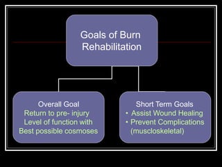
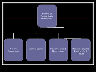
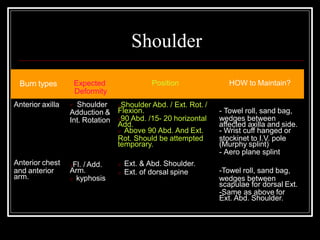
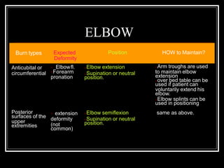
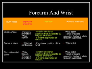
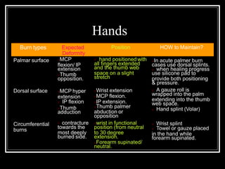
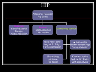
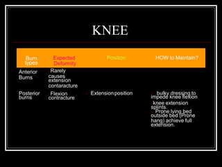
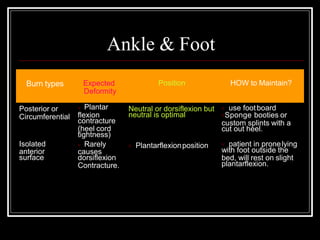
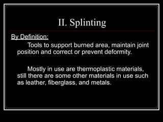
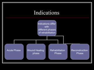
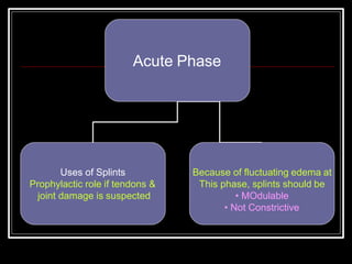
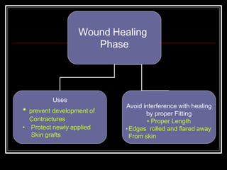
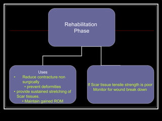
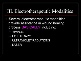
Goals of Burn
Rehabilitation
Overall Goal
Return to pre- injury
Level of function with
Best possible cosmoses
Short Term Goals
• Assist Wound Healing
• Prevent Complications
(muscloskeletal)
I. Positioning
 By definition positioning is:
The proper alignment and adjustment of
body parts.
 Positioning is a fundamental portion of burn
rehabilitation.
Benefits of
Positioning in
Burn Rehab.
Prevents
Contracture
Controls Edema Prevent Localized
Neuropathies
Maintain elongated
Position of soft
Tissues
 Burn patient has tendency to assume flexed
adducted position (Fetal position) most probably
as a reaction to pain.
 Positioning program is maintained and/ or
modified according to:
 Patient medical condition.
 ROM
 Skin condition.
 Positioning program should be individualized.
However, generally speaking, body parts should be
positioned as to maintain burned tissue in their
elongated state.
 Typically limbs should be positioned in extension-
abduction alignments.
 Positioning is maintained using splints, pillows,
and/ or foam wedges.
Specific Burn Sites
Body Segment
Anterior or
Circumferential
burns
Asymmetrical neck
burn
Head Burn that
Includes the ear
Posterior neck Burn
Ear not involvd
NECK BURNS
Burn types Expected
Deformity
Position HOW to Maintain?
Anterior or
Circumferential
burns
Flexion
Contracture
Extension/
Hyperextension
- Towel under shoulders or
between scapulae
- Foam cervical collar
Asymmetrical
neck burn
Lat. Fl.
Towards
burned side
Mid line
Or rotated away
--Towel roll, sand bag,
wedges on affected side.
- Prone lying head rotated
opposite side.
Head burns that
include the ear
Folding of the
Helix and
condritis
Avoid any
pressure over
the ear
- Foam or gel filled bag is
used to elevate the ear
from the bed.
Posterior neck
burns- Ear not
involved
Hyperextension
of the neck
Head in midline - Pillows are used to
elevate the head and
lengthen posterior tissues.
Trunk burns
Burn types Expected
Deformity
Position HOW to Maintain?
Clavicular &
pectoral shoulder retraction
From pectoral
region to below
umblicus
Same as above with upper
Back hyperextension
- A square towel or
blanket between
scapulae.
- Fig. of 8 wrapping
Same as above
with towel extended
downwards.
Burns of the
lower back
shoulder girdle
protraction and
glenohumeral
adduction
same as above
plus
kyphosis
Exaggerated
lordosis Midline position
Using pillows under
knee to flatten back
Lateral trunk
burn
Scoliosis concave
to burned side Maintain trunk straight
Towel roll, sand
bag, wedges on
affected side
Shoulder
Burn types Expected
Deformity
Position HOW to Maintain?
Anterior axilla  Shoulder
Adduction &
Int. Rotation
Shoulder Abd. / Ext. Rot. /
Flexion.
90 Abd. /15- 20 horizontal
Add.
 Above 90 Abd. And Ext.
Rot. Should be attempted
temporary.
- Towel roll, sand bag,
wedges between
affected axilla and side.
- Wrist cuff hanged or
stockinet to I.V. pole
(Murphy splint)
- Aero plane splint
Anterior chest
and anterior
arm.
Fl. / Add.
Arm.
 kyphosis
 Ext. & Abd. Shoulder.
 Ext. of dorsal spine -Towel roll, sand bag,
wedges between
scapulae for dorsal Ext.
-Same as above for
Ext. Abd. Shoulder.
ELBOW
Burn types Expected
Deformity
Position HOW to Maintain?
Anticubital or
circumferential
 Elbowfl.
Forearm
pronation
Elbow extension
Supination or neutral
position.
Posterior
surfaces of the
upper
extremities
 extension
deformity
(not
common)
Elbow semiflexion
Supination or neutral
position.
Arm troughs are used
to maintain elbow
extension
over bed table can be
used if patient can
voluntarily extend his
elbow.
Elbow splints can be
used in positioning
same as above.
Forearm And Wrist
Burn types Expected
Deformity
Position HOW to Maintain?
Volar surface Forearm
pronation
Wrist
flexion
 Wrist splint
 Towel or gauze
placed in the hand while
forearm supinated.
Dorsal surface  Wristext.
contracture
wrist in functional
position (from neutral to 30
degree extension.
Forearm supinated or
neutral.
Functional position of the
wrist
 Wristsplint
Circumferential
burns
Wrist
flexion.
Forearm
pronation
wrist in functional
position (from neutral to 30
degree extension.
Forearm supinated or
neutral.
 Wrist splint
 Towel or gauze
placed in the hand while
forearm supinated.
Hands
Burn types Expected
Deformity
Position HOW to Maintain?
Palmar surface MCP
flexion/ IP
extension
Thumb
opposition.
 hand positionedwith
all fingers extended
and the thumb web
space on a slight
stretch
Dorsal surface MCP hyper
extension
 IP flexion
Thumb
adduction
In acute palmer burn
cases use dorsal splints.
 when healing progress
use silicone pad to
provide both positioning
& pressure.
 A gauze roll is
wrapped into the palm
extending into the thumb
web space.
 Hand splint (Volar)
Circumferential
burns
 contracture
towards the
most deeply
burned side.
Wrist extension
MCP flexion.
IP extension.
Thumb palmer
abduction or
opposition
wrist in functional
position (from neutral
to 30 degree
extension.
Forearm supinated/
neutral.
 Wrist splint
 Towel or gauze placed
in the hand while
forearm supinated.
HIP
Anterior or Posterior
Hip Burns
Deformity
Flexion/ External
Rotation
And or Adduction
Position
• Slight Abduction
• Mid rotation
Maintaining position
Towel roll or sand
bag lat. To Thigh
For neutral rotation
▲ foam wedge
Blanket between legs
For hip abduction
Prone lying
minimize
Hip flexion
Knee ext. splint
Reduce hip flexion
With prone lying
KNEE
Burn
types
Expected
Deformity
Position HOW to Maintain?
Anterior
Burns
Posterior
burns
Rarely
causes
extension
contaracture
Flexion
contracture
 Extensionposition  bulky dressing to
impede knee flexion
knee extension
splints.
 Prone lying bed
outside bed (Prone
hang) achieve full
extension.
Ankle & Foot
Burn types Expected
Deformity
Position HOW to Maintain?
Posterior or
Circumferential
Neutral or dorsiflexion but
neutral is optimal
 use footboard
Sponge booties or
custom splints with a
cut out heel.
Isolated
anterior
surface
 Plantar
flexion
contracture
(heel cord
tightness)
 Rarely
causes
dorsiflexion
Contracture.
 Plantarflexionposition  patient in pronelying
with foot outside the
bed, will rest on slight
plantarflexion.
II. Splinting
By Definition:
Tools to support burned area, maintain joint
position and correct or prevent deformity.
Mostly in use are thermoplastic materials,
still there are some other materials in use such
as leather, fiberglass, and metals.
Indications
Indications differ
with
different phases
of rehabilitation
Acute Phase Wound Healing
phase
Rehabilitation
Phase
Reconstruction
Phase
Acute Phase
Uses of Splints
Prophylactic role if tendons &
joint damage is suspected
Because of fluctuating edema at
This phase, splints should be
• MOdulable
• Not Constrictive
Wound Healing
Phase
Uses
• prevent development of
Contractures
• Protect newly applied
Skin grafts
Avoid interference with healing
by proper Fitting
• Proper Length
•Edges rolled and flared away
From skin
Rehabilitation
Phase
Uses
• Reduce contracture non
surgically
• prevent deformities
• provide sustained stretching of
Scar tissues.
• Maintain gained ROM
If Scar tissue tensile strength is poor
Monitor for wound break down
Reconstructive
Phase
Uses
• For fixation following release of
Contractures or reconstruction
surgery
Monitor for wound Maceration
Examples Of Splints In Use
Region
Cervical
Ear
Mouth
Axilla and anterior chest
Splints
 Soft neck collar (foam)
 Philadelphia collar
 Molded neck splint
 Watusi collar (plastic tubes)
 Halo- neck collar
 Semi- rigid oxygen mask
 mouth spreader
 External traction hook
 Axilla air plane splint
 Clavicle figure of eight splint
Region
Elbow And Knee
Hip
Ankle
Wrist & Hand
Splints
 Gutter or trough splint
 Airslpint
 hip spica
 Abduction splint
 Spreader Bar
 Posterior foot drop
 High top gym shoe
 Anterior & posterior ankle conformer
 Wrist splint
 Thumb spica
 Thumb web spacer
III. Electrotherapeutic Modalities
 Several electrotherapeutic modalities
provide assistance in wound healing
process BASICALLY including:
 HVPGS.
 US THERAPY.
 ULTRAVIOLET RADIATIONS
 LASER
HVPGS
 There are several possible explanations of its
effect on wound healing:
1 Positive electrical stimulation stimulates repair
process.
2 Negative pole stimulation will destroy any
bacteria.
3 Increasing superficial circulation hastens
healing
Application
Parameters
Intensity
According to
patient tolerance.
Rate setting
Continuous
•Surged Pulse
rate 80 pulse/sec.
Electrodes
•Active (Usually
Anode) cover
treatment area.
•Dispersive (~
Cathode.) on the
back
Treatment Time
Time of treatment
20-30 minutes.
ULTRASOUND THERAPY
 Effects of US on wound healing include:
1- Promotion of formation of granulation tissue.
2- Accelerated re- epithelization.
3 It reduces wound infection, through improving
circulation (?!).
4 It improves scar pliability ( thus used in
hypertrophic scars).
5 Phonophoresis can be used to introduce
wound healing medications.
APPLICATION
IN CONTACT
•Using coupling media as
Paraffin oil, aquassonic gel,
Or aquasonic gel pad.
•Usually applied at wound
edges
SUB- AQUATIC
•Using suitably sized water
container and previously
boiled water.
• Usually applied to wound bed.
• Distance 1-5 cm from skin
.
ULTRAVIOLET RADIATIONS
UVR
1 Accelerates healing through facilitating mitosis
in the germinal layers of the skin.
2 Help in maintaining sterility through destroying
surface bacteria.
N.B.: High doses should be avoided at growing
wound edges as it may induce more skin
damage.
Notice
 If the condition shows wound infection high
exposure doses would be initially implemented.
 Avoid UVR in early stages of burn rehabilitation
(inflammatory stage of healing) as it may
aggravate the burn insult
LASER
Enhance
Fibroplasia
Enhance immune
Cells to attack
Pathogens
IncreaseATP
Synthesis
Quicken Collagen
Synthesis
Increase
Prostaglandins
EFFECTS
“ Bio-stimulation”
Types Of Laser In Use For Wound Healing
Types
Helium- Neon
(He-Ne)
632.8 nm
Galium- Aresnide
(Ga As)
Or Infrared Laser
(IR)
904 nm
Argon
(Ar)
488 – 514 nm
Carbon Dioxide
(CO2)
10.6 nm

Burns_and_rehabilitation.pptx

  • 1.
    Goals of Burn Rehabilitation OverallGoal Return to pre- injury Level of function with Best possible cosmoses Short Term Goals • Assist Wound Healing • Prevent Complications (muscloskeletal)
  • 2.
    I. Positioning  Bydefinition positioning is: The proper alignment and adjustment of body parts.  Positioning is a fundamental portion of burn rehabilitation.
  • 3.
    Benefits of Positioning in BurnRehab. Prevents Contracture Controls Edema Prevent Localized Neuropathies Maintain elongated Position of soft Tissues
  • 4.
     Burn patienthas tendency to assume flexed adducted position (Fetal position) most probably as a reaction to pain.  Positioning program is maintained and/ or modified according to:  Patient medical condition.  ROM  Skin condition.
  • 5.
     Positioning programshould be individualized. However, generally speaking, body parts should be positioned as to maintain burned tissue in their elongated state.  Typically limbs should be positioned in extension- abduction alignments.  Positioning is maintained using splints, pillows, and/ or foam wedges.
  • 6.
    Specific Burn Sites BodySegment Anterior or Circumferential burns Asymmetrical neck burn Head Burn that Includes the ear Posterior neck Burn Ear not involvd
  • 7.
    NECK BURNS Burn typesExpected Deformity Position HOW to Maintain? Anterior or Circumferential burns Flexion Contracture Extension/ Hyperextension - Towel under shoulders or between scapulae - Foam cervical collar Asymmetrical neck burn Lat. Fl. Towards burned side Mid line Or rotated away --Towel roll, sand bag, wedges on affected side. - Prone lying head rotated opposite side. Head burns that include the ear Folding of the Helix and condritis Avoid any pressure over the ear - Foam or gel filled bag is used to elevate the ear from the bed. Posterior neck burns- Ear not involved Hyperextension of the neck Head in midline - Pillows are used to elevate the head and lengthen posterior tissues.
  • 8.
    Trunk burns Burn typesExpected Deformity Position HOW to Maintain? Clavicular & pectoral shoulder retraction From pectoral region to below umblicus Same as above with upper Back hyperextension - A square towel or blanket between scapulae. - Fig. of 8 wrapping Same as above with towel extended downwards. Burns of the lower back shoulder girdle protraction and glenohumeral adduction same as above plus kyphosis Exaggerated lordosis Midline position Using pillows under knee to flatten back Lateral trunk burn Scoliosis concave to burned side Maintain trunk straight Towel roll, sand bag, wedges on affected side
  • 9.
    Shoulder Burn types Expected Deformity PositionHOW to Maintain? Anterior axilla  Shoulder Adduction & Int. Rotation Shoulder Abd. / Ext. Rot. / Flexion. 90 Abd. /15- 20 horizontal Add.  Above 90 Abd. And Ext. Rot. Should be attempted temporary. - Towel roll, sand bag, wedges between affected axilla and side. - Wrist cuff hanged or stockinet to I.V. pole (Murphy splint) - Aero plane splint Anterior chest and anterior arm. Fl. / Add. Arm.  kyphosis  Ext. & Abd. Shoulder.  Ext. of dorsal spine -Towel roll, sand bag, wedges between scapulae for dorsal Ext. -Same as above for Ext. Abd. Shoulder.
  • 10.
    ELBOW Burn types Expected Deformity PositionHOW to Maintain? Anticubital or circumferential  Elbowfl. Forearm pronation Elbow extension Supination or neutral position. Posterior surfaces of the upper extremities  extension deformity (not common) Elbow semiflexion Supination or neutral position. Arm troughs are used to maintain elbow extension over bed table can be used if patient can voluntarily extend his elbow. Elbow splints can be used in positioning same as above.
  • 11.
    Forearm And Wrist Burntypes Expected Deformity Position HOW to Maintain? Volar surface Forearm pronation Wrist flexion  Wrist splint  Towel or gauze placed in the hand while forearm supinated. Dorsal surface  Wristext. contracture wrist in functional position (from neutral to 30 degree extension. Forearm supinated or neutral. Functional position of the wrist  Wristsplint Circumferential burns Wrist flexion. Forearm pronation wrist in functional position (from neutral to 30 degree extension. Forearm supinated or neutral.  Wrist splint  Towel or gauze placed in the hand while forearm supinated.
  • 12.
    Hands Burn types Expected Deformity PositionHOW to Maintain? Palmar surface MCP flexion/ IP extension Thumb opposition.  hand positionedwith all fingers extended and the thumb web space on a slight stretch Dorsal surface MCP hyper extension  IP flexion Thumb adduction In acute palmer burn cases use dorsal splints.  when healing progress use silicone pad to provide both positioning & pressure.  A gauze roll is wrapped into the palm extending into the thumb web space.  Hand splint (Volar) Circumferential burns  contracture towards the most deeply burned side. Wrist extension MCP flexion. IP extension. Thumb palmer abduction or opposition wrist in functional position (from neutral to 30 degree extension. Forearm supinated/ neutral.  Wrist splint  Towel or gauze placed in the hand while forearm supinated.
  • 13.
    HIP Anterior or Posterior HipBurns Deformity Flexion/ External Rotation And or Adduction Position • Slight Abduction • Mid rotation Maintaining position Towel roll or sand bag lat. To Thigh For neutral rotation ▲ foam wedge Blanket between legs For hip abduction Prone lying minimize Hip flexion Knee ext. splint Reduce hip flexion With prone lying
  • 14.
    KNEE Burn types Expected Deformity Position HOW toMaintain? Anterior Burns Posterior burns Rarely causes extension contaracture Flexion contracture  Extensionposition  bulky dressing to impede knee flexion knee extension splints.  Prone lying bed outside bed (Prone hang) achieve full extension.
  • 15.
    Ankle & Foot Burntypes Expected Deformity Position HOW to Maintain? Posterior or Circumferential Neutral or dorsiflexion but neutral is optimal  use footboard Sponge booties or custom splints with a cut out heel. Isolated anterior surface  Plantar flexion contracture (heel cord tightness)  Rarely causes dorsiflexion Contracture.  Plantarflexionposition  patient in pronelying with foot outside the bed, will rest on slight plantarflexion.
  • 16.
    II. Splinting By Definition: Toolsto support burned area, maintain joint position and correct or prevent deformity. Mostly in use are thermoplastic materials, still there are some other materials in use such as leather, fiberglass, and metals.
  • 17.
    Indications Indications differ with different phases ofrehabilitation Acute Phase Wound Healing phase Rehabilitation Phase Reconstruction Phase
  • 18.
    Acute Phase Uses ofSplints Prophylactic role if tendons & joint damage is suspected Because of fluctuating edema at This phase, splints should be • MOdulable • Not Constrictive
  • 19.
    Wound Healing Phase Uses • preventdevelopment of Contractures • Protect newly applied Skin grafts Avoid interference with healing by proper Fitting • Proper Length •Edges rolled and flared away From skin
  • 20.
    Rehabilitation Phase Uses • Reduce contracturenon surgically • prevent deformities • provide sustained stretching of Scar tissues. • Maintain gained ROM If Scar tissue tensile strength is poor Monitor for wound break down
  • 21.
    Reconstructive Phase Uses • For fixationfollowing release of Contractures or reconstruction surgery Monitor for wound Maceration
  • 22.
    Examples Of SplintsIn Use Region Cervical Ear Mouth Axilla and anterior chest Splints  Soft neck collar (foam)  Philadelphia collar  Molded neck splint  Watusi collar (plastic tubes)  Halo- neck collar  Semi- rigid oxygen mask  mouth spreader  External traction hook  Axilla air plane splint  Clavicle figure of eight splint
  • 23.
    Region Elbow And Knee Hip Ankle Wrist& Hand Splints  Gutter or trough splint  Airslpint  hip spica  Abduction splint  Spreader Bar  Posterior foot drop  High top gym shoe  Anterior & posterior ankle conformer  Wrist splint  Thumb spica  Thumb web spacer
  • 24.
    III. Electrotherapeutic Modalities Several electrotherapeutic modalities provide assistance in wound healing process BASICALLY including:  HVPGS.  US THERAPY.  ULTRAVIOLET RADIATIONS  LASER
  • 25.
    HVPGS  There areseveral possible explanations of its effect on wound healing: 1 Positive electrical stimulation stimulates repair process. 2 Negative pole stimulation will destroy any bacteria. 3 Increasing superficial circulation hastens healing
  • 26.
    Application Parameters Intensity According to patient tolerance. Ratesetting Continuous •Surged Pulse rate 80 pulse/sec. Electrodes •Active (Usually Anode) cover treatment area. •Dispersive (~ Cathode.) on the back Treatment Time Time of treatment 20-30 minutes.
  • 27.
    ULTRASOUND THERAPY  Effectsof US on wound healing include: 1- Promotion of formation of granulation tissue. 2- Accelerated re- epithelization. 3 It reduces wound infection, through improving circulation (?!). 4 It improves scar pliability ( thus used in hypertrophic scars). 5 Phonophoresis can be used to introduce wound healing medications.
  • 28.
    APPLICATION IN CONTACT •Using couplingmedia as Paraffin oil, aquassonic gel, Or aquasonic gel pad. •Usually applied at wound edges SUB- AQUATIC •Using suitably sized water container and previously boiled water. • Usually applied to wound bed. • Distance 1-5 cm from skin .
  • 29.
    ULTRAVIOLET RADIATIONS UVR 1 Accelerateshealing through facilitating mitosis in the germinal layers of the skin. 2 Help in maintaining sterility through destroying surface bacteria. N.B.: High doses should be avoided at growing wound edges as it may induce more skin damage.
  • 30.
    Notice  If thecondition shows wound infection high exposure doses would be initially implemented.  Avoid UVR in early stages of burn rehabilitation (inflammatory stage of healing) as it may aggravate the burn insult
  • 31.
    LASER Enhance Fibroplasia Enhance immune Cells toattack Pathogens IncreaseATP Synthesis Quicken Collagen Synthesis Increase Prostaglandins EFFECTS “ Bio-stimulation”
  • 32.
    Types Of LaserIn Use For Wound Healing Types Helium- Neon (He-Ne) 632.8 nm Galium- Aresnide (Ga As) Or Infrared Laser (IR) 904 nm Argon (Ar) 488 – 514 nm Carbon Dioxide (CO2) 10.6 nm