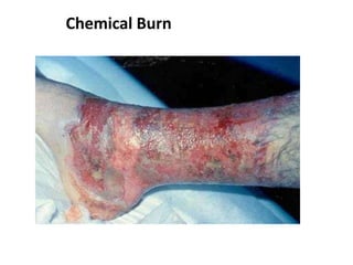
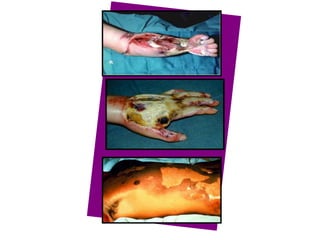
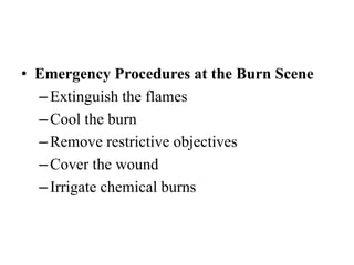
1. A burn is an injury to the skin or flesh caused by heat, electricity, chemicals, friction or radiation. The severity depends on the temperature and duration of exposure.
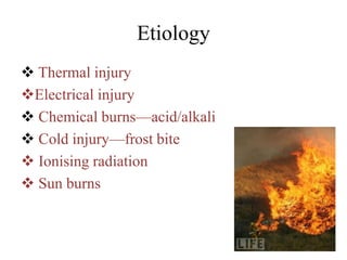
2. About 2.4 million people suffer burns annually in the US, with 700,000 cases requiring medical treatment. The main causes are thermal, electrical, chemical and radiation burns.
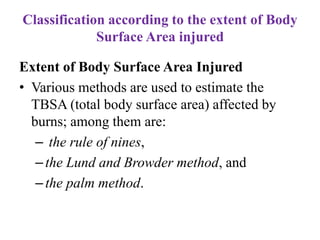
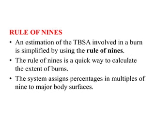
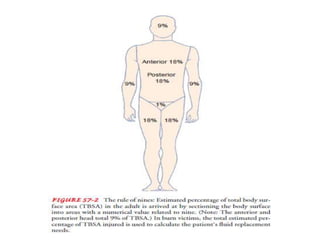
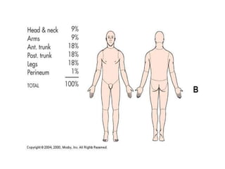
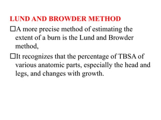
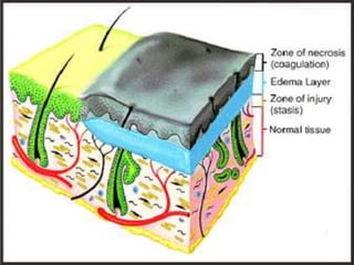
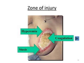
3. Burns are classified by depth and extent of the affected body surface area. Depth is classified as superficial, partial-thickness, or full-thickness. Extent is classified using methods like the Rule of Nines or Lund and Browder chart.
A burn is a type of injury to skin, or other tissues, caused by heat, cold, electricity, chemicals, friction, or radiation. Most burns are due to heat from hot liquids, solids, or fire. While rates are similar for males and females the underlying causes often differ.
BURN - Presented By Mohammed Haroon Rashid Haroon Rashid
Subject is Medical Surgical Nursing - II & Topic is Burn, Presented by Mohammed Haroon Rashid Basci B.Sc Nursing 3rd Year in Florence College of Nursing
medical surgical nursing , nursing management of burn patients, it includes definition, classification of burn injury, clinical manifestaion, assessment of burn injury , management of patient with burn, care given to the patient.
Lung Cancer: Artificial Intelligence, Synergetics, Complex System Analysis, S...Oleg Kshivets
RESULTS: Overall life span (LS) was 2252.1±1742.5 days and cumulative 5-year survival (5YS) reached 73.2%, 10 years – 64.8%, 20 years – 42.5%. 513 LCP lived more than 5 years (LS=3124.6±1525.6 days), 148 LCP – more than 10 years (LS=5054.4±1504.1 days).199 LCP died because of LC (LS=562.7±374.5 days). 5YS of LCP after bi/lobectomies was significantly superior in comparison with LCP after pneumonectomies (78.1% vs.63.7%, P=0.00001 by log-rank test). AT significantly improved 5YS (66.3% vs. 34.8%) (P=0.00000 by log-rank test) only for LCP with N1-2. Cox modeling displayed that 5YS of LCP significantly depended on: phase transition (PT) early-invasive LC in terms of synergetics, PT N0—N12, cell ratio factors (ratio between cancer cells- CC and blood cells subpopulations), G1-3, histology, glucose, AT, blood cell circuit, prothrombin index, heparin tolerance, recalcification time (P=0.000-0.038). Neural networks, genetic algorithm selection and bootstrap simulation revealed relationships between 5YS and PT early-invasive LC (rank=1), PT N0—N12 (rank=2), thrombocytes/CC (3), erythrocytes/CC (4), eosinophils/CC (5), healthy cells/CC (6), lymphocytes/CC (7), segmented neutrophils/CC (8), stick neutrophils/CC (9), monocytes/CC (10); leucocytes/CC (11). Correct prediction of 5YS was 100% by neural networks computing (area under ROC curve=1.0; error=0.0).
CONCLUSIONS: 5YS of LCP after radical procedures significantly depended on: 1) PT early-invasive cancer; 2) PT N0--N12; 3) cell ratio factors; 4) blood cell circuit; 5) biochemical factors; 6) hemostasis system; 7) AT; 8) LC characteristics; 9) LC cell dynamics; 10) surgery type: lobectomy/pneumonectomy; 11) anthropometric data. Optimal diagnosis and treatment strategies for LC are: 1) screening and early detection of LC; 2) availability of experienced thoracic surgeons because of complexity of radical procedures; 3) aggressive en block surgery and adequate lymph node dissection for completeness; 4) precise prediction; 5) adjuvant chemoimmunoradiotherapy for LCP with unfavorable prognosis.
Pulmonary Thromboembolism - etilogy, types, medical- Surgical and nursing man...VarunMahajani
Disruption of blood supply to lung alveoli due to blockage of one or more pulmonary blood vessels is called as Pulmonary thromboembolism. In this presentation we will discuss its causes, types and its management in depth.
New Directions in Targeted Therapeutic Approaches for Older Adults With Mantl...i3 Health
i3 Health is pleased to make the speaker slides from this activity available for use as a non-accredited self-study or teaching resource.
This slide deck presented by Dr. Kami Maddocks, Professor-Clinical in the Division of Hematology and
Associate Division Director for Ambulatory Operations
The Ohio State University Comprehensive Cancer Center, will provide insight into new directions in targeted therapeutic approaches for older adults with mantle cell lymphoma.
STATEMENT OF NEED
Mantle cell lymphoma (MCL) is a rare, aggressive B-cell non-Hodgkin lymphoma (NHL) accounting for 5% to 7% of all lymphomas. Its prognosis ranges from indolent disease that does not require treatment for years to very aggressive disease, which is associated with poor survival (Silkenstedt et al, 2021). Typically, MCL is diagnosed at advanced stage and in older patients who cannot tolerate intensive therapy (NCCN, 2022). Although recent advances have slightly increased remission rates, recurrence and relapse remain very common, leading to a median overall survival between 3 and 6 years (LLS, 2021). Though there are several effective options, progress is still needed towards establishing an accepted frontline approach for MCL (Castellino et al, 2022). Treatment selection and management of MCL are complicated by the heterogeneity of prognosis, advanced age and comorbidities of patients, and lack of an established standard approach for treatment, making it vital that clinicians be familiar with the latest research and advances in this area. In this activity chaired by Michael Wang, MD, Professor in the Department of Lymphoma & Myeloma at MD Anderson Cancer Center, expert faculty will discuss prognostic factors informing treatment, the promising results of recent trials in new therapeutic approaches, and the implications of treatment resistance in therapeutic selection for MCL.
Target Audience
Hematology/oncology fellows, attending faculty, and other health care professionals involved in the treatment of patients with mantle cell lymphoma (MCL).
Learning Objectives
1.) Identify clinical and biological prognostic factors that can guide treatment decision making for older adults with MCL
2.) Evaluate emerging data on targeted therapeutic approaches for treatment-naive and relapsed/refractory MCL and their applicability to older adults
3.) Assess mechanisms of resistance to targeted therapies for MCL and their implications for treatment selection
Explore natural remedies for syphilis treatment in Singapore. Discover alternative therapies, herbal remedies, and lifestyle changes that may complement conventional treatments. Learn about holistic approaches to managing syphilis symptoms and supporting overall health.
Report Back from SGO 2024: What’s the Latest in Cervical Cancer?bkling
Are you curious about what’s new in cervical cancer research or unsure what the findings mean? Join Dr. Emily Ko, a gynecologic oncologist at Penn Medicine, to learn about the latest updates from the Society of Gynecologic Oncology (SGO) 2024 Annual Meeting on Women’s Cancer. Dr. Ko will discuss what the research presented at the conference means for you and answer your questions about the new developments.
Factory Supply Best Quality Pmk Oil CAS 28578–16–7 PMK Powder in Stockrebeccabio
Factory Supply Best Quality Pmk Oil CAS 28578–16–7 PMK Powder in Stock
Telegram: bmksupplier
signal: +85264872720
threema: TUD4A6YC
You can contact me on Telegram or Threema
Communicate promptly and reply
Free of customs clearance, Double Clearance 100% pass delivery to USA, Canada, Spain, Germany, Netherland, Poland, Italy, Sweden, UK, Czech Republic, Australia, Mexico, Russia, Ukraine, Kazakhstan.Door to door service
Hot Selling Organic intermediates
Title: Sense of Taste
Presenter: Dr. Faiza, Assistant Professor of Physiology
Qualifications:
MBBS (Best Graduate, AIMC Lahore)
FCPS Physiology
ICMT, CHPE, DHPE (STMU)
MPH (GC University, Faisalabad)
MBA (Virtual University of Pakistan)
Learning Objectives:
Describe the structure and function of taste buds.
Describe the relationship between the taste threshold and taste index of common substances.
Explain the chemical basis and signal transduction of taste perception for each type of primary taste sensation.
Recognize different abnormalities of taste perception and their causes.
Key Topics:
Significance of Taste Sensation:
Differentiation between pleasant and harmful food
Influence on behavior
Selection of food based on metabolic needs
Receptors of Taste:
Taste buds on the tongue
Influence of sense of smell, texture of food, and pain stimulation (e.g., by pepper)
Primary and Secondary Taste Sensations:
Primary taste sensations: Sweet, Sour, Salty, Bitter, Umami
Chemical basis and signal transduction mechanisms for each taste
Taste Threshold and Index:
Taste threshold values for Sweet (sucrose), Salty (NaCl), Sour (HCl), and Bitter (Quinine)
Taste index relationship: Inversely proportional to taste threshold
Taste Blindness:
Inability to taste certain substances, particularly thiourea compounds
Example: Phenylthiocarbamide
Structure and Function of Taste Buds:
Composition: Epithelial cells, Sustentacular/Supporting cells, Taste cells, Basal cells
Features: Taste pores, Taste hairs/microvilli, and Taste nerve fibers
Location of Taste Buds:
Found in papillae of the tongue (Fungiform, Circumvallate, Foliate)
Also present on the palate, tonsillar pillars, epiglottis, and proximal esophagus
Mechanism of Taste Stimulation:
Interaction of taste substances with receptors on microvilli
Signal transduction pathways for Umami, Sweet, Bitter, Sour, and Salty tastes
Taste Sensitivity and Adaptation:
Decrease in sensitivity with age
Rapid adaptation of taste sensation
Role of Saliva in Taste:
Dissolution of tastants to reach receptors
Washing away the stimulus
Taste Preferences and Aversions:
Mechanisms behind taste preference and aversion
Influence of receptors and neural pathways
Impact of Sensory Nerve Damage:
Degeneration of taste buds if the sensory nerve fiber is cut
Abnormalities of Taste Detection:
Conditions: Ageusia, Hypogeusia, Dysgeusia (parageusia)
Causes: Nerve damage, neurological disorders, infections, poor oral hygiene, adverse drug effects, deficiencies, aging, tobacco use, altered neurotransmitter levels
Neurotransmitters and Taste Threshold:
Effects of serotonin (5-HT) and norepinephrine (NE) on taste sensitivity
Supertasters:
25% of the population with heightened sensitivity to taste, especially bitterness
Increased number of fungiform papillae
HOT NEW PRODUCT! BIG SALES FAST SHIPPING NOW FROM CHINA!! EU KU DB BK substit...GL Anaacs
Contact us if you are interested:
Email / Skype : kefaya1771@gmail.com
Threema: PXHY5PDH
New BATCH Ku !!! MUCH IN DEMAND FAST SALE EVERY BATCH HAPPY GOOD EFFECT BIG BATCH !
Contact me on Threema or skype to start big business!!
Hot-sale products:
NEW HOT EUTYLONE WHITE CRYSTAL!!
5cl-adba precursor (semi finished )
5cl-adba raw materials
ADBB precursor (semi finished )
ADBB raw materials
APVP powder
5fadb/4f-adb
Jwh018 / Jwh210
Eutylone crystal
Protonitazene (hydrochloride) CAS: 119276-01-6
Flubrotizolam CAS: 57801-95-3
Metonitazene CAS: 14680-51-4
Payment terms: Western Union,MoneyGram,Bitcoin or USDT.
Deliver Time: Usually 7-15days
Shipping method: FedEx, TNT, DHL,UPS etc.Our deliveries are 100% safe, fast, reliable and discreet.
Samples will be sent for your evaluation!If you are interested in, please contact me, let's talk details.
We specializes in exporting high quality Research chemical, medical intermediate, Pharmaceutical chemicals and so on. Products are exported to USA, Canada, France, Korea, Japan,Russia, Southeast Asia and other countries.
Couples presenting to the infertility clinic- Do they really have infertility...Sujoy Dasgupta
Dr Sujoy Dasgupta presented the study on "Couples presenting to the infertility clinic- Do they really have infertility? – The unexplored stories of non-consummation" in the 13th Congress of the Asia Pacific Initiative on Reproduction (ASPIRE 2024) at Manila on 24 May, 2024.
2. Definition
• A burn is a type of injury to the flesh or skin which can
be caused by heat, electricity, chemicals, friction or
radiation
3. • Human skin can tolerate temperatures up to 42-440 C
(107-1110 F) but above these, the higher the
temperature the more severe the tissue destruction
• Below 450 C (1130 F), resulting changes are
reversible but >450 C, protein damage exceeds the
capacity of the cell to repair
4. Incidence
• About 2.4 million people suffer burns annually
• Account for an estimated 700,000 cases visits per year and
45,000 require hospitalizations
• Between 8,000-12,000 burn patients die, and approximately
one million will sustain substantial or permanent disabilities
7. CLASSIFICATION OF BURNS BY
CAUSATIVE AGENTS
Thermal Burns
–Caused by flame, flash, scald, or contact
with hot objects
–It is the most common type of burn
Chemical Burns
• Result from tissue injury and destruction
from necrotizing substances (chemicals)
• Most commonly caused by acids
8. Smoke Inhalation Injuries
Result from inhalation of hot air or noxious chemicals
Cause damage to respiratory tract
Important determinant of mortality in fire victims
Eg :Carbon monoxide (CO) poisoning
– CO is produced by the incomplete combustion of burning
materials
– Inhaled CO displaces oxygen 200 x more powerful than
oxygen
– CO is colorless, odorless and tasteless
9. Signs and Symptoms
Presence of facial burns
Singed nasal hair
Hoarseness,
painful swallowing
Darkened oral and nasal membranes
Wheezing on auscultation
Edema is the nose and airways
Flushing
Nausea/vomiting
Syncope, coma, death
10. Electrical Burns
• Intense heat generated from an electrical
current
• May result from direct damage to nerves and
vessels causing tissue anoxia and death
• Severity of injury depends on the amount of
voltage, tissue resistance, current pathways,
surface area, and on the length of time of the
flow
12. Cold Thermal Injury (Frostbite)
– Usually affects fingers, toes, nose, and ears
– Numbness, pallor, severe pain, swelling,
edema
• resulting from prolonged exposure to
freezing or subfreezing temperatures.
13. Classification according to the depth of
tissue destruction
1. Superficial partial-thickness injuries (first
degree burn):
In a superficial partial-thickness burn,
the epidermis is destroyed or injured and
a portion of the dermis may be injured.
The damaged skin may be painful and
appear red and dry, as in sunburn, or it may
blister (very minimal).
14. Typical Characteristics for Superficial
thickness burn
–Mild to severe erythema (pink to red)
–NO BLISTERS
–Skin blanches
–Painful, tingling
–Pain responds well to cooling
–Lasts about 48 hours; healing in 3-7 days
15. 2. Deep partial-thickness injuries (second
degree burn):
• A deep partial-thickness burn involves
destruction of the epidermis and upper layers
of the dermis and injury to deeper portions of
the dermis.
• The wound is painful, appears red, and exudes
fluid.
• Capillary refill follows tissue blanching. Hair
follicles remain intact.
• Deep partial-thickness burns take longer to
heal and are more likely to result in
hypertrophic scars.
16. Typical Characteristics for deep partial
thickness burn
–Large blisters over an extensive area
–Edema
–Red base with broken epidermis
–Wet, shiny and weeping
–Sensitive to cold air
–Healing in 2-3 weeks
–Grafts MAY be needed
18. 3. Ful-thickness injuries (third degree burn):
• A full-thickness burn involves total destruction of
epidermis and dermis and, in some cases,
underlying tissue as well.
• Wound color ranges widely from white to red,
brown, or black. The burned area is painless
because nerve fibers are destroyed.
• The wound appears leathery; hair follicles and
sweat glands are destroyed
19. Typical Characteristics for Full-thickness burn
• Deep, red, black, white, yellow, or brown area
• Edema
• Tissue open with fat exposed
• Little to no pain
• Requires removal of eschar and skin grafting
• Scarring and contractures are likely
• Takes weeks to months to heal
20.
21. Classification according to the extent of Body
Surface Area injured
Extent of Body Surface Area Injured
• Various methods are used to estimate the
TBSA (total body surface area) affected by
burns; among them are:
– the rule of nines,
–the Lund and Browder method, and
–the palm method.
22. RULE OF NINES
• An estimation of the TBSA involved in a burn
is simplified by using the rule of nines.
• The rule of nines is a quick way to calculate
the extent of burns.
• The system assigns percentages in multiples of
nine to major body surfaces.
23.
24.
25. LUND AND BROWDER METHOD
A more precise method of estimating the
extent of a burn is the Lund and Browder
method,
It recognizes that the percentage of TBSA of
various anatomic parts, especially the head and
legs, and changes with growth.
26.
27. PALM METHOD
• In patients with scattered burns, a method to
estimate the percentage of burn is the palm
method.
• The size of the patient’s palm is approximately
1% of TBSA.
28. Criteria for Classifying the Extent of Burn
Injury(American Burn Association)
Minor Burn Injury
• Second-degree burn of less than 15% total body
surface area(TBSA) in adults or less than 10%
TBSA in children
• Third-degree burn of less than 2% TBSA not
involving special care areas (eyes, ears, face,
hands, feet, perineum, joints)
• Excludes electrical injury, inhalation injury,
concurrent trauma, all poor-risk patients (eg,
extremes of age, concurrent disease)
29. Moderate, Uncomplicated Burn Injury
• Second-degree burns of 15%–25% TBSA in
adults or10%–20% in children
• Third-degree burns of less than 10% TBSA not
involving special care areas
• Excludes electrical injury, inhalation injury,
concurrent trauma, all poor-risk patients (eg,
extremes of age, concurrent disease)
30. Major Burn Injury
• Second-degree burns exceeding 25% TBSA in
adults or 20% in children
• All third-degree burns exceeding 10% TBSA
• All burns involving eyes, ears, face, hands,
feet, perineum, joints
• All inhalation injury, electrical injury,
concurrent trauma, all poor-risk patients
31.
32. 32
Blister may ↑size because continuous
exudation and collection of tissue fluid
35. Burn Photos
Electrical Burns
Entrance Wounds
Electrical Burns
Exit Wounds
Entrance wound of electrical
burns from an overheated tool
Severe swelling
peaks 24-72 hrs after
Electrical burns mummified
1st 2 fingers later removed
38. Pathologic Features
• Zone of coagulation (necrosis): Superficial area of
coagulation necrosis and cell death on exposure to
temperatures >450 (primary injury)
• Zone of stasis (vascular thrombosis): Local capillary
circulation is sluggish, depending on the adequacy of the
resuscitation, can either remain viable or proceed to cell
death (secondary injury)
• Zone of hyperemia (increased capillary permeability)
42. LOCAL AND SYSTEMIC RESPONSES
TO BURNS
• Burns that do not exceed 25% TBSA produce a
primarily local response.
• Burns that exceed 25% TBSA may produce
both a local and a systemic response and are
considered major burn injuries.
• These systemic responses are due to the
release of cytokines and other mediators into
the systemic circulation and include the
following:
43. Effects of a severe burn
1. Local response include tissue edema
effects on fluid, electrolytes and blood volume
2. Cardiovascular
3. Respiratory
4. Immune
5. Integumentary
6. Gastrointestinal
7. Urinary
43
44. Cardiovascular system
• Blood pressure falls-fluid leaks from intravascular
to interstitial (sodium and protein)
• Hypotension
• tachycardia
• Blood flow in intravascular is concentrated and
cause static.
• Cardiac output ↓,
• Due to that tissue perfusion ↓,
45. Hematologic changes
• Thrombocytopenia, abnormal platelet function,
depressed fibrinogen levels, deficit plasma
clotting factors.
• Life span ↓RBC.
• Blood loss during diagnostic and therapeutic
procedure.
46. Respiration system
• Majority of deaths from fire are due to smoke
inhalation.
• Pulmonary damage can be from direct inhalation
injury or systemic respond to the injury.
• Damage to cilia and cell in the airway-
inflammation.
• Mucociliary transport mechanism not
functioning-bronchial congestion and infection.
• Pulmonary edema, fluids escape to interstitial.
• Airway obstruction
47.
48. Gastrointestinal
• Burn >20% experience ↓peristalsis, gastric
distention and ↑risk of aspiration.
• Paralytic ileus due to secondary to burn trauma.
• Stress ulcer (stomach/duodenum) due to burn
injury.
• Indication of stress ulcer-malena stool or
hematemesis.
• These signs suggest gastric or duodenal erosion
(Curling`s ulcer)
• Gastric distention and nausea may lead to
vomiting.
49. CURLING’S ULCER
• Acute ulcerative gastro duodenal disease
• Occur within 24 hours after burn
• Due to reduced GI blood flow and
mucosal damage
• Treat clients with H2 blockers,
mucoprotectants, and early enteral
nutrition
• Watch for sudden drop in hemoglobin
4/1/2011 49
50. Immunologic changes
• Skin barrier to invading organisms s destroyed,
circulating levels of immunoglobulins are ↓
• Changes in WBC both quantitative and
qualitative.
• Depression of neutrophil, phagocytic and
bactericidal activity is found after burn injury.
• All this changes in the immune system can
make the burn patient more susceptible to
infection
52. Diagnosis
• A detailed history and physical
examination is the first step. The
physician will evaluate the type, duration, and
timing of the burn; the burn location and
severity; and associated dehydration,
disfigurement, and infection.
• Fires in enclosed spaces should raise the
suspicion for smoke–inhalation injury.
53. Lab Tests
• Routine blood work for
a patient with a burn
injury includes a complete
blood count, platelet
count, clotting studies,
liver function studies, and
carboxyhemoglobin,
electrolyte, blood urea
nitrogen, glucose and
creatinine levels.
• Urinalysis may reveal
myoglobinuria and
hemoglobinuria.
• If pt. is 35 or older,
he’ll also need an
electrocardiogram.
• Chest x-rays and
arterial blood gas levels
allow the evaluation of
alveolar function.
54. MANAGEMENT OF THE BURN INJURY
Phases of Management
• Burn care then proceeds through three phases:
– Emergent/resuscitative phase (on-the-scene care),
– Acute/intermediate phase, and
– Rehabilitation phase.
55. Phase Duration Priorities
Emergent or
immediate
resuscitative
From onset of injury to
completion
of fluid resuscitation
First aid
Prevention of shock
Prevention of respiratory distress
Detection and treatment of
concomitant injuries
Wound assessment and initial care
Acute From beginning of diuresis
to near
completion of wound
closure
Wound care and closure
Prevention or treatment of
complications, including infection
Nutritional support
Rehabilitation From major wound closure
to return
to individual’s optimal level
of physical
and psychosocial
adjustment
Prevention of scars and
contractures
Physical, occupational, and
vocational rehabilitation
Functional and cosmetic
reconstruction
Psychosocial counseling
57. • Emergency Procedures at the Burn Scene
–Extinguish the flames
–Cool the burn
–Remove restrictive objectives
–Cover the wound
–Irrigate chemical burns
58. • Emergency Medical Management
• The patient is transported to the nearest
emergency department.
• The hospital nurses (staff) and physician are
alerted that the patient is in route to the
emergency department so that life-saving
measures can be initiated immediately by a
trained team.
• Initial priorities in the emergency department
remain airway, breathing, and circulation.
59. • For mild pulmonary injury, inspired air is
humidified and the patient is encouraged to cough
so that secretions can be removed by suctioning.
• For more severe situations, it is necessary to
remove secretions by bronchial suctioning and to
administer bronchodilators and mucolytic agents.
• If edema of the airway develops, endotracheal
intubation may be necessary.
60. • Continuous positive airway pressure and
mechanical ventilation may also be required to
achieve adequate oxygenation.
• A large-bore (16- or 18-gauge) intravenous
catheter should be inserted in a non-burned
area (if not inserted earlier).
61. Management of fluid loss and shock
Fluid Replacement Therapy:
• The total volume and rate of intravenous fluid
replacement are gauged by the patient’s response.
• The adequacy of fluid resuscitation is determined
by:
– Output totals of 30 to 50 mL/hour
– systolic blood pressure exceeding 100 mm Hg
and/or
– pulse rate less than 110/minute.
63. Fluid Requirements:
• The projected fluid requirements for the first 24
hours are calculated by the clinician based on
the extent of the burn injury.
• Some combination of fluid categories may be
used:
–Colloids (whole blood, plasma, and plasma
expanders) and
– Crystalloids/electrolytes (physiologic sodium
chloride or lactated Ringer’s solution).
64. Fluid Requirements:
• Adequate fluid resuscitation results in slightly
decreased blood volume levels during the first
24 post-burn hours and restores plasma levels
to normal by the end of 48 hours.
• Oral resuscitation can be successful in adults
with less than 20% TBSA and children with
less than 10% to 15% TBSA.
65. Guidelines and Formulas for Fluid
Replacement in Burn Patients
Consensus Formula
• Lactated Ringer’s solution (or other balanced
saline solution): 2–4 mL× kg body weight × %
total body surface area (TBSA) burned.
• Half to be given in first 8 hours; remaining
half to be given over next 16 hours.
66. • The following example illustrates use of the
formula in a management of a 70-kg patient
with a 50% TBSA burn:
• Steps
–1, Consensus formula: 2 to 4 mL/kg/% TBSA
–2, 2 × 70 × 50 = 7,000 mL/24 hours
–3, Plan to administer: First 8 hours = 3,500
mL, or 437 mL/ hour; next 16 hours = 3,500
mL, or 219 mL/hour
67. Evans Formula
• 1. Colloids: 1 mL × kg body weight × % TBSA burned
• 2. Electrolytes (saline): 1 mL × body weight × % TBSA
burned
• 3. Glucose (5% in water): 2,000 mL for insensible loss
• Day 1: Half to be given in first 8 hours; remaining half
over next 16 hours
• Day 2: Half of previous day’s colloids and electrolytes;
all of insensible fluid replacement
• Maximum of 10,000 mL over 24 hours. Second- and
third-degree
• (partial- and full-thickness) burns exceeding 50% TBSA
are calculated
• on the basis of 50% TBSA.
68. Brooke Army Formula
• 1. Colloids: 0.5 mL × kg body weight × %
TBSA burned
• 2. Electrolytes (lactated Ringer’s solution): 1.5
mL × kg body weight × % TBSA burned
• 3. Glucose (5% in water): 2,000 mL for
insensible loss
69. Parkland/Baxter Formula
• Lactated Ringer’s solution: 4 mL × kg body
weight × % TBSA burned
• Day 1: Half to be given in first 8 hours; half to
be given over next16 hours
• Day 2: Varies. Colloid is added.
70. Hypertonic Saline Solution
• Concentrated solutions of sodium chloride (NaCl)
and lactate with concentration of 250–300 mEq of
sodium per liter, administered at a rate sufficient
to maintain a desired volume of urinary output.
• Do not increase the infusion rate during the first 8
post burn hours.
• Serum sodium levels must be monitored closely.
• Goal: Increase serum sodium level and osmolality
to reduce edema and prevent pulmonary
complications.
72. ACUTE PHASE
- Begins when the client is hemodynamically
stable, capillary permeability is restored, and
diuresis has begun
- Usually begins 48 - 72 hours after the time of
injury
73. - Emphasis during this phase is placed on
restorative therapy, and the phase continues
until wound closure is achieved
- The focus is on infection control, wound care,
wound closure, nutritional support, pain
management, and physical therapy
74. Pathophysiology
• Diuresis from fluid mobilization occurs, and
the patient is no longer grossly edematous
• Bowel sounds return
• Healing begins
• Formation of granulation tissue
• A partial-thickness burn wound will heal from
the edges
• Full-thickness burns must be covered by skin
grafts
75. • Wound Care
• Daily observation
• Assessment
• Cleansing
• Debridement
• Appropriate coverage of the graft:
– Fine-mesh gauze next to the graft followed by
middle and outer dressings
– Sheet skin grafts must be kept free of
blebs (small blisters)
76. Excision and Grafting
• Eschar is removed down to the subcutaneous
tissue or fascia
• Cultured Epithelial Autographs (CEA): CEA is
grown from biopsies obtained from the
patient’s own skin
• Artificial Skin: used when life-threatening full-
thickness or deep partial-thickness wounds
where conventional autograft is not available
or advisable
77. Pain Management
• Opioid every 1 to 3 hours for pain
• Several drugs in combination
• Morphine with haloperidol
• Nonpharmacologic strategies
• Relaxation tapes
• Visualization, guided imagery
• Meditation
82. REHABILITATION PHASE
• The rehabilitation phase is defined as
beginning when the patient’s burn wounds are
covered with skin or healed and the patient is
able to resume a level of self-care activity
• Complications
– Skin and joint contractures
– Hypertrophic scarring
82
83. Rehabilitation Phase
• Both patient and family actively learn how to
care for healing wounds
• Cosmetic surgery is often needed following
major burns
• Role of exercise cannot be overemphasized
• Constant encouragement and reassurance
• Address spiritual and cultural needs
• Maintain a high-calorie, high-protein diet
• Occupational therapy 83
84.
85. Definitions
Graft
• A skin graft is a tissue of epidermis and varying
amounts of dermis that is detached from its own
blood supply and placed in a new area with a new
blood supply.
• Skin graft is a segment of skin separated from the
donor site and transplanted to the recipient site
devoid of its blood supply
86. Indications for Grafts
• Extensive wounds.
• Burns.
• Specific surgeries that may require skin grafts
for healing to occur.
• Areas of prior infection with extensive skin
loss.
• Cosmetic reasons in reconstructive surgeries.
87. Classification of Grafts
1. Autografts – A tissue transferred from one
part of the body to another.
2. Homografts/Allograft – tissue transferred
from a genetically different individual of the
same species.
3. Iso grafts: genetically identical donor-twins
4. Xenografts – a graft transferred from an
individual of one species to an individual of
another species.
88. Types of Grafts
Grafts are typically described in terms of thickness or
depth.
Split Thickness(Partial): Contains 100% of the epidermis
and a portion of the dermis. Split thickness grafts are
further classified as thin or thick.
Full Thickness: Contains 100% of the epidermis and
dermis.
89. Split Thickness
Used when cosmetic appearance is not a
primary issue or when the size of the wound
is too large to use a full thickness graft.
1. Chronic Ulcers
2. Temporary coverage
3. Correction of pigmentation disorders
4. Burns
90. Split thickness Skin Graft
• Advantages
– Can tolerate less than ideal conditions for survival
– Used to resurface large wounds, line cavities,
resurface mucosal deficits, close donor sites of
flaps, and resurface muscle flaps
– Can be used to achieve temporary closure
– Donor sites heal spontaneously
– Donor sites may be reharvested
91. Split thickness Skin Graft
• Disadvantages
– More fragile, especially over areas with little underlying
bulk
– They contract more during healing
– Do not grow with the individual
– Tend to be smoother, thinner & shinier than normal skin
– They tend to be abnormally pigmented & lack hair
growth
– more functional than cosmetic.
– produce an undesirable masklike appearance on the face
– The wound created at the donor site is often more
painful than the recipient site
92. Full Thickness
Indications for full thickness skin grafts include:
1. If adjacent tissue has premalignant or malignant
lesions and precludes the use of a flap.
2. Specific locations that lend themselves well to
FTSGs include the nasal tip, forehead, eyelids,
medial canthus, concha, and digits.
93. Full Thickness Skin Graft
• Advantages
– Nearly all qualities of normal skin (color, texture and
thickness)
– Resistant to trauma
– Sensation is good
– Esthetically is good
• Ideal for visible areas of the face
– Undergo less contraction while healing
• This is important on the face as well as on the hands and
over mobile joint surfaces.
– In children are more likely to grow with the individual
94. Full Thickness Skin Graft
• Disadvantages
– limited to relatively small, uncontaminated, well-
vascularized wounds
– Do not have as wide a range of application as
split-thickness grafts
– Donor sites must be closed primarily or resurfaced
with a split-thickness graft from another site
95. Type of Graft Advantages Disadvantages
Thin Split
Thickness
-Best Survival
-Heals Rapidly
-Least resembles original skin.
-Least resistance to trauma.
-Poor Sensation
-Maximal Secondary
Contraction
Thick Split
Thickness
-More qualities of normal
skin.
-Less Contraction
-Looks better
-Fair Sensation
-Lower graft survival
-Slower healing.
Full
Thickness
-Most resembles normal
skin.
-Minimal Secondary
contraction
-Resistant to trauma
-Good Sensation
-Aesthetically pleasing
-Poorest survival.
-Donor site must be closed
surgically.
-Donor sites are limited.
96. Donor Sites selection
The ideal donor site would provide skin that is
identical to the skin surrounding the recipient area.
- Colour
- Thickness
- Hair
- Texture
Acute phase of burns-- STSG
97. Donor site selection
• Split-thickness skin grafts
– are commonly harvested from the thigh, buttocks,
abdominal wall, or scalp
– The method of harvesting the split-thickness skin graft
depends primarily on the size and thickness needed for
coverage
• Full-thickness skin grafts
– taken from the supraclavicular pre- or postauricular areas
provide a suitable color match for defects of the face
– Taken from body creases allowing primary closure and
hidden scars
98. Harvesting Tools
• Razor Blades
• Grafting Knives (Blair, Ferris, Smith, Humbly, Goulian)
• Manual Drum Dermatomes (Padgett, Reese)
• **Electric/Air Powered Dermatomes (Brown,
Padgett, Hall)
Electric & Air Powered tools are most commonly used.
106. NURSING MANAGEMENT
• Maintain patent airway/respiratory function.
• Restore hemodynamic stability/circulating
volume.
• Alleviate pain.
• Prevent complications.
• Provide emotional support for patient/significant
other (SO).
• Provide information about condition, prognosis,
and treatment.
107. • Provide humidified oxygen, and monitor
arterial blood gases (ABGs), pulse oximetry,
and carboxyhemoglobin levels.
• Assess breath sounds and respiratory rate,
rhythm, depth, and symmetry; monitor for
hypoxia.
108. • Monitor vital signs and urinary output (hourly),
central venous pressure (CVP), pulmonary artery
pressure, and cardiac output.
• Note and report signs of hypovolemia or fluid
overload.
• Maintain IV lines and regular fluids at appropriate
rates, as prescribed. Document intake, output,
and daily weight.
• Elevate the head of bed and burned extremities.
109. • Administer IV opioid analgesics as prescribed,
and assess response to medication; observe
for respiratory depression in patient who is
not mechanically ventilated.
• Provide emotional support, reassurance, and
simple explanations about procedures.
110. • Assess wound status.
• Support patient during distressing and painful
wound care.
• Monitor IV and oral fluid intake; use IV
infusion pumps.
• Measure intake and output and daily weight.
• Report changes (e.g., blood pressure, pulse
rate) to physician.
111. • Support and address the verbal and nonverbal
concerns of the patient and family.
• Instruct family in ways to support patient.
• Make psychological or social work referrals as
needed.
112. NURSING DIAGNOSIS
• Impaired gas exchange related to carbon
monoxide poisoning, smoke inhalation, and
upper airway obstruction.
• Ineffective airway clearance related to edema
and effects of smoke inhalation.
• Fluid volume deficit related to increased capillary
permeability and evaporative losses from burn
wound.
• Hypothermia related to loss of skin
microcirculation and open wounds.
113. • Pain related to tissue and nerve injury.
• Anxiety related to fear and the emotional
impact of burn injury.