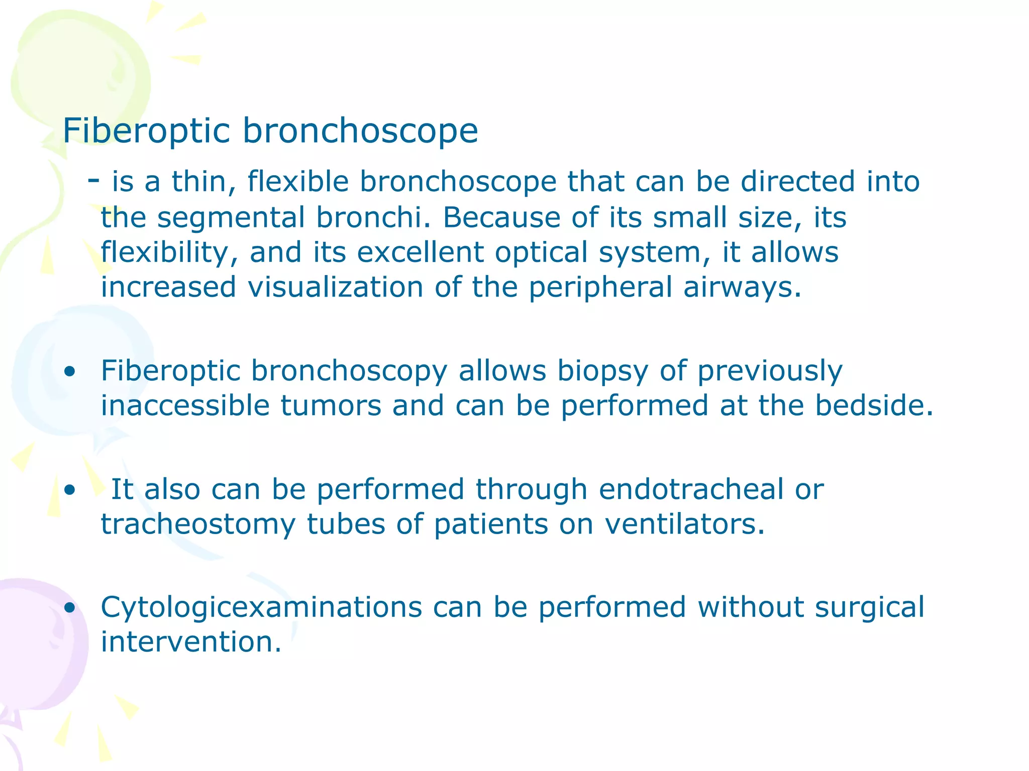
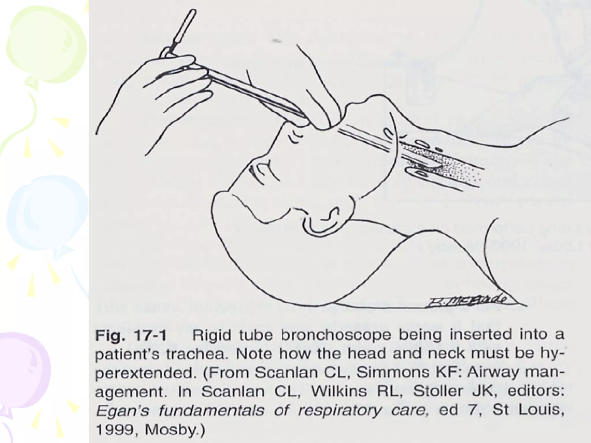
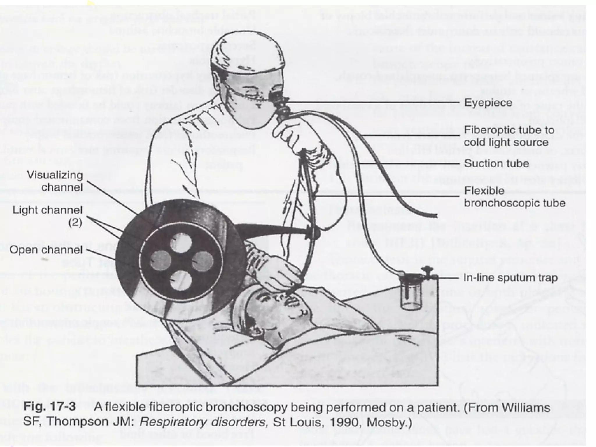
BronchoscopyBronchoscopy
DefinitionDefinition
• Bronchoscopy
– is the direct inspection and examination of the larynx,
trachea, and bronchi through either a flexible fiberoptic
bronchoscope or a rigid bronchoscope. The fiberoptic
scope is used more frequently in current practice.
PurposesPurposes
• Diagnostic bronchoscopy is used to:
(1) to examine tissues or collect secretions,
(2) to determine if a tumor can be resected surgically, and
(3) to diagnose bleeding sites (source of hemoptysis).
• Therapeutic bronchoscopy is used to:
(1) remove foreign bodies from the tracheobronchial tree,
(2) remove secretions obstructing
(3) treat postoperative atelectasis,
EquipmentEquipment
• A bronchoscope is an instrument about 3ft long
and 0.5 ins or smaller in diameter that combines
four narrow chambers into one tube
– One lumen contains a fiber-optic light source so that
structures can be viewed effectively
– 2nd
chamber lumen is attached to a suction device &
airway secretions can be removed
– 3rd
chamber has tiny metallic alligator forceps that can
be extended past the proximal end for tissue biopsies
– 4th
chamber lumen allows passage of a small wire brush
that can be passed vigorously over airway structures for
collection of tissue cells for microscopic evaluation
Fiberoptic bronchoscope
- is a thin, flexible bronchoscope that can be directed into
the segmental bronchi. Because of its small size, its
flexibility, and its excellent optical system, it allows
increased visualization of the peripheral airways.
• Fiberoptic bronchoscopy allows biopsy of previously
inaccessible tumors and can be performed at the bedside.
• It also can be performed through endotracheal or
tracheostomy tubes of patients on ventilators.
• Cytologicexaminations can be performed without surgical
intervention.
Rigid bronchoscope
- is a hollow metal tube with a light at its end.
- used mainly for removing foreign substances,
Investigating the source of massive hemoptysis,
Performing endobronchial surgical procedures.
• Rigid bronchoscopy is performed in the operating room, not
at the bedside.
Before procedureBefore procedure
• A signed consent form is obtained from the patient.
• Food and fluids are withheld for 6 hours before the test to reduce
the risk of aspiration.
• Nurse explains the procedure to the patient to reduce fear and
anxiety and administers pre operative medication.
• The patient must remove dentures and other oral prostheses.
• The examination is usually performed.
• A topical anesthetic such as lidocaine (Xylocaine) may be sprayed
on the pharynx or dropped on the epiglottis and vocal cords and
into the trachea to suppress the cough reflex and minimize
discomfort.
• Sedatives or opioids are administered intravenously as prescribed
to provide moderate sedation.
During ProcedureDuring Procedure
• Topical anesthetic (lidocaine) is administered to control
gag/cough reflex and prevent bronchospasm
• Intubation preferred but not required. Intubation will not
allow visualization of the vocal cords
• Scope is inserted and the airways viewed
• O2 needs to be provided to patient via mask or by
removing one prong of the nasal cannula from the nose to
allow for insertion of the scope
• Diagnostic and/or therapeutic procedures are
performed
• Intubated patients on vents need special adapters
for advancement of the scope. Adapter should
allow for:
– No loss of ventilating pressures
– No loss of PEEP
• Continuous monitoring of EKG and O2 saturation
by pulse oximeter is recommended
• Equipment is cleaned by decontamination with
alkaline glutaraldehyde
After procedureAfter procedure
• Patient takes nothing by mouth until the cough reflex returns,
because the preoperative sedation and local anesthesia impair the
protective laryngeal reflex and swallowing for several hours.
• Once the patient demonstrates a cough reflex, the nurse may
offer ice chips and eventually fluids.
• Nurse assesses for confusion and lethargy in the elderly.
• Nurse also monitors the patient’s respiratory status and observes
for hypoxia, hypotension, tachycardia, dysrhythmias, hemoptysis,
and dyspnea.
• The patient is not discharged from the recovery area until
adequate cough reflex and respiratory status are present.
• Nurse instructs the patient and family caregivers to report any
shortness of breath or bleeding immediately.
Hazards and complicationsHazards and complications
• Most common complication is mild epistaxis
• Bronchospasm or laryngospasm
– From irritation of the airway
• Bleeding
• Hypoxemia
• Arrhythmias
– From vagal stimulation. Monitor ECG and remove scope
until cardiac status is stabilized
• Seizures
• Aspiration
• Pneumothorax
• Respiratory depression
ContraindicationsContraindications
• Hypoxemia
• Bleeding disorders
• Cardiovascular instability
• Status asthmaticus
• Marked hypercpanea
 Bronchoscopy, Diagnostic technique

Bronchoscopy, Diagnostic technique

  • 1.
  • 2.
    DefinitionDefinition • Bronchoscopy – isthe direct inspection and examination of the larynx, trachea, and bronchi through either a flexible fiberoptic bronchoscope or a rigid bronchoscope. The fiberoptic scope is used more frequently in current practice.
  • 4.
    PurposesPurposes • Diagnostic bronchoscopyis used to: (1) to examine tissues or collect secretions, (2) to determine if a tumor can be resected surgically, and (3) to diagnose bleeding sites (source of hemoptysis). • Therapeutic bronchoscopy is used to: (1) remove foreign bodies from the tracheobronchial tree, (2) remove secretions obstructing (3) treat postoperative atelectasis,
  • 5.
    EquipmentEquipment • A bronchoscopeis an instrument about 3ft long and 0.5 ins or smaller in diameter that combines four narrow chambers into one tube – One lumen contains a fiber-optic light source so that structures can be viewed effectively – 2nd chamber lumen is attached to a suction device & airway secretions can be removed – 3rd chamber has tiny metallic alligator forceps that can be extended past the proximal end for tissue biopsies – 4th chamber lumen allows passage of a small wire brush that can be passed vigorously over airway structures for collection of tissue cells for microscopic evaluation
  • 8.
    Fiberoptic bronchoscope - isa thin, flexible bronchoscope that can be directed into the segmental bronchi. Because of its small size, its flexibility, and its excellent optical system, it allows increased visualization of the peripheral airways. • Fiberoptic bronchoscopy allows biopsy of previously inaccessible tumors and can be performed at the bedside. • It also can be performed through endotracheal or tracheostomy tubes of patients on ventilators. • Cytologicexaminations can be performed without surgical intervention.
  • 10.
    Rigid bronchoscope - isa hollow metal tube with a light at its end. - used mainly for removing foreign substances, Investigating the source of massive hemoptysis, Performing endobronchial surgical procedures. • Rigid bronchoscopy is performed in the operating room, not at the bedside.
  • 12.
    Before procedureBefore procedure •A signed consent form is obtained from the patient. • Food and fluids are withheld for 6 hours before the test to reduce the risk of aspiration. • Nurse explains the procedure to the patient to reduce fear and anxiety and administers pre operative medication. • The patient must remove dentures and other oral prostheses. • The examination is usually performed. • A topical anesthetic such as lidocaine (Xylocaine) may be sprayed on the pharynx or dropped on the epiglottis and vocal cords and into the trachea to suppress the cough reflex and minimize discomfort. • Sedatives or opioids are administered intravenously as prescribed to provide moderate sedation.
  • 13.
    During ProcedureDuring Procedure •Topical anesthetic (lidocaine) is administered to control gag/cough reflex and prevent bronchospasm • Intubation preferred but not required. Intubation will not allow visualization of the vocal cords • Scope is inserted and the airways viewed • O2 needs to be provided to patient via mask or by removing one prong of the nasal cannula from the nose to allow for insertion of the scope
  • 14.
    • Diagnostic and/ortherapeutic procedures are performed • Intubated patients on vents need special adapters for advancement of the scope. Adapter should allow for: – No loss of ventilating pressures – No loss of PEEP • Continuous monitoring of EKG and O2 saturation by pulse oximeter is recommended • Equipment is cleaned by decontamination with alkaline glutaraldehyde
  • 16.
    After procedureAfter procedure •Patient takes nothing by mouth until the cough reflex returns, because the preoperative sedation and local anesthesia impair the protective laryngeal reflex and swallowing for several hours. • Once the patient demonstrates a cough reflex, the nurse may offer ice chips and eventually fluids. • Nurse assesses for confusion and lethargy in the elderly. • Nurse also monitors the patient’s respiratory status and observes for hypoxia, hypotension, tachycardia, dysrhythmias, hemoptysis, and dyspnea. • The patient is not discharged from the recovery area until adequate cough reflex and respiratory status are present. • Nurse instructs the patient and family caregivers to report any shortness of breath or bleeding immediately.
  • 17.
    Hazards and complicationsHazardsand complications • Most common complication is mild epistaxis • Bronchospasm or laryngospasm – From irritation of the airway • Bleeding • Hypoxemia • Arrhythmias – From vagal stimulation. Monitor ECG and remove scope until cardiac status is stabilized • Seizures • Aspiration • Pneumothorax • Respiratory depression
  • 18.
    ContraindicationsContraindications • Hypoxemia • Bleedingdisorders • Cardiovascular instability • Status asthmaticus • Marked hypercpanea