Breast Cancer Detection and Classification using Ultrasound and Ultrasound Elastography Images

This document summarizes a research paper that aims to classify breast lesions as benign or malignant using ultrasound and ultrasound elastography images. It presents a methodology for preprocessing images, extracting textural features using discrete wavelet transform and gray level co-occurrence matrix, performing feature reduction with principal component analysis, and classifying images using support vector machine and k-nearest neighbor classifiers. The methodology is applied to a dataset of ultrasound and elastography images and classifiers are evaluated based on accuracy, sensitivity, and specificity metrics derived from confusion matrices. The results show high performance for both classifiers on the elastography images and slightly better performance for the k-nearest neighbor classifier on the ultrasound images.
![International Research Journal of Engineering and Technology (IRJET) e-ISSN: 2395 -0056
Volume: 04 Issue: 07 | July-2017 www.irjet.net p-ISSN: 2395-0072
© 2017, IRJET | Impact Factor value: 5.181 | ISO 9001:2008 Certified Journal | Page 597
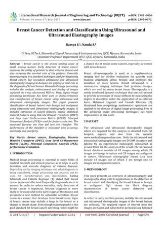
by feature extraction. Statistical textural features are
extracted using discrete wavelet transform (DWT) and
GLCM (gray level co-occurrence matrix). Feature
reduction is done by principal component analysis
(PCA).The features are then given to the classifiers
namely support vector machine (SVM), and KNN(k nearest
neighbor) which labels the breast lesions as benign or
malignant.
Fig-1: Block Diagram of Breast Cancer Detection and
Classification.
3.1. Pre Processing
Preprocessing is one of the primary procedures in the
image processing technique. The aim of preprocessing is
the improvisation of the image data by reducing the noise
or by suppressing the undesired distortions. This also
helps in improving certain features in the image which can
be used for further operations. In this work median filter
is used for preprocessing.
3.1.1 Median filter
Median filtering is widely used in digital image
processing because, under certain conditions, it preserves
edges while removing noise. The median filter is a method
where in it can effectively distinguish out-of-range isolated
noise from legitimate image features such as edges and
lines. Specifically, the median filter replaces a pixel by the
median, instead of the average, of all pixels in a
neighborhood .
y[m, n] = {x [ i , j ] , ( i , j ) € }
(1)
where represents a neighborhood defined by the user,
centered around location [m, n] in the image.
3.2 Feature Extraction
Feature extraction is a technique where in the
characteristic attributes of an image is extracted and
generates a set of meaning full descriptors for an image. In
this paper mainly the statistical features are extracted
using discrete wavelet transform and GLCM.
3.2.1 Discrete Wavelet Transform
The discrete wavelet transform (DWT) is one of the
efficient tools for image compression. The principle of this
transform is to hierarchically decompose a image into a
multi resolution pyramid, where the image is split into a
coarse approximation. The advantage of DWT is that it
provides very good compaction properties for many
classes of images. In this work dB8 is used for
decomposition. GLCM feature values have been computed
from the horizontal, vertical, and diagonal components of
first level of decomposition.
3.2.2 Gray Level Co-Occurrence Matrix
Gray-level co-occurrence matrix is one of the most known
texture analysis methods that estimates image properties
related to second-order statistics by considering the
spatial relationship between two neighboring pixels,
where the first pixel is the reference pixel and the second
pixel is the neighbor pixel. The GLCM is computed based
on two parameters, which are the relative distance d
between the pixel pair measured in pixel number and their
relative orientation φ. Here, relative distance d=1 and
relative orientation φ=0.
Computation of Gray-Level Co-occurrence Matrix
To describe the computation of GLCM matrix for breast
ultrasound images, let us consider a two-dimensional
image I(x, y), (x = 1, ...,n , y = 1,...,m), having G gray-levels. A
co-occurrence matrix C(i , j) is, mathematically, defined
over a n x m image I, parameterized by an offset ( x, y)
as given in equation below.
(i,j)= (2)
Here, the ( x, y) parameterization makes the co-
occurrence matrix sensitive to rotation. (i, j) denotes
INPUT IMAGE
PREPROCESSING
FEATURE
EXTRACTION
FEATURE
REDUCTION
CLASSIFICATION
(SVM) and (KNN)
BENIGN MALIGNANT](data:image/gif;base64,R0lGODlhAQABAIAAAAAAAP///yH5BAEAAAAALAAAAAABAAEAAAIBRAA7)
Recommended
Recommended
More Related Content
What's hot
What's hot (20)
Similar to Breast Cancer Detection and Classification using Ultrasound and Ultrasound Elastography Images
Similar to Breast Cancer Detection and Classification using Ultrasound and Ultrasound Elastography Images (20)
More from IRJET Journal
More from IRJET Journal (20)
Recently uploaded
Recently uploaded (20)
Breast Cancer Detection and Classification using Ultrasound and Ultrasound Elastography Images
- 1. International Research Journal of Engineering and Technology (IRJET) e-ISSN: 2395 -0056 Volume: 04 Issue: 07 | July-2017 www.irjet.net p-ISSN: 2395-0072 © 2017, IRJET | Impact Factor value: 5.181 | ISO 9001:2008 Certified Journal | Page 596 Breast Cancer Detection and Classification Using Ultrasound and Ultrasound Elastography Images Ramya S.1, Nanda S.2 1IV Sem, M.Tech, Biomedical Signal Processing & Instrumentation, SJCE, Mysuru, Karnataka, India 2Assistant Professor, Department Of IT, SJCE, Mysuru, Karnataka, India ---------------------------------------------------------------------***--------------------------------------------------------------------- Abstract - Breast cancer is the second leading cause of death among women. Early detection of breast cancer improves the ability of patients to deal with the disease and also increases the survival rate of the patients. Generally mammography is a standard technique used for diagnosing breast cancer, but nowadays ultrasound and ultrasound elastography imaging techniques are playing a vital role in the diagnosis of breast lesions. Biomedical image processing includes the analysis, enhancement and display of images captured via x-ray, ultrasound, MRI etc. Here, digital image processing techniques has been used in characterization and classification of breast cancer using ultrasound and ultrasound elastography images. This paper presents classification of breast lesion’s into benign and malignant using ultrasound and ultrasound elastrography images. It includes extraction of salient features, mainly statistical textural features using Discrete Wavelet Transform (DWT) and Gray Level Co-Occurrence Matrix (GLCM). Principal Component Analysis (PCA)is used for feature reduction. The extracted features are then fed into the classifier. The performance of the classifier is evaluated with accuracy, sensitivity and specificity. Key Words: Breast cancer, Elastography, Discrete Wavelet Transform (DWT), Gray Level Co-Occurrence Matrix (GLCM), Principal Component Analysis (PCA), performance evaluation. 1. INTRODUCTION Medical image processing is essential in many fields of medical research and clinical practice as it helps in early detection and accurate diagnosis of various diseases. Depending on the imaging technique and what diagnosis is being considered, image processing and analysis can be used for characterization and classification. Fatima Eddaoudi and Fakhita Regragui (1) stated that breast cancer represents the most frequently diagnosed cancer in women. In order to reduce mortality, early detection of breast cancer is important, because diagnosis is more likely to be successful in the early stages of the disease and presented a new method for automatic detection. Breast cancer is a cancer that develops from breast tissue. Signs of breast cancer may include a lump in the breast or a change in breast shape. Even though Mammography is the gold standard for breast cancer screening, there might be a chance that it misses some cancers, especially in women with dense breasts. Breast ultrasonography is used as a supplementary imaging tool for further evaluation for patients with mammo graphically dense breasts and improves the detection of mass lesions. Breast ultrasound is a noninvasive imaging technique that produces images, which are used to assess breast tissue. Elastography is a newly developed dynamic technique that uses ultrasound to provide an estimate of tissue stiffness by measuring the degree of distortion under the application of an external force. Mohamed Lagzouli and Youssfi Elkettani (2) illustrated how morphology mathematics operations are applied in the domain of digital image processing. Few of the advanced image processing techniques has been implemented in this work. 2 DATASET The ultrasound and ultrasound elastography images which are required for the analysis is obtained from JSS hospital, mysuru and also from the website www.medicalimagesonline.com . Both the ultrasound and ultrasound elastography images are DICOM in nature and labeled by an experienced radiologists considered as ground truth for the analysis of the result. The ultrasound breast database consists of 61 images among which 33 images are benign in nature and 28 images are malignant in nature. Ultrasound elastography breast data base include 13 images out of which 3 are benign and 10 images are malignant in nature. 3. METHODOLOGY This work presents an overview of ultrasonography and elastography along with its applications in the detection of breast cancer and classifying the breast lesions as benign or malignant. Fig1. shows the block diagram representation of breast cancer detection and classification. In order to carry out this process, initially the ultrasound and ultrasound elastography images of the breast lesions are collected. The required region of interest from the images are taken and subjected to preprocessing followed
- 2. International Research Journal of Engineering and Technology (IRJET) e-ISSN: 2395 -0056 Volume: 04 Issue: 07 | July-2017 www.irjet.net p-ISSN: 2395-0072 © 2017, IRJET | Impact Factor value: 5.181 | ISO 9001:2008 Certified Journal | Page 597 by feature extraction. Statistical textural features are extracted using discrete wavelet transform (DWT) and GLCM (gray level co-occurrence matrix). Feature reduction is done by principal component analysis (PCA).The features are then given to the classifiers namely support vector machine (SVM), and KNN(k nearest neighbor) which labels the breast lesions as benign or malignant. Fig-1: Block Diagram of Breast Cancer Detection and Classification. 3.1. Pre Processing Preprocessing is one of the primary procedures in the image processing technique. The aim of preprocessing is the improvisation of the image data by reducing the noise or by suppressing the undesired distortions. This also helps in improving certain features in the image which can be used for further operations. In this work median filter is used for preprocessing. 3.1.1 Median filter Median filtering is widely used in digital image processing because, under certain conditions, it preserves edges while removing noise. The median filter is a method where in it can effectively distinguish out-of-range isolated noise from legitimate image features such as edges and lines. Specifically, the median filter replaces a pixel by the median, instead of the average, of all pixels in a neighborhood . y[m, n] = {x [ i , j ] , ( i , j ) € } (1) where represents a neighborhood defined by the user, centered around location [m, n] in the image. 3.2 Feature Extraction Feature extraction is a technique where in the characteristic attributes of an image is extracted and generates a set of meaning full descriptors for an image. In this paper mainly the statistical features are extracted using discrete wavelet transform and GLCM. 3.2.1 Discrete Wavelet Transform The discrete wavelet transform (DWT) is one of the efficient tools for image compression. The principle of this transform is to hierarchically decompose a image into a multi resolution pyramid, where the image is split into a coarse approximation. The advantage of DWT is that it provides very good compaction properties for many classes of images. In this work dB8 is used for decomposition. GLCM feature values have been computed from the horizontal, vertical, and diagonal components of first level of decomposition. 3.2.2 Gray Level Co-Occurrence Matrix Gray-level co-occurrence matrix is one of the most known texture analysis methods that estimates image properties related to second-order statistics by considering the spatial relationship between two neighboring pixels, where the first pixel is the reference pixel and the second pixel is the neighbor pixel. The GLCM is computed based on two parameters, which are the relative distance d between the pixel pair measured in pixel number and their relative orientation φ. Here, relative distance d=1 and relative orientation φ=0. Computation of Gray-Level Co-occurrence Matrix To describe the computation of GLCM matrix for breast ultrasound images, let us consider a two-dimensional image I(x, y), (x = 1, ...,n , y = 1,...,m), having G gray-levels. A co-occurrence matrix C(i , j) is, mathematically, defined over a n x m image I, parameterized by an offset ( x, y) as given in equation below. (i,j)= (2) Here, the ( x, y) parameterization makes the co- occurrence matrix sensitive to rotation. (i, j) denotes INPUT IMAGE PREPROCESSING FEATURE EXTRACTION FEATURE REDUCTION CLASSIFICATION (SVM) and (KNN) BENIGN MALIGNANT
- 3. International Research Journal of Engineering and Technology (IRJET) e-ISSN: 2395 -0056 Volume: 04 Issue: 07 | July-2017 www.irjet.net p-ISSN: 2395-0072 © 2017, IRJET | Impact Factor value: 5.181 | ISO 9001:2008 Certified Journal | Page 598 the cardinality of the set of pairs of points that have gray- level values of i and j, for a displacement vector d ( x, y) =(p, q) n x m. The characteristics of benign and malignant breast ultrasound images computed from GLCMs are energy, entropy, correlation, kurtosis, skewness. 3.2.2.1 Energy Energy or the angular second moment measures the textural uniformity of an image . The larger value of energy indicates more homogeneous the image. Energy = (3) 3.2.2.2 Entropy It is a measure of randomness of intensity image. Entropy measures the disorder or randomness of an image and it achieves its largest value when all elements in image matrix are equal. Entropy= (4) 3.2.2.3 Correlation Correlation measures how correlated a pixel is to its neighborhood. It can also be described as a measure of linear dependencies among neighboring pixels in an image. Correlation = (5) where, I (i, j) is the gray-level value for each pixel in the region of interest, N is the total number of pixels in the region of interest and are means and standard deviations of I (i, j). 3.2.2.4 Kurtosis kurtosis is a measure of the "tailedness" of the probability distribution of a real-valued random variable. kurtosis is a descriptor of the shape of a probability distribution and there are different ways of quantifying it for a theoretical distribution and corresponding ways of estimating it from a sample from a population. 3.2.2.5 Skew ness Skew ness is a measure of the asymmetry of the probability distribution of a real-valued random variable about its mean. 3.3 Feature Reduction Feature reduction or dimensionality reduction is basically done to reduce the dimensionality of the feature set and also to identify new meaningful underlying variables. Principal component analysis (PCA) is used for feature reduction purpose 3.3.1 Principal Component Analysis Principal component analysis (PCA) is a very popular technique for dimensionality reduction. Given a set of data on n dimensions, PCA aims to find a linear subspace of dimension d lower than n such that the data points lie mainly on this linear subspace. Such a reduced subspace attempts to maintain most of the variability of the data. The linear subspace can be specified by d orthogonal vectors that form a new coordinate system, called the ‘principal components’. The principal components are orthogonal, linear transformations of the original data points. However, the hope is that only d < n principal components are needed to approximate the space spanned by the n original axes. Here, In this work a single principal component is considered. 3.4 Classification The reduced feature set obtained after the feature reduction process is fed into classifier directly. Support Vector Machine and K-Nearest Neighbor classifiers are used for classifying the breast lesions as benign or malignant. 3.4.1 Support Vector Machine (SVM) Based on the statistical learning theory and structural risk minimization, SVM makes its extensive use as an effective algorithm in dealing classification and regression problems. To derive a hyper plane by maximizing the margin between two classes is the main idea of SVM. Mapping an input data into a higher dimension space is done by Kernel function which can be linear, quadratic, polynomial, radial base function and multiple layer perceptron. The decision function which provides the solution to the classification is given by (6) Where is the positive Lagrange multiplier, is the support vector and the function for convolution of the kernel of the decision function is . 3.4.2 k-Nearest Neighbor (kNN) The k-nearest neighbor is a non-parametric method used for classification. In this case, the input consists of the k closest training examples in the feature space. In k-NN classification, the output is a class membership. An object is classified by a majority vote of its neighbors, with the object being assigned to the class most common
- 4. International Research Journal of Engineering and Technology (IRJET) e-ISSN: 2395 -0056 Volume: 04 Issue: 07 | July-2017 www.irjet.net p-ISSN: 2395-0072 © 2017, IRJET | Impact Factor value: 5.181 | ISO 9001:2008 Certified Journal | Page 599 among its k nearest neighbors (k is a positive integer, typically small). If k = 1, then the object is simply assigned to the class of that single nearest neighbor. Euclidean distance is generally used to find the distance between the k-nearest neighbor and is given by Euclidean= (7) Where x and y represent the training and testing samples. 3.5 Performance Evaluation Accuracy, specificity and sensitivity are the parameters used to measure the performance of the classifiers. These parameters are calculated in terms of True positive (TP), False positive (FP), True negative (TN), False negative (FN) obtained from the confusion matrix of the classifiers. Accuracy measures the global performance of the classifier about the correct decision. Sensitivity measures the reliability of the system at making positive identifications where as specificity measures the reliability of the system at making negative identifications. The equation of the performance parameters are as mentioned below. Accuracy = (7) Sensitivity = (8) Specificity = (9) 4. RESULTS The ultrasound and ultrasound elastography images are obtained while performing the ultrasound examinations. They are labeled by an experienced radiologists considered as ground truth for the analysis of the result. Region of interest from the breast ultrasound images is selected, and the selected ROI (region of interest) is taken as an input for further processing. The accuracy of the classifier is then evaluated using performance evaluation. Fig- 2 shows the input ultrasound breast images Fig-3 shows the ultrasound elastography images. Fig-4 shows the image after applying median filter. Fig- 2: Benign and Malignant breast ultrasound images. Fig- 3: Benign and Malignant breast ultrasound elastography images. Fig-4: Extracting the required ROI The range of GLCM feature values obtained benign and malignant images of both ultrasound and ultrasound elastography images is shown in table 1 and table 2 respectively. Table-1: Range of feature values obtained for Breast Ultrasound benign and malignant images. Table-2: Range of feature values obtained for Breast Ultrasound elastography benign and malignant images.
- 5. International Research Journal of Engineering and Technology (IRJET) e-ISSN: 2395 -0056 Volume: 04 Issue: 07 | July-2017 www.irjet.net p-ISSN: 2395-0072 © 2017, IRJET | Impact Factor value: 5.181 | ISO 9001:2008 Certified Journal | Page 600 In this work 70:30 ratio is taken for training and testing. The ultrasound breast image dataset includes 33 benign images out of which 23 images are given for training and 10 for testing. 28 malignant ultrasound images are included in the image dataset, where 20 are given for training and 8 for testing. The ultrasound elastography breast image dataset includes 3 benign images out of which 2 images are given for training and 1 for testing. 10 malignant ultrasound images are included in the image dataset, where 6 are given for training and 4 for testing. Table-3: Confusion matrix for ultrasound image dataset Table-4: Confusion matrix for ultrasound elastography image dataset In ultrasound breast images, according to SVM classifier, all the 8 malignant images are classified as malignant and out of 10 benign images 1 is miss classified as malignant. As per k-NN classifier 8 are classified as benign and 2 are classified as malignant out of 10 benign and all malignant images are correctly classified as malignant. For breast ultrasound elastography dataset, both in SVM and k-NN classifier benign and malignant images are correctly classified. Table-5: Performance of the classifier for ultrasound dataset Table-6: Performance of the classifier for ultrasound elastography dataset. Receiver operating curve helps in accessing the performance of an imaging system. Fig 5 and Fig 6 shows the ROC plot of SVM and k-NN classifier for ultrasound images. Fig 7 and Fig 8 shows the ROC plot SVM and k-NN classifier for ultrasound elastography images. Fig-5: Receiver Operating Curve for SVM Fig-6: Receiver Operating Curve for k-NN
- 6. International Research Journal of Engineering and Technology (IRJET) e-ISSN: 2395 -0056 Volume: 04 Issue: 07 | July-2017 www.irjet.net p-ISSN: 2395-0072 © 2017, IRJET | Impact Factor value: 5.181 | ISO 9001:2008 Certified Journal | Page 601 Fig-7: Receiver Operating Curve for SVM Fig-8: Receiver Operating Curve for k-NN 5. CONCLUSIONS This method is helpful in the detection of breast cancer and classification of breast lesion as benign or malignant.DWT and GLCM are used for textural features. The obtained features are then passed through PCA and the classifiers SVM and k-NN. For ultrasound dataset SVM gives an accuracy of 94.74% and KNN classifier gives an accuracy of 89.47%. For ultrasound elastography dataset both SVM and KNN classifiers give 100% accuracy. This work can be extended in order to distinguish the type of breast cancer. From this work better results can be obtained when ultrasound elastography images are considered for the analysis when compared to the ultrasound images. And also it shows that SVM classifier gives a better accuracy compared to that of a k-NN classifier. REFERENCES [1] Fatima Eddaoudi and Fakhita Regragui “Microcalcifications detection in mammographic images using texture coding”, Applied Mathematical Sciences, 2011 [2] Mohamed Lagzouli and Youssfi Elkettani. “A New Morphology Algorithm for ultrasound Images”. International Journal of Engineering Research & Technology, 2014 [3] Kumm T.R., Szabunio M.M. “Elastography for the characterization of breast lesions”. initial clinical experience Cancer Control. 2010. [4] Yerli H., Yilmaz T., Kaskati T., Gulay H. “Qualitative and semiquantitative evaluations of solid breast lesions by sonoelastography”. J Ultrasound Med. 2011 [5] Tozaki M., Isobe S., Yamaguchi M., Ogawa Y., Homma K., Saito M. “Ultrasonographic elastography of the breast using acoustic radiation force impulse technology”. preliminary study. Jpn J Radiol. 2011. [6] Meng W., Zhang G., Wu C., Wu G., Song Y., Lu “Preliminary results of acoustic radiation force impulse (ARFI) ultrasound imaging of breast lesions”. Ultrasound Med Biol. 2011. [7] Bai M., Du L., Gu J., Li F., Jia X. “Virtual touch tissue quantification using acoustic radiation force impulse technology” initial clinical experience with solid breast masses. J Ultrasound Med. 2012. [8] Tozaki M., Fukuma E. “Pattern classification of shear wave elastography images for differential diagnosis between benign and malignant solid breast masses”. Acta Radiol. December 2011. [9] Evans A., Whelehan P., Thomson K., McLean D., Brauer K., Purdie C. “Quantitative shear wave ultrasound elastography: initial experience in solid breast masses”. Breast Cancer. 2010. [10] Keramidas E.G., Iakovidis D.K., Maroulis D. and Karkanis S., “Efficient and Effective Ultrasound Image Analysis Scheme for Thyroid Nodule Detection”. 2007. [11] Saiti F., Naini A.A., Shoorehdeli M. A. and Teshnehlab M., “Thyroid Disease Diagnosis Based on Genetic Algorithms using k-NN and SVM”. 2009. [12] Tsantis S., Cavouras D., Kalatzis I., Piliouras N., Dimitropoulos N. and Nikiforidis G. “Development of A Support Vector Machine-Based Image Analysis system for assessing the Thyroid Nodule malignancy risk on ultrasound”. 2005. [13] Dogantekin E., Dogantekin A. “An expert system based on Generalized Discriminant Analysis and Wavelet Support Vector Machine for diagnosis of thyroid diseases”. 2011