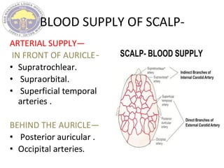
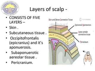
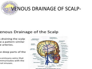
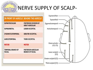
CRANIOTOMY FLAPS ,INCISION AND
CLOSURE.
ANATOMIC AND NEUROVASCULAR
CONSIDERATION-
• EXTENT --The scalp
extends from the top
of the forehead in
front to the superior
nuchal line behind.
• Laterally it projects
down to the zygomatic
arch and external
acoustic meatus.
BLOOD SUPPLY OF SCALP-
ARTERIAL SUPPLY—
IN FRONT OF AURICLE—
• Supratrochlear.
• Supraorbital.
• Superficial temporal
arteries .
BEHIND THE AURICLE—
• Posterior auricular .
• Occipital arteries.
Layers of scalp -
• CONSISTS OF FIVE
LAYERS –
• Skin .
• Subcutaneous tissue .
• Occipitofrontalis
(epicranius) and it’s
aponuerosis.
• Subaponuerotic
aereolar tissue .
• Pericranium.
VENOUS DRAINAGE OF SCALP-
NERVE SUPPLY OF SCALP-
BASIC SURGICAL PRINCIPLES OF
CRANIOTOMY--
• PREOPERATIVE REVIEW OF PATIENT AND SCANS.
• PREPARATION OF SCALP.
• PROPER POSITIONING OF PATIENT.
• SCALP TOILETING.
• PROPER DRAPPING.
• SKIN FLAPS .
• PLACING OF BURR HOLES , CRANIOTOMY SIZE.
• DUROTOMY .
• CLOSURES .
PRINCIPLES FOR BONE FLAPS -
• Most direct access to target.
• For cerebral convexity directly centered over
the lesion.
• Number of burr holes varies.
• Separating of underlying dura .
• If dura is lacerated during cutting ,saw should
be turned off and removed backwards via
entrance hole.
• Air cells opened : –
• Remove the mucosa. Pack with betadine
soaked gelfoam. Pack with bone wax. Cover it
up with vascularized tissue.
• Proposed bony cuts over venous sinuses
should be done last .
• Cut sinus can be sewn or tamponade .
• Bony bleeds are stopped with bone wax.
• Penfield’s dissector used to separate dura.
• Epidural tacking sutures to control epidural
bleeding before opening dura.
OPENING OF DURAMATER --
• Manually palpate the dura.
• Dura opened as straight, curved or flap like
incisions.
• Flaps based towards sinuses.
• Opened with sharp hook and knife.
• Incision further opened with dural scissors.
• Placement of cottonoid along the intended
incision.
• Suitable cuff of dura around the bone for suturing
later.
CLOSURE OF WOUNDS-
• Closure in layers.
• Check for BP, valsalva maneuver.
• Hitch suture.
• Water tight but not tension.
• Bone flap replacement.
• Skin closed in two layers.
TYPES OF CRANIOTOMY-
1-TREPHINE CRANIOTOMY – PROVIDES A
CIRCULAR OPENING .
• LESS TIME CONSUMING.
• IDEAL FOR EVACUATING POST TRAUMATIC EDH .
2- FLAP CRANIOTOMY –
• A) FREE BONE FLAP CRANIOTOMY .
• B)- OSTEOPLASTIC CRANIOTOMY- Bone flap is
elevated along with its musculofascial
attachment. Believed to be less prone to
infection due to intact blood supply of the bone
flap.
SKIN FLAPS AND CRANIOTOMIES-
ANTERIOR SKIN FLAPS AND CRANIOTOMIES-
1- SUPRA ORBITAL KEYHOLE CRANIOTOMY-
2- THE LYNCH HOWARTH INCISON.
3- ORBITOTOMY.
SUPRAORBITAL KEY HOLE
CRANIOTOMY-
• In 1900 Krause first demonstrated supra-orbital, subfrontal
approach on cadaver, then eight years later he reported
the first resection of skull base meningioma through this
approach.
• In 1913, Frazier advocated a supraorbital ridge resection,
which was found useful in surgery for pituitary adenomas.
• In the 1990, Perneczky and colleagues popularized the
keyhole concept and the technique commonly used today,
the supraorbital keyhole craniotomy. The evolution,
however, began with Fedor Krause's .
• More recently, Jane and Delashaw , described a
supraorbital craniotomy in the approach to orbital tumors.
Other variants have been proposed by Al-Mefty et al.
INDICATIONS-
• Aneurysm of Anterior circulation except those
of distal anterior cerebral artery.
• For high positioned basilar bifurcation of
basilar-Superior cerebellar artery aneurysm.
• Tumor of anterior cranial fossa, sphenoid ridge.
• Pathologies of sellar and suprasellar region.
POSITIONING-
• Head is elevated.
• Head is extended as
it allows relaxation
of frontal lobe.
SKIN INCISION-
• Type-Superciliary,
transciliary,
transpalpebral.
• The incision is made
in the lateral 2/3rd
of eyebrow.
• CRANIOTOMY –
• Supraorbital craniotomy,
bone flap with shape of “D”.
• Dura is opened in semilunar
fashion with base at orbital
rim.
• The limitation with the
lighting of microscope deep
down a narrow corridor can
be overcome by Endoscope ,
which can be held by
assistant or retractor arm.
• A second look with
endoscope can also allow
visualization of gross
resection of lesion.
COMPLICATIONS DUE TO INCISION:--
• 1. Transient loss of supraorbital
sensation(7.5%) .
• 2. Frontal paresis (5.5%).
• 3. Opening of frontal sinus can cause CSF
leak(4%) .
• 4. Burning of eyebrow due to microscopic
light on 100% intensity. (Not seen with
intensity below 70%).
• 5. Bone flap resorption.
VARIANTS OF SUPRAORBITAL
CRANIOTOMY-
• Lateral variation:- To partially remove the lesser
sphenoid wing exposing the frontal and temporal
dura mater. Also on removing clinoid process,
paraclinoid segment of ICA can be visualised.
• Medial Modification: For surgical view of
suprasellar and interhemispheric structure. With
possibility of interhemispheric and subfrontal
dissection.
• Basal variation: To gain more oblique view of
deep seated prepontine and interpeduncular
region via subfrontal exposure after removing
orbital rim and partial removal of orbital roof.
LYNCH HOWARTH FLAP-
• Incision made mid way
between the nasal dorsum
and medial canthus aong the
naso facial crease.
• Used in
transetmosphenoidal
approach for sellar mass.
• Common extracranial
approach for repair of CSF
fistulas of cribriform
,ethmoid and sphenoid
region.
ORBITOTMY-
• Orbitotomy refers to surgical approach for an
orbital mass lesion.
• ANTERIOR ORBITOTOMY.
• LATERAL ORBITOTOMY.
• TRANSFRONTAL ORBITOTOMY.
• TEMPOROFRONTAL ORBITOTMY.
ANTERIOR ORBITOTOMY-
• It is indicated only
when the lesion is
readily palpable
through the eyelids
and is judged to be
mainly in front of the
equator of eyeball.
• Aproached by superior,
inferior,medial and
lateral incision.
LATERAL ORBITOTMY-
• Indictions:
• Laterally placed extraconal tumours.
• Intraconal tumours lateral or inferior to the
optic nerve.
• It is useful for lacrimal gland tumours,
retrobulbar lesions, such as cavernomas and
can be extended for posterior lesions.
• Advantages: • Good exposure, Well-tolerated
procedure.
• Disadvantages: • Visible but minimal scar.
TRANSFRONTAL ORBTITOTOMY
• Indications:
• Superiorly and medially placed moderate and
small-sized tumours .
• Intraconal tumours medial to optic nerve.
• In this technique, orbit is opened through its
roof.
• In this procedure supratrochlear nerve is
preserved. The approach to the tumour should
be preferably between the superior rectus and
medial rectus muscles,to avoid any injury to the
branches of the oculomotor nerve. This
procedure is particularly useful for tumour to the
optic nerve.
TEMPOROFRONTAL ORBITOTOMY
Indications:
• All tumours with middle fossa extension.
• All tumours with infratemporal extension.
• This approach provides an access to the orbit
(through its roof) and anterior and middle cranial
fossa simultaneously.
• Advantages: Minimally invasive, particularly for
retention cysts.
• Disadvantages: • Visible scar, • Risk of infection,
• Limited indications.
FRONTAL FLAPS-
• UNILATERAL FRONTAL FLAPS.
• BICORONAL/BIFRONTAL/SOUTTAR FLAP.
• FRONTOTEMPORAL FLAP.
UNILATERAL FRONTAL FLAP -
• Exposes anterior
frontal lobe.
• Begins along coronal
suture and curves
anteriorly along the
midline preferably
ending at hair line.
• Earlier names were
Fergusson flap or Mc
kissock flap.
Bicoronal/Souttar flap
• Large exposures of anterior cranial fossa and
sella.
• Frontotemporal lesions and cranial base.
• Superior to zygomatic arch 1cm anterior to
tragus,extends over the bregma to the
corresponding site on the opposite side .
• Reflect up to orbit rim.
• Based on Supraorbital/trochlear vessels.
• Suitable for frontal lobe,subfrontal approaches to
anterior skullbase and transcortical access to
ventricles.
• An extended frontal or
bifrontal craniotomies for
exposure of sella,
anterior cranial base.
• Supine with head
extended .
• Holes placed on either
sides of saggital sinus and
intervening bone is
removed with roneguers
or drill .
• Either removed as single
piece or conversion of
Frontal flap to bifrontal
flap.
Advantages of bifrontal flap--
• Incision is posterior to hair line so has better
cosmetic outcome.
• Provide widest exposure of the skull on both
sides simultaneously.
FRONTO- TEMPORAL FLAP
(PTERIONAL CRANIOTOMY)
• Walter dandy concieved the
idea of pterional approach .
• Popularized by Yasargil.
• Supine position with head end
elevated to 30 degrees and
rotated by the same to
opposite side.
• Extends from zygoma to 1 to 2
cm off the frontal midline
following a curve behind the
natural hair line.
• The pterion is located in the temporal fossa,
approximately 2.6 cm behind and 1.3 cm
above the posterolateral margin of
the frontozygomatic suture.
• It is the junction between four bones:
• Parietal bone, squamous part of temporal
bone, greater wing of sphenoid bone, frontal
bone.
• These bones are typically joined by
five cranial sutures:
• the sphenoparietal suture joins the sphenoid
and parietal bones
• the coronal suture joins the frontal bone to
the sphenoid and parietal bones
• the squamous suture joins the temporal
bone to the sphenoid and parietal bones
• the sphenofrontal suture joins the sphenoid
and frontal bones
• the sphenosquamosal suture joins the
sphenoid and temporal bones
• For aneurysms of anterior circulation, basilar top, for
tumors of retro orbital, parasellar and subfrontal areas.
• Bone flap centered over the pterion.
• Further bone may be removed from the inferior
temporal squama and to improve vision, drilling of the
sphenoid ridge can be done .
• Addition of orbito-zygomatic craniotomy (FTOZ) will
allow for a more lower and anterior approach.
• Early visualisation of optic nerve and carotid artery .
TEMPORAL FLAPS AND
CRANIOTOMIES-
• QUESTION MARK TEMPORAl FLAP.
• HORSE SHOE SHAPED TEMPORAL FLAP.
• CUSHINGS LINEAR TEMPORAL FLAP.
QUESTION MARK TEMPORAL FLAP-
• Based on zygoma anterior
to tragus and curves up
and posteriorly over the
ear and extends
anteriorly and parallel to
superior temporal line to
the end of the hair line.
• To access the anterior
temporal region and the
subtemporal region.
HORSE SHOE TEMPORAL FLAP-
• Inverted “U” shape with
base directed towards
vascular supply.
• With the tragus at the
centre.
• Used for approaching
posterior temporal
region .
CUSHING’S LINEAR FLAP-
• Straight incision marked 1
cm in front of tragus and
extended upwards.
• Used in evacuation of
temporal extra dural
hematomas and for
temporal lesions.
OCCIPITAL FLAP -
• HORSE SHOE FLAP .
• MITRE FLAP.
OCCIPITAL HORSE SHOE FLAP -
• Inverted U shape
incision based on
occipital artery.
• For occipital lobe
lesions and
hematomas.
MITRE FLAP -
• Traditional, ceremonial head-
dress of bishops.
• Inion to vertex vertical limb.
• Upper limb then falls over
posterior parietal region
towards the ear.
• Blood supply from the occipital
artery.
• Occipital lobe, posterior falx
and superior tentorial surface.
Question mark skin flap (Trauma flap)
• Cranial trauma, for
hemisphere decompression.
• Exposure to whole
hemisphere.
• Based on zygoma.
• Blood supply from superior
temporal and supraorbital
vessels.
• Curves around 3.5cm
posterior to external auditory
meatus.
• Anterior limb extends to hair
line .
MIDLINE INCISION FOR
TRANSCALLOSSAL APPROACH-
• Incision begin in
midline at the
hairline ,then
upwards with 2/3rd
of incision infront of
coronal suture.
• There after incision
curved towards the
pinna.
INFRATENTORIAL FLAPS AND
CRANIOTOMIES-
• Midline suboccipital craniotomy.
• Lateral suboccipital craniotomy.
• Retromastoid suboccipital craniotomy.
MIDLINE SUBOCCIPITAL
CRANIOTOMY--
• INDICATIONS
• Cerebellar stroke ,Chiari
malformations
(symptomatic, large syrinx) ,
Tumors , Vascular lesions
(aneurysms, cavernous
malformations, AVMs) ,
Infections .
• POSITION
• Prone on chest rolls, arms
placed by sides .
• Head in 3-point Mayfield
head frame / horseshoe
head rest , flexed at neck .
• INCISION: midline, from inion to C2 spinous process .
• Expose midline keel, lower part of inion, foramen magnum,
C1 lamina.
• Craniotomy is done with care to preserve underlying dura.
• Burr holes can be placed close to transverse sinus or
sigmoid sinus just below the superior nuchal line.
• C1 LAMINECTOMY -
• Foramen magnum and posterior arch of C1 are exposed till
entire width of dura
• To reduce risk of vertebral artery injury, electrocautery is
not used more than 15 mm lateral to midline when
performing subperiosteal dissection of superior posterior
ring of C1.
• By leaving muscle
attachments and laminae of
C2 intact, postoperative pain
and potential spinal
instability (→ cervical
kyphosis) are minimized.
• Dura is opened in Y or I
manner .
• Watertight closure of dura
should be done to prevent
formation of
psuedomeningocoele.
LATERAL SUBOCCIPITAL
CRANIOTOMY-
• INDICATIONS
• Exposure of the lateral cerebellar hemisphere,
anterolateral brainstem, posterior aspect of petrous
bone, craniovertebral junction, and upper cervical
cord.
• Building block for more extensive procedures, such as
the transcondylar, far lateral, extreme lateral, and
posterior petrosal approach.
• Provides vertebral artery (VA) control, VA can be
mobilized from the vertebral sulcus and foramen
transversarium of C1.
• It provides access to the lateral and sometimes ventral
brainstem and cerebellum with minimal retraction.
• TECHNIQUE ----
• Lateral oblique positioning,
head flexed until nuchal muscles
become firm
• J-shaped or "hockey stick"
incision; starts at upper
mastoid; leave muscle insertion
cuff at superior nuchal line for
later closure.
• VA lays in the floor of the
suboccipital triangle (superior
oblique, inferior oblique, and
rectus capitis posterior major
muscles); transverse process of
C1 vertebra can be used as a
localizing landmark for the VA.
RETROMASTOID SUBOCCIPITAL
CRANIOTOMY-
• INDICATIONS ---CP angle lesions (acoustic neuroma,
MVD for trigeminal neuralgia etc).
• POSITIONS-
• PARK BENCH POSITION --The head is flexed until the
chin is 3CM from the sternum, rotated contra-laterally
to the lesion, and flexed 30-degree laterally toward the
contralateral shoulder, allowing to increase the angle
between the atlas and foramen magnum.
• SEMISITTING POSITION with neck flexed and face
rotated away from side of surgery (ipsilateral
tentorium parallel to floor) .
• ASTERION – sigmoid-
transverse sinus junction
most commonly (but not
always) lies at anteriorly-
superiorly (so it is safe to
drill here if targeting
venous sinus angle) .
• Landmark- on the line from
root of zygoma to inion
where it is intersected with
vertical line just behind
mastoid process.
• INCISIONS-
• Vertical or slightly curvilinear
(lazy-S) vertically behind
auricle, 1-2 fingerbreadths
behind hairline central incision
third behind ear (1/3 of
incision is above transverse-
sigmoid junction and 2/3 is
below) .
• Dr. Graham uses vertical
incision just beyond digastric
groove.
• Dr. Broaddus uses lazy-C
incision and lower portion
goes rather low on neck.
• Lazy lambda” just above
asterion:
• CRANIOTOMY
• FOR MVD - small oval retrosigmoid craniectomy
• FOR VESTIBULAR SCHWANNOMA – larger
craniotomy, extend along sigmoid sinus and
posteriorly to allow cerebellar retraction.
• EXTENDED RETROSIGMOID CRANIOTOMY
• Adding bony skeletonization of the sigmoid and
transverse sinuses with additional
mastoidectomy.
• Permits access to areas that are difficult to access
with the classic approach—ventral to the
brainstem and near the tentorium.
CRANIOTOMY FLAPS ,INCISION AND CLOSURE.pdf

CRANIOTOMY FLAPS ,INCISION AND CLOSURE.pdf

  • 1.
  • 2.
    ANATOMIC AND NEUROVASCULAR CONSIDERATION- •EXTENT --The scalp extends from the top of the forehead in front to the superior nuchal line behind. • Laterally it projects down to the zygomatic arch and external acoustic meatus.
  • 3.
    BLOOD SUPPLY OFSCALP- ARTERIAL SUPPLY— IN FRONT OF AURICLE— • Supratrochlear. • Supraorbital. • Superficial temporal arteries . BEHIND THE AURICLE— • Posterior auricular . • Occipital arteries.
  • 4.
    Layers of scalp- • CONSISTS OF FIVE LAYERS – • Skin . • Subcutaneous tissue . • Occipitofrontalis (epicranius) and it’s aponuerosis. • Subaponuerotic aereolar tissue . • Pericranium.
  • 5.
  • 6.
  • 7.
    BASIC SURGICAL PRINCIPLESOF CRANIOTOMY-- • PREOPERATIVE REVIEW OF PATIENT AND SCANS. • PREPARATION OF SCALP. • PROPER POSITIONING OF PATIENT. • SCALP TOILETING. • PROPER DRAPPING. • SKIN FLAPS . • PLACING OF BURR HOLES , CRANIOTOMY SIZE. • DUROTOMY . • CLOSURES .
  • 8.
    PRINCIPLES FOR BONEFLAPS - • Most direct access to target. • For cerebral convexity directly centered over the lesion. • Number of burr holes varies. • Separating of underlying dura . • If dura is lacerated during cutting ,saw should be turned off and removed backwards via entrance hole.
  • 9.
    • Air cellsopened : – • Remove the mucosa. Pack with betadine soaked gelfoam. Pack with bone wax. Cover it up with vascularized tissue. • Proposed bony cuts over venous sinuses should be done last . • Cut sinus can be sewn or tamponade . • Bony bleeds are stopped with bone wax. • Penfield’s dissector used to separate dura. • Epidural tacking sutures to control epidural bleeding before opening dura.
  • 10.
    OPENING OF DURAMATER-- • Manually palpate the dura. • Dura opened as straight, curved or flap like incisions. • Flaps based towards sinuses. • Opened with sharp hook and knife. • Incision further opened with dural scissors. • Placement of cottonoid along the intended incision. • Suitable cuff of dura around the bone for suturing later.
  • 11.
    CLOSURE OF WOUNDS- •Closure in layers. • Check for BP, valsalva maneuver. • Hitch suture. • Water tight but not tension. • Bone flap replacement. • Skin closed in two layers.
  • 12.
    TYPES OF CRANIOTOMY- 1-TREPHINECRANIOTOMY – PROVIDES A CIRCULAR OPENING . • LESS TIME CONSUMING. • IDEAL FOR EVACUATING POST TRAUMATIC EDH . 2- FLAP CRANIOTOMY – • A) FREE BONE FLAP CRANIOTOMY . • B)- OSTEOPLASTIC CRANIOTOMY- Bone flap is elevated along with its musculofascial attachment. Believed to be less prone to infection due to intact blood supply of the bone flap.
  • 13.
    SKIN FLAPS ANDCRANIOTOMIES- ANTERIOR SKIN FLAPS AND CRANIOTOMIES- 1- SUPRA ORBITAL KEYHOLE CRANIOTOMY- 2- THE LYNCH HOWARTH INCISON. 3- ORBITOTOMY.
  • 14.
    SUPRAORBITAL KEY HOLE CRANIOTOMY- •In 1900 Krause first demonstrated supra-orbital, subfrontal approach on cadaver, then eight years later he reported the first resection of skull base meningioma through this approach. • In 1913, Frazier advocated a supraorbital ridge resection, which was found useful in surgery for pituitary adenomas. • In the 1990, Perneczky and colleagues popularized the keyhole concept and the technique commonly used today, the supraorbital keyhole craniotomy. The evolution, however, began with Fedor Krause's . • More recently, Jane and Delashaw , described a supraorbital craniotomy in the approach to orbital tumors. Other variants have been proposed by Al-Mefty et al.
  • 15.
    INDICATIONS- • Aneurysm ofAnterior circulation except those of distal anterior cerebral artery. • For high positioned basilar bifurcation of basilar-Superior cerebellar artery aneurysm. • Tumor of anterior cranial fossa, sphenoid ridge. • Pathologies of sellar and suprasellar region.
  • 16.
    POSITIONING- • Head iselevated. • Head is extended as it allows relaxation of frontal lobe. SKIN INCISION- • Type-Superciliary, transciliary, transpalpebral. • The incision is made in the lateral 2/3rd of eyebrow.
  • 17.
    • CRANIOTOMY – •Supraorbital craniotomy, bone flap with shape of “D”. • Dura is opened in semilunar fashion with base at orbital rim. • The limitation with the lighting of microscope deep down a narrow corridor can be overcome by Endoscope , which can be held by assistant or retractor arm. • A second look with endoscope can also allow visualization of gross resection of lesion.
  • 18.
    COMPLICATIONS DUE TOINCISION:-- • 1. Transient loss of supraorbital sensation(7.5%) . • 2. Frontal paresis (5.5%). • 3. Opening of frontal sinus can cause CSF leak(4%) . • 4. Burning of eyebrow due to microscopic light on 100% intensity. (Not seen with intensity below 70%). • 5. Bone flap resorption.
  • 19.
    VARIANTS OF SUPRAORBITAL CRANIOTOMY- •Lateral variation:- To partially remove the lesser sphenoid wing exposing the frontal and temporal dura mater. Also on removing clinoid process, paraclinoid segment of ICA can be visualised. • Medial Modification: For surgical view of suprasellar and interhemispheric structure. With possibility of interhemispheric and subfrontal dissection. • Basal variation: To gain more oblique view of deep seated prepontine and interpeduncular region via subfrontal exposure after removing orbital rim and partial removal of orbital roof.
  • 20.
    LYNCH HOWARTH FLAP- •Incision made mid way between the nasal dorsum and medial canthus aong the naso facial crease. • Used in transetmosphenoidal approach for sellar mass. • Common extracranial approach for repair of CSF fistulas of cribriform ,ethmoid and sphenoid region.
  • 21.
    ORBITOTMY- • Orbitotomy refersto surgical approach for an orbital mass lesion. • ANTERIOR ORBITOTOMY. • LATERAL ORBITOTOMY. • TRANSFRONTAL ORBITOTOMY. • TEMPOROFRONTAL ORBITOTMY.
  • 22.
    ANTERIOR ORBITOTOMY- • Itis indicated only when the lesion is readily palpable through the eyelids and is judged to be mainly in front of the equator of eyeball. • Aproached by superior, inferior,medial and lateral incision.
  • 23.
    LATERAL ORBITOTMY- • Indictions: •Laterally placed extraconal tumours. • Intraconal tumours lateral or inferior to the optic nerve. • It is useful for lacrimal gland tumours, retrobulbar lesions, such as cavernomas and can be extended for posterior lesions. • Advantages: • Good exposure, Well-tolerated procedure. • Disadvantages: • Visible but minimal scar.
  • 25.
    TRANSFRONTAL ORBTITOTOMY • Indications: •Superiorly and medially placed moderate and small-sized tumours . • Intraconal tumours medial to optic nerve. • In this technique, orbit is opened through its roof. • In this procedure supratrochlear nerve is preserved. The approach to the tumour should be preferably between the superior rectus and medial rectus muscles,to avoid any injury to the branches of the oculomotor nerve. This procedure is particularly useful for tumour to the optic nerve.
  • 27.
    TEMPOROFRONTAL ORBITOTOMY Indications: • Alltumours with middle fossa extension. • All tumours with infratemporal extension. • This approach provides an access to the orbit (through its roof) and anterior and middle cranial fossa simultaneously. • Advantages: Minimally invasive, particularly for retention cysts. • Disadvantages: • Visible scar, • Risk of infection, • Limited indications.
  • 29.
    FRONTAL FLAPS- • UNILATERALFRONTAL FLAPS. • BICORONAL/BIFRONTAL/SOUTTAR FLAP. • FRONTOTEMPORAL FLAP.
  • 30.
    UNILATERAL FRONTAL FLAP- • Exposes anterior frontal lobe. • Begins along coronal suture and curves anteriorly along the midline preferably ending at hair line. • Earlier names were Fergusson flap or Mc kissock flap.
  • 31.
    Bicoronal/Souttar flap • Largeexposures of anterior cranial fossa and sella. • Frontotemporal lesions and cranial base. • Superior to zygomatic arch 1cm anterior to tragus,extends over the bregma to the corresponding site on the opposite side . • Reflect up to orbit rim. • Based on Supraorbital/trochlear vessels. • Suitable for frontal lobe,subfrontal approaches to anterior skullbase and transcortical access to ventricles.
  • 32.
    • An extendedfrontal or bifrontal craniotomies for exposure of sella, anterior cranial base. • Supine with head extended . • Holes placed on either sides of saggital sinus and intervening bone is removed with roneguers or drill . • Either removed as single piece or conversion of Frontal flap to bifrontal flap.
  • 33.
    Advantages of bifrontalflap-- • Incision is posterior to hair line so has better cosmetic outcome. • Provide widest exposure of the skull on both sides simultaneously.
  • 34.
    FRONTO- TEMPORAL FLAP (PTERIONALCRANIOTOMY) • Walter dandy concieved the idea of pterional approach . • Popularized by Yasargil. • Supine position with head end elevated to 30 degrees and rotated by the same to opposite side. • Extends from zygoma to 1 to 2 cm off the frontal midline following a curve behind the natural hair line.
  • 35.
    • The pterionis located in the temporal fossa, approximately 2.6 cm behind and 1.3 cm above the posterolateral margin of the frontozygomatic suture. • It is the junction between four bones: • Parietal bone, squamous part of temporal bone, greater wing of sphenoid bone, frontal bone. • These bones are typically joined by five cranial sutures: • the sphenoparietal suture joins the sphenoid and parietal bones • the coronal suture joins the frontal bone to the sphenoid and parietal bones • the squamous suture joins the temporal bone to the sphenoid and parietal bones • the sphenofrontal suture joins the sphenoid and frontal bones • the sphenosquamosal suture joins the sphenoid and temporal bones
  • 36.
    • For aneurysmsof anterior circulation, basilar top, for tumors of retro orbital, parasellar and subfrontal areas. • Bone flap centered over the pterion. • Further bone may be removed from the inferior temporal squama and to improve vision, drilling of the sphenoid ridge can be done . • Addition of orbito-zygomatic craniotomy (FTOZ) will allow for a more lower and anterior approach. • Early visualisation of optic nerve and carotid artery .
  • 37.
    TEMPORAL FLAPS AND CRANIOTOMIES- •QUESTION MARK TEMPORAl FLAP. • HORSE SHOE SHAPED TEMPORAL FLAP. • CUSHINGS LINEAR TEMPORAL FLAP.
  • 38.
    QUESTION MARK TEMPORALFLAP- • Based on zygoma anterior to tragus and curves up and posteriorly over the ear and extends anteriorly and parallel to superior temporal line to the end of the hair line. • To access the anterior temporal region and the subtemporal region.
  • 39.
    HORSE SHOE TEMPORALFLAP- • Inverted “U” shape with base directed towards vascular supply. • With the tragus at the centre. • Used for approaching posterior temporal region .
  • 40.
    CUSHING’S LINEAR FLAP- •Straight incision marked 1 cm in front of tragus and extended upwards. • Used in evacuation of temporal extra dural hematomas and for temporal lesions.
  • 41.
    OCCIPITAL FLAP - •HORSE SHOE FLAP . • MITRE FLAP.
  • 42.
    OCCIPITAL HORSE SHOEFLAP - • Inverted U shape incision based on occipital artery. • For occipital lobe lesions and hematomas.
  • 43.
    MITRE FLAP - •Traditional, ceremonial head- dress of bishops. • Inion to vertex vertical limb. • Upper limb then falls over posterior parietal region towards the ear. • Blood supply from the occipital artery. • Occipital lobe, posterior falx and superior tentorial surface.
  • 44.
    Question mark skinflap (Trauma flap) • Cranial trauma, for hemisphere decompression. • Exposure to whole hemisphere. • Based on zygoma. • Blood supply from superior temporal and supraorbital vessels. • Curves around 3.5cm posterior to external auditory meatus. • Anterior limb extends to hair line .
  • 45.
    MIDLINE INCISION FOR TRANSCALLOSSALAPPROACH- • Incision begin in midline at the hairline ,then upwards with 2/3rd of incision infront of coronal suture. • There after incision curved towards the pinna.
  • 46.
    INFRATENTORIAL FLAPS AND CRANIOTOMIES- •Midline suboccipital craniotomy. • Lateral suboccipital craniotomy. • Retromastoid suboccipital craniotomy.
  • 47.
    MIDLINE SUBOCCIPITAL CRANIOTOMY-- • INDICATIONS •Cerebellar stroke ,Chiari malformations (symptomatic, large syrinx) , Tumors , Vascular lesions (aneurysms, cavernous malformations, AVMs) , Infections . • POSITION • Prone on chest rolls, arms placed by sides . • Head in 3-point Mayfield head frame / horseshoe head rest , flexed at neck .
  • 48.
    • INCISION: midline,from inion to C2 spinous process . • Expose midline keel, lower part of inion, foramen magnum, C1 lamina. • Craniotomy is done with care to preserve underlying dura. • Burr holes can be placed close to transverse sinus or sigmoid sinus just below the superior nuchal line. • C1 LAMINECTOMY - • Foramen magnum and posterior arch of C1 are exposed till entire width of dura • To reduce risk of vertebral artery injury, electrocautery is not used more than 15 mm lateral to midline when performing subperiosteal dissection of superior posterior ring of C1.
  • 49.
    • By leavingmuscle attachments and laminae of C2 intact, postoperative pain and potential spinal instability (→ cervical kyphosis) are minimized. • Dura is opened in Y or I manner . • Watertight closure of dura should be done to prevent formation of psuedomeningocoele.
  • 50.
    LATERAL SUBOCCIPITAL CRANIOTOMY- • INDICATIONS •Exposure of the lateral cerebellar hemisphere, anterolateral brainstem, posterior aspect of petrous bone, craniovertebral junction, and upper cervical cord. • Building block for more extensive procedures, such as the transcondylar, far lateral, extreme lateral, and posterior petrosal approach. • Provides vertebral artery (VA) control, VA can be mobilized from the vertebral sulcus and foramen transversarium of C1. • It provides access to the lateral and sometimes ventral brainstem and cerebellum with minimal retraction.
  • 51.
    • TECHNIQUE ---- •Lateral oblique positioning, head flexed until nuchal muscles become firm • J-shaped or "hockey stick" incision; starts at upper mastoid; leave muscle insertion cuff at superior nuchal line for later closure. • VA lays in the floor of the suboccipital triangle (superior oblique, inferior oblique, and rectus capitis posterior major muscles); transverse process of C1 vertebra can be used as a localizing landmark for the VA.
  • 52.
    RETROMASTOID SUBOCCIPITAL CRANIOTOMY- • INDICATIONS---CP angle lesions (acoustic neuroma, MVD for trigeminal neuralgia etc). • POSITIONS- • PARK BENCH POSITION --The head is flexed until the chin is 3CM from the sternum, rotated contra-laterally to the lesion, and flexed 30-degree laterally toward the contralateral shoulder, allowing to increase the angle between the atlas and foramen magnum. • SEMISITTING POSITION with neck flexed and face rotated away from side of surgery (ipsilateral tentorium parallel to floor) .
  • 54.
    • ASTERION –sigmoid- transverse sinus junction most commonly (but not always) lies at anteriorly- superiorly (so it is safe to drill here if targeting venous sinus angle) . • Landmark- on the line from root of zygoma to inion where it is intersected with vertical line just behind mastoid process.
  • 55.
    • INCISIONS- • Verticalor slightly curvilinear (lazy-S) vertically behind auricle, 1-2 fingerbreadths behind hairline central incision third behind ear (1/3 of incision is above transverse- sigmoid junction and 2/3 is below) . • Dr. Graham uses vertical incision just beyond digastric groove. • Dr. Broaddus uses lazy-C incision and lower portion goes rather low on neck. • Lazy lambda” just above asterion:
  • 56.
    • CRANIOTOMY • FORMVD - small oval retrosigmoid craniectomy • FOR VESTIBULAR SCHWANNOMA – larger craniotomy, extend along sigmoid sinus and posteriorly to allow cerebellar retraction. • EXTENDED RETROSIGMOID CRANIOTOMY • Adding bony skeletonization of the sigmoid and transverse sinuses with additional mastoidectomy. • Permits access to areas that are difficult to access with the classic approach—ventral to the brainstem and near the tentorium.