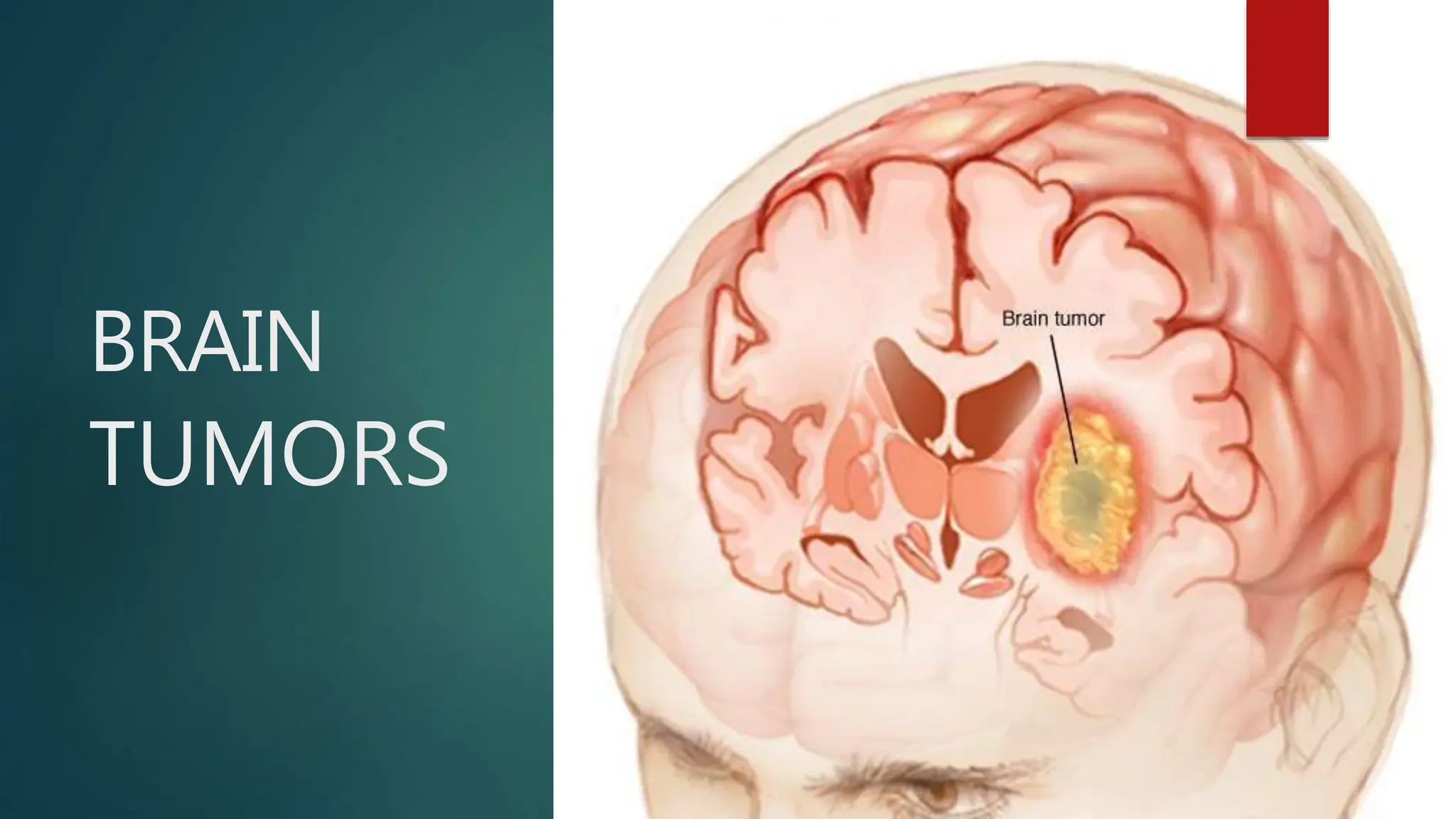
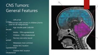
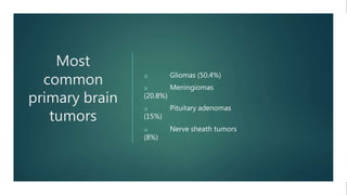
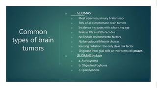
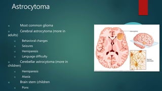
BRAIN
TUMORS
When most normal cells grow old or get damaged, they die, and new cells take their place.
Sometimes, this process goes wrong.
New cells form when the body doesn't need them, and old or damaged cells don't die as they
should.
The build-up of extra cells often forms a mass of tissue called a growth or tumor
Brain tumour : Defination
u known as an intracranial tumour,
u is an abnormal mass of tissue in which cells grow and multiply
uncontrollably, seemingly unchecked by the mechanisms that control
normal cells
u CNS Tumors Tumors of the
nervous system may arise from
u Cells of the coverings –
meninges, bone, subcutaneous
u Cells intrinsic to the brain
u Other cell populations w
i
t
h
i
n
the skull
u Metastases
(spread f
r
o
melsewhere in
the body )
Classification
u Based on source
u Primary – originates from C
N
S
u Secondary / metastatic – originates elsewhere
in body
u Based on location
u Supratentorial m/c in adults
u Infratentorial m/c in pediatric age g
r
o
u
p
u Tissue of
origin
u Glial :
u Meningial :
u Embryonal;
u Mixed
u Vascular
u Bony origin
Tissue of origin Children Adults
Astrocytes Pilocytic
Astrocytoma (PCA)
Glioblastoma
Multiforme (GBM)
Oligodendrocytes Oligodendroglioma
Ependyma Ependymoma
Neurons Medulloblastoma
Meninges Meningioma
u Intra cranial/ intra axial
u Extra dural
u Intra dural (but outside parenchymal)
u Intra parenchymal
u Intra ventricular
u Extra cranial
u Calvarial bony tumours
u Tumour arising from Skin and subcutaneous t
i
s
s
u
e
.
Benign brain tumours
u Benign brain tumours do not contain cancer
cells : ‒
u Usually, benign tumours can be removed and the rarely grow back. ‒
brain tumours have an obvious border or edge.
u They dont spread to other parts of the body
u They don't invade tissues around them
u However, benign tumours can press on sensitive area of brain and can c
a
u
s
e
serious neurological deficits.
u Unlike benign tumour of other parts of the body, benign tumour of the b
r
a
i
n
are sometimes life threatening
u with time benign brain tumours can become malignant.
Malignant brain tumors.
u contain cancer
cells :
u More serious and often are threat to life
u Rapid Growth
u Invade or crowd nearby healthy brain tissue
u Cancer cell may spread to other parts of the brain or to the spinal c
o
r
d
u rarely spread to other parts of the body.
Metastatic Brain Tumor
u Originates from malignancies outside of the CNS and spread to the
brain,typically through arterial circulation.
u Approx. 25% of individual with systemic cancer develop metastatic
brain
tumor approx.
u 80% in cerebral hemisphere and 20% in the posterior
fossa.
u 1/3rd of brain metastases orginate in Lungs→ Breast→ Skin→ GI
tract→
Kidney
u Frontal lobe is the most
common site
u Average survival with the treatment is approx. 6 months but varies wi
d
e
l
y
by the extent of other systemic metastases.
CNS Tumors:
General Features
u 10% of all
tumors.
u
Commonest solid cancers in children.(2nd to
Leuk for all malignancies)
u Age: double peak 1st& 6th
decade
u Adults - 70% supratentorial
u Children - 70% infratentorial
u No/very rare extra neural
spread
u Metastasis most common
u Adults M/C location
supratentorial
u Children M/C location
infratentorial
CLASSIFICATION OF CNS TUMOURS
1. TUMOURS OF THE GLIAL TISSUE – (GLIOMAS)
2. TUMOURS OF NEURONS
3. MIXED TUMOURS WITH GLIAL & NEURONAL COMPONENTS
4. POORLY DIFFERENTIATED AND EMBRYONAL TUMOURS
5. TUMOURS OF THE MENINGES
6. PERIPHERAL NERVE SHEATH TUMOURS
7. METASTATIC TUMOURS
8. PRIMARY CNS LYMPHOMA
9. Miscellaneous TUMOURS: angioma (vascular origin), pituitary adenoma,
WHO Classification
Basis
u Increased
cellularity
u Nuclear atypia
u Endothelial
proliferation
u Necrosis
RISK FACTOR
u A risk factor is something that may increase the chance of
getting a disease.
u Ionizing Radiations : especially from high dose x-rays and other
sourcescan cause cell damage that leads to a tumor. most common types
are meningioma or glioma.
u Family History : It is rare for brain tumors to run in a family. only a
very f
e
w
number of families have several member with brain tumors
Most
common
primary brain
tumors
u Gliomas (50.4%)
u Meningiomas
(20.8%)
u Pituitary adenomas
(15%)
u Nerve sheath tumors
(8%)
Common
types of brain
tumors
u GLIOMAS
u Most common primary brain tumor
u 50% of all symptomatic brain tumors
u Incidence increases with advancing age
u Peak in 8th and 9th decades
u No known environmental factors
u No behavioural lifestyle choices
u Ionizing radiation: the only clear risk factor
u Originate from glial cells or their stem cell precursors
u GLIOMAS Include
u a. Astrocytoma
u b. Oligodendroglioma
u c. Ependymoma
Astrocytoma
u Most common glioma
u Cerebral astrocytoma (more in
adults)
u Behavioral changes
u Seizures
u Hemiparesis
u Language difficulty
u Cerebellar astrocytoma (more in
children)
u Hemiparesis
u Ataxia
u Brain stem (children
u Pons
WHO Grading
of
Astrocytoma
u Grade 1
u Pilocystic astrocytoma
u Bening cytological feactures
u Survival >10 years
u Grade 2
u Low grade astrocytoma
u Moderate cellularity – no aplasia or increase mitotic aactivity
u Survival >5 years
u Grade 3
u Anaplastic astrocytoma
u Increased cellularity, aplasia and mitotic activity
u Survival 3 years
u Grade 4
u Glioblastoma multiforme
u Fertures of grade 3 and microvascular proliferation and
necrosis
u Survival <1years
u Radiological and
pathological
Oligodendroglioma
u Derived from oligodendrocytes or their precursors
u Oligodendrocytes produce the white matter in the brain
u 5-7% of all intracranial gliomas
u Most often in the 3rd and 4th decades
u Males:females = 2:1
u Found primarily in cerebral hemispheres, within the brain
parenchyma
u Highly infiltrative
u
May metastasize distantly in ventricular & subarachnoid spaces like the
GBM (CSF seeding
u Round regular “fried-egg” cells on
histology
Prognosis of
Oligodendroglioma
Median Survival
u Low-grade
oligodendrogliomas: 8-16 years
u Anaplastic
oligodendrogliomas: 5 years
u Tumours that have
1p/19qLOH—best prognosis
u Many pts die from
malignant
transformation of the tumour
Ependymoma
u Arise from ependymal cells (an intraventricular
tumor)
u More common in children
u 10% pediatric intracranial tumors
u 5% of adult intracranial tumors
u Most common in the 4th ventricle
u Ataxia, vertigo, increased ICP
u May grow in brain parenchyma without
obvious
attachment to the ventricular system
u Spinal lesions more common in adults
u Intracranial ependymomas predominate in
children
Prognosis
u 5-year survival:
40-50
u 10-year survival:
47-68
u Better prognosis
u Young age
u Infratentorial
u Gross total excision
u Low-grade histology
MENINGIOMA
u Second most common primary brain tumor
u Originate from arachnoid cells (meningoepithelial cap cells normally seen in
arachnoid villi
u 20% of all intracranial tumors (with asymptomatic cases—40% or more
u 7% of all posterior fossa tumors
u 3-12% of cerebellopontine angle tumors
u Most diagnosed in 6th % 7th decades
u Female: Male—3:2 to 2:1
u Multiple in 5-15% (NF-2
u 90% intracranial
u 10% intraspinal
u Spinal meningioma: 10x in women
u All familial meningiomas occur with NF-2
u Rare in children (more in boys
u Rare without dural attachments
u Usually Intraventricular or posterior fossa
u Commonly with sarcomatous changes
u Frequently with NF-2
Etiology of
Meningioma
u Progesterone receptors
u Expressed in 80% of
women with
meningiomas
u Expressed in 40% of
men with meningiomas
Sites of predilection
u Cerebral
convexity
(Sylvian & parasagittal
areas
u Falxcerebri
u Skull base
u Olfactory
groove
u Sphenoid
ridge
u CP angle
u
Tuberculumsella
Pathology
u Nodular tumors occasionally
meningiomas en
plaque(sheer-like formation)
u Highly vascular
u Encapsulated and attached in the dura
(blood
supply from external carotid artery)
u Hyperostosis of adjacent bone (bone
proliferation
u Histological Characteristics
u Benign
u Typical features: Whorls of arachnoid cells
surrounding a central hyaline material that
eventually calcifies to form PSAMMOMABODIES
Signs and symptoms
u Depend on tumor size, type and
location.
u Symptoms may be caused when a tumor presses on the nerve or
harms the
part of the brain
u Most common symptoms of brain tumors
are :
u Headaches (usually worse in the morning)
u Nausea and vomiting
u Changes in speech, vision, or hearing
u Problems balancing or walking
u Changes in mood, personality, or ability to
concentrate
u Problems with memory
u Muscle jerking or twitching (seizures or convulsions)
u Numbness or tingling in the arms or legs
Headache
u presenting symptom in 30 % of the cases & devlops during the course
of t
h
edisease in 70% of the cases
u It is important to identify the specific nature of the headaches, because
certainfeatures often indicate the presence of a brain tumor. these feature
include :
u 1. The headache that interrupts sleep or is worse on waking and
improves
throughout the day
u 2. The headache that is elicited by postural changes, coughing, or exercise
u 3. The headache of recent onset that is more severe or of a different type
than usual
u 4. The new onset of headache in a previously asymptomatic person
u 5. The headache associated with nausea and vomiting,
papilledema, or focal neurological signs
Seizure
u Presenting symptom in 1/3rd of cases and is present in 50%-70% of
cases
at some stage of the disease.
u Approx 10%-20% adults with new onset seizure activity have brain
tumors.
u Seizures produce by glioma in
u Frontal Lobe (59%)
u Parietal Lobe (42%)
u Temporal Lobe (35%)
u Occipital Lobe (33%)
Altered Mental Status
u The initial symptom in 15% to 20% of individuals.
u Slight changes in concentration, memory, affect, personality,
initiative,&
abstract reasoning to severe cognitive problems & confusion
Papilledema (Swelling of optic nerve)
u less frequent now-a- days because brain tumors are being
diagnosed earlier with the use of sensitive imaging techniques.
u It is more common in children with slow growing tumors and
posterior
fossa tumors.
u Other less common symptoms are vomiting and frank positional
vertigo, usually accompanying tumors found in the posterior fossa
Specific Signs & Symptoms
u Few Clinical features are related to functional areas of the brain thus
h
a
v
e
a specific localizing value in medically diagnosing a brain tumor.
u FRONTAL LOBE Functions : Motor functioning, initiation of
action, interpretation of emotion, including motor speech. motor
praxis,
attention, cognition, emotions, intellegence, judgement, motivation and
memory.
u Disorders : Hemiparesis, Seizures, Aphasia & Gait
Difficulties.
u With gowth of tumor there may be personality changes like
Disinhibition, Irritability, Impaired Judgement, & Lack of Initiation
u PARITAL LOBE Functions :
u General Condition : Contralateral Sensory loss &
hemiparesis,
Homonymous visual deficits or neglect, agnosia's, apraxia's & visual-spatial
disorders.
u If Dominant Parietal Lobe is involved, Aphasia & Seizures may be
present.
u If Non-Dominant Parietal Lobe is Involved, Contralateral
neglect &
decreased awareness of impairments can commonly be found.
u OCCIPITAL LOBE Functions :
u It is primary processing area of visual information.
u General Condition : Dysfunction of the eye movement &
Homonymous hemianopsia.
u If parieto- occipital junction is involved, visual agnosia &
agraphia are
often present.
u Bilateral tumour may cause Cortical Blindness
u TEMPORAL LOBE Functions :
u Auditory and limbic processing.
u Condition :
u Ant. lesion - clinically silent until they become very large and causing
seizes.
u
Lateral Side - Auditory and perceptual changes Medial Side - Changes in
cognitive integration, long-term memory, learning, and emotions may be seen.
u Dominant Temporal lobe - Aphasia Left Temporal lobe lesion - Anomia,
agraphia, acalculia, Wernicke aphasia (Fluent, nonsensical speech)
u Bitemporal involvement - It is rare and causes memory deficits &
possible dementia.
u CEREBELLUM
Function :
u Coordination & Equilibrium.
u
Common Symptoms : In adults, headache, nausea and vomiting present in 40% of condition &
ataxia in 25% of Cases.
u Lesion of midline - Truncal & Gait Ataxia
u Lesion of Hemispheres - Uni. Appendicular ataxia mostly in UE
u Lesion of either Hemisphere - Ipsilateral dysmetria, dysdiadochokinesia, and intention
tremor.
u
Lesion in cerebellopontine angle - hearing loss, headache, ataxia, dizziness, tinnitus and facial
may occur.
u If tumor invades meninges the at foramen magnum causes cerebellar tonsil herniation,
nuchal
rigidity and head tilt away from lesion may be seen.
u ** As cerebellum is located in an extremely confined space, even minimal increases in
pressure can cause death from cerebellar tonsil herniation.
u BRAIN STEM Function :
u It communicates information to and from the cerebral cortex via
fiber tracts, control basic life function. Reticular formation specifically
controls consciousness and attention.
u Symptoms : Tumor have an insidious onset and may include
g
a
i
t disturbances, diplopia, focal weakness, headache, vomiting,
facial numbness and weakness, and perrsonality changes.
u Dorsal Midbrain - Parinaud Syndrome(Loss of upward gaze,
pupillary
areflexia to light, and loss of convergence)
u Reticular System of Pons & Medulla - Apnea, hypo- or hyper-
ventilation, orthostic hypotension or syncope
u PITUITARY GLAND Functions :
u It secretes hormones that regulate many bodily process
u Condition : Tumors are typically large and affect pitutary function by
compressing i
t
sstructure or hypersecreting hormones.
u Enlarging tumor decreases the hormone production resulting in Pituitary
disorders are
specific to type of hormones involved e.g. Cushings disease, hypothyroidism, Addison
disease, diabetes etc.
u As tumor enlarge it compresses nearby area :
u Lateral Extension –
u 3rd & 4th cranial nerve - Diplopia
u 5th Nerve - Ipsilateral Facial Numbeness
u Internal Carotid artery occlusion - Cerebral Infarction
u Upward Extension - Compresses Optic Chiasma &
Hypothalamus
u Downward Extension - Compresses Sphenoid Sinus
Diagnosis
u A clinical diagnosis consist of information the physician gathers
during a comprehensive examination.
u Medical History including the specific nature of S&S
u Neurological Examination - Testing of reflexes & assess visual,
cognitive, sensory, and motor function.
u Opthalmic examination – papiloedema, pupillary light reflex, visual
acuity a
n
d
Visual Feild
u After clinical diagnosis suspects the tumor the next diagnostic step is
TumorImaging
Radiological Diagnosis
u Static neurological imaging includes CT and MRI,
u Noninvasive techniques
u Provide accurate anatomical and functional analysis of intracranial structures.
u CT Scan :
u CT uses ionizing radiation, thin bands of x-rays, to produce images of slices of brain
tissue.
u It was the first brain imaging technique to allow determination of tumor size.
u Contrast enhancement helps to identify isodense tumor from surrounding parenchyma,
hypodense lesions in edematous areas, and optimal sites for tumor biopsy.
u After surgical intervention, CT can be used to confirm the proper tissue biopsy site a
n
d
determine the success of tumor resection.
u Although MRI has become the preferred method, CT scanning offers lower cost, a shorter
scanning time, and a more sensitive method to detect calcification and bony involvement.
u Magnetic Resonance Imaging :
u MRI is the imaging procedure of
choice
u MRI uses magnetic fields
u MRI is superior to CT in detecting & localizing tumor as well as
evaluating
edema, hydrocephalus or hemorrhage.
u MRI is more sensitive imaging modality
u Contrast enhancement with gadolinium sharpens the definition of
lesion
u MRI enhanced with gadolinium can distinguish between edema and
tumor
u MRI also cannot accurately predict tumor type or grade of
malignancy, f
o
rwhich we need to biopsy the lesion
u Dynamic Imaging.
u Positron emission tomography (PET)
u Single photon emission CT (SPECT)
u Magnetic Resonance Spectroscopy (MRS)
u Functional MRI PET Scan
u It is non-invasive and uses cyclotron and specific isotopes to obtain info a
b
o
u
t
metabolism and physiology of the tumor and surrounding tissue.
u It uses radioactive markers to measure glucose metabolism which is useful to
determine the grade of primary brain tumor.
u It also helps in study of metabolic effect of chemotherrapy, Radiation
therapy and
steroids on the tumor.
u It is expensive & less reliable in patient with heavy dose of chemo
therapy
u SPECT Scan :
u It is functional imaging technique evolved from PET scan & uses isotopes w
/
o
cyclotron to assess cerebral blood flow and determining tumor location.
u It is used to identify high- & low- grade tumor to differentiate between t
u
m
o
r
recurrence and radiation necrosis.
u It is used pre-op with static imaging to localize highest metabolic area of t
u
m
o
r
for biopsy.
u SPECT is less sensitive method to obtain physiological information on t
u
m
o
r
s
.
u It is more readily available and less expensive.
u Magnetic Resonance Spectroscopy :
u It is a non-invasive technique used in conjunction with static
MRI t
omeasure the metabolism of brain tumors.
u It has been proved to differentiate successfully normal brain
f
r
o
m
malignant tumor and recurrent tumor from radiation necrosis.
u It also has been used to document early treatment response and
provideinformation regarding histological grade of astrocytomas.
u Magnetic resonance angiography (MRA) generates images of
blood vessels without dye or ionizing radiation to evaluate the blood
flow and position of vessels leading to the brain tumor.
u Functional Magnetic Resonance Imaging :
u It uses a conventional MRI scanner fitted with echo planar
technology t
omap cerebral blood flow at the capillary level.
u Its intended purpose is to provide information regarding the
diffusion o
fcontrast into tumor, resulting in better resolution of tumor
and edema.
u It can also be used to identify the motor, sensory, and
language areas of the brain or the functional eloquent cortex
u Biopsy
u Surgical biopsy is performed to obtain tumor tissue as part of
tumor
resection or as a separate diagnostic procedure.
u Stereotactic biopsy is a computer-directed needle
biopsy.
u When guided by advanced imaging tools, stereotactic biopsy yields
the l
o
w
e
s
tsurgical morbidity and highest degree of diagnostic information.
u This technique is frequently used with deep-seated tumors in functionally
important or inaccessible areas of the brain in order to preserve function.
Laboratory Diagnosis
u Laboratory testing is often used to further assess focal deficits
during t
h
ediagnosis and management of brain tumors.
u Perimetry is the measurement of visual fields used when evaluating
tumorsnear the optic chiasm.
u Electroencephalography (EEG) is used to monitor brain activity and detect
seizures but has limited value during screening because EEG findings are often
normal in clients with brain tumors.
u Lumbar puncture is used to analyze CSF, which is useful in the
diagnosis a
n
d
detection of dissemination of certain brain tumors.
u Audiometry and vestibular testing are useful for diagnosing tumors
in t
h
ecerebellopontine angle.
u Endocrine testing is used to examine endocrine abnormalities with
tumors i
nthe pituitary gland and hypothalamus.
TREATMENT
u Medical & Surgical Management
u The ultimate goal of tumor management are to improve quality of
life a
n
d
extend survival, by improving body function & structures
u Treatment techniques are determined by histological type, location,
grade,
and size of tumor; age of onset; and medical history of the patient.
u Four Type of Treatment are discussed :
u 1. Traditional Surgery
u 2. Chemotherapy
u 3. Radiation Therapy
u 4. Stereotactic Radiosurgery
u TRADITIONAL SURGERY :
u Primary Goal : Maximal tumor resection with the least amount of
damageto neural or supporting structures.
u The purposes of surgery in the management of brain tumors
include the
following :
u 1. Biopsy to establisha diagnosis
u 2. Partial resectionto decrease the tumor massto be treated by other m
e
t
h
o
d
s
u 3. Completeresectionof the tumor
u 4. Provisionof access for adjuvanttreatmenttechniques
u Biopsies areperformedthroughopen,needle, and stereotacticneedle techniques.
u ‒Open biopsies involve exposure of the tumor followed by removal of a sample
through surgical excision.
u ‒Needle biopsies involve insertion a needle into the tumor through a hole in the skull
and t
h
e
excision of the tissue sample drawn through the needle.
u ‒Stereotactic needle biopsies use computers and MRI or CT scanning equipment to
assisst in
directing the needle into the tumor.
u Craniotomy
u Partial & Complete Resections are accomplished through craniotomy. –Craniotomy
involves
removal of a portion of the skull and seperation of the dura mater to expose the tumor.
u –Stereotactic craniotomy uses technologyto guide neurosurgeon duing the
procedure.
u –Awake craniotomy allows for intra-op brain mapping.
CHEMOTHERAPY :
u It can be used independently or as an adjuvant to surgery or
radiation.
u Chemotherapy can be administered in a number of different
ways.
u
Most agents are given intravenously through a peripheral intravenous line or through a
catheter such as a peripherally inserted central catheter (PICC).
u Chemotherapy drugs impede cellular replication of the tumor cells, interfering
with their
ability to copy deoxyribonucleic acid (DNA) and reproduce.
u Methotrexate (Highly neuro-toxic) is admnistered with Leucovorin
(Antidote)
u Temozolomide is orally available chemotherapeutic agent for the Rx of
Gliomas.
u
The antiangiogenesis monoclonal antibody Avastin (bevacizumab) improved the
progression- free survival and the tumor images on MRIs of patients with glioblastoma.
The drug targets vascular endothelial growth factor (VEGF) and is administered
intravenously.
RADIATION THERAPY :
u It can be used alone or in conjunction with surgery or
chemotherapy t
otreat malignant brain tumors.
u It is typically chosen as a treatment option for tumors that are too large
or inaccessible for surgical resection and to eradicate residual neoplastic
cells after a surgical debulking.
u Radiotherapy consists of the delivery of high-powered photons,
w
i
t
h
energies in a much greater range than that of standard x-rays, as
an external beam directly at the tumor site.
Radiosurgery
u Radiosurgery involves relatively high-dose hypofractionated
radiation beams directed at small tumor areas through the use of
computer imaging. This type of treatment includes the Gamma Knife,
linear accelerators, and the cyberknife.
HAVE A NICE DAY !!
Thank you for
your Attention …

brain tumors-.power point brain diseases

  • 1.
  • 2.
    When most normalcells grow old or get damaged, they die, and new cells take their place. Sometimes, this process goes wrong. New cells form when the body doesn't need them, and old or damaged cells don't die as they should. The build-up of extra cells often forms a mass of tissue called a growth or tumor
  • 3.
    Brain tumour :Defination u known as an intracranial tumour, u is an abnormal mass of tissue in which cells grow and multiply uncontrollably, seemingly unchecked by the mechanisms that control normal cells
  • 4.
    u CNS TumorsTumors of the nervous system may arise from u Cells of the coverings – meninges, bone, subcutaneous u Cells intrinsic to the brain u Other cell populations w i t h i n the skull u Metastases (spread f r o melsewhere in the body )
  • 5.
    Classification u Based onsource u Primary – originates from C N S u Secondary / metastatic – originates elsewhere in body u Based on location u Supratentorial m/c in adults u Infratentorial m/c in pediatric age g r o u p u Tissue of origin u Glial : u Meningial : u Embryonal; u Mixed u Vascular u Bony origin Tissue of origin Children Adults Astrocytes Pilocytic Astrocytoma (PCA) Glioblastoma Multiforme (GBM) Oligodendrocytes Oligodendroglioma Ependyma Ependymoma Neurons Medulloblastoma Meninges Meningioma
  • 6.
    u Intra cranial/intra axial u Extra dural u Intra dural (but outside parenchymal) u Intra parenchymal u Intra ventricular u Extra cranial u Calvarial bony tumours u Tumour arising from Skin and subcutaneous t i s s u e .
  • 7.
    Benign brain tumours uBenign brain tumours do not contain cancer cells : ‒ u Usually, benign tumours can be removed and the rarely grow back. ‒ brain tumours have an obvious border or edge. u They dont spread to other parts of the body u They don't invade tissues around them u However, benign tumours can press on sensitive area of brain and can c a u s e serious neurological deficits. u Unlike benign tumour of other parts of the body, benign tumour of the b r a i n are sometimes life threatening u with time benign brain tumours can become malignant.
  • 8.
    Malignant brain tumors. ucontain cancer cells : u More serious and often are threat to life u Rapid Growth u Invade or crowd nearby healthy brain tissue u Cancer cell may spread to other parts of the brain or to the spinal c o r d u rarely spread to other parts of the body.
  • 9.
    Metastatic Brain Tumor uOriginates from malignancies outside of the CNS and spread to the brain,typically through arterial circulation. u Approx. 25% of individual with systemic cancer develop metastatic brain tumor approx. u 80% in cerebral hemisphere and 20% in the posterior fossa. u 1/3rd of brain metastases orginate in Lungs→ Breast→ Skin→ GI tract→ Kidney u Frontal lobe is the most common site u Average survival with the treatment is approx. 6 months but varies wi d e l y by the extent of other systemic metastases.
  • 10.
    CNS Tumors: General Features u10% of all tumors. u Commonest solid cancers in children.(2nd to Leuk for all malignancies) u Age: double peak 1st& 6th decade u Adults - 70% supratentorial u Children - 70% infratentorial u No/very rare extra neural spread u Metastasis most common u Adults M/C location supratentorial u Children M/C location infratentorial
  • 11.
    CLASSIFICATION OF CNSTUMOURS 1. TUMOURS OF THE GLIAL TISSUE – (GLIOMAS) 2. TUMOURS OF NEURONS 3. MIXED TUMOURS WITH GLIAL & NEURONAL COMPONENTS 4. POORLY DIFFERENTIATED AND EMBRYONAL TUMOURS 5. TUMOURS OF THE MENINGES 6. PERIPHERAL NERVE SHEATH TUMOURS 7. METASTATIC TUMOURS 8. PRIMARY CNS LYMPHOMA 9. Miscellaneous TUMOURS: angioma (vascular origin), pituitary adenoma,
  • 12.
    WHO Classification Basis u Increased cellularity uNuclear atypia u Endothelial proliferation u Necrosis
  • 13.
    RISK FACTOR u Arisk factor is something that may increase the chance of getting a disease. u Ionizing Radiations : especially from high dose x-rays and other sourcescan cause cell damage that leads to a tumor. most common types are meningioma or glioma. u Family History : It is rare for brain tumors to run in a family. only a very f e w number of families have several member with brain tumors
  • 14.
    Most common primary brain tumors u Gliomas(50.4%) u Meningiomas (20.8%) u Pituitary adenomas (15%) u Nerve sheath tumors (8%)
  • 15.
    Common types of brain tumors uGLIOMAS u Most common primary brain tumor u 50% of all symptomatic brain tumors u Incidence increases with advancing age u Peak in 8th and 9th decades u No known environmental factors u No behavioural lifestyle choices u Ionizing radiation: the only clear risk factor u Originate from glial cells or their stem cell precursors u GLIOMAS Include u a. Astrocytoma u b. Oligodendroglioma u c. Ependymoma
  • 16.
    Astrocytoma u Most commonglioma u Cerebral astrocytoma (more in adults) u Behavioral changes u Seizures u Hemiparesis u Language difficulty u Cerebellar astrocytoma (more in children) u Hemiparesis u Ataxia u Brain stem (children u Pons
  • 17.
    WHO Grading of Astrocytoma u Grade1 u Pilocystic astrocytoma u Bening cytological feactures u Survival >10 years u Grade 2 u Low grade astrocytoma u Moderate cellularity – no aplasia or increase mitotic aactivity u Survival >5 years u Grade 3 u Anaplastic astrocytoma u Increased cellularity, aplasia and mitotic activity u Survival 3 years u Grade 4 u Glioblastoma multiforme u Fertures of grade 3 and microvascular proliferation and necrosis u Survival <1years
  • 18.
  • 19.
    Oligodendroglioma u Derived fromoligodendrocytes or their precursors u Oligodendrocytes produce the white matter in the brain u 5-7% of all intracranial gliomas u Most often in the 3rd and 4th decades u Males:females = 2:1 u Found primarily in cerebral hemispheres, within the brain parenchyma u Highly infiltrative u May metastasize distantly in ventricular & subarachnoid spaces like the GBM (CSF seeding u Round regular “fried-egg” cells on histology
  • 20.
    Prognosis of Oligodendroglioma Median Survival uLow-grade oligodendrogliomas: 8-16 years u Anaplastic oligodendrogliomas: 5 years u Tumours that have 1p/19qLOH—best prognosis u Many pts die from malignant transformation of the tumour
  • 21.
    Ependymoma u Arise fromependymal cells (an intraventricular tumor) u More common in children u 10% pediatric intracranial tumors u 5% of adult intracranial tumors u Most common in the 4th ventricle u Ataxia, vertigo, increased ICP u May grow in brain parenchyma without obvious attachment to the ventricular system u Spinal lesions more common in adults u Intracranial ependymomas predominate in children
  • 22.
    Prognosis u 5-year survival: 40-50 u10-year survival: 47-68 u Better prognosis u Young age u Infratentorial u Gross total excision u Low-grade histology
  • 23.
    MENINGIOMA u Second mostcommon primary brain tumor u Originate from arachnoid cells (meningoepithelial cap cells normally seen in arachnoid villi u 20% of all intracranial tumors (with asymptomatic cases—40% or more u 7% of all posterior fossa tumors u 3-12% of cerebellopontine angle tumors u Most diagnosed in 6th % 7th decades u Female: Male—3:2 to 2:1 u Multiple in 5-15% (NF-2 u 90% intracranial u 10% intraspinal u Spinal meningioma: 10x in women u All familial meningiomas occur with NF-2 u Rare in children (more in boys u Rare without dural attachments u Usually Intraventricular or posterior fossa u Commonly with sarcomatous changes u Frequently with NF-2
  • 24.
    Etiology of Meningioma u Progesteronereceptors u Expressed in 80% of women with meningiomas u Expressed in 40% of men with meningiomas
  • 25.
    Sites of predilection uCerebral convexity (Sylvian & parasagittal areas u Falxcerebri u Skull base u Olfactory groove u Sphenoid ridge u CP angle u Tuberculumsella
  • 26.
    Pathology u Nodular tumorsoccasionally meningiomas en plaque(sheer-like formation) u Highly vascular u Encapsulated and attached in the dura (blood supply from external carotid artery) u Hyperostosis of adjacent bone (bone proliferation u Histological Characteristics u Benign u Typical features: Whorls of arachnoid cells surrounding a central hyaline material that eventually calcifies to form PSAMMOMABODIES
  • 27.
    Signs and symptoms uDepend on tumor size, type and location. u Symptoms may be caused when a tumor presses on the nerve or harms the part of the brain u Most common symptoms of brain tumors are : u Headaches (usually worse in the morning) u Nausea and vomiting u Changes in speech, vision, or hearing u Problems balancing or walking u Changes in mood, personality, or ability to concentrate u Problems with memory u Muscle jerking or twitching (seizures or convulsions) u Numbness or tingling in the arms or legs
  • 28.
    Headache u presenting symptomin 30 % of the cases & devlops during the course of t h edisease in 70% of the cases u It is important to identify the specific nature of the headaches, because certainfeatures often indicate the presence of a brain tumor. these feature include : u 1. The headache that interrupts sleep or is worse on waking and improves throughout the day u 2. The headache that is elicited by postural changes, coughing, or exercise u 3. The headache of recent onset that is more severe or of a different type than usual u 4. The new onset of headache in a previously asymptomatic person u 5. The headache associated with nausea and vomiting, papilledema, or focal neurological signs
  • 29.
    Seizure u Presenting symptomin 1/3rd of cases and is present in 50%-70% of cases at some stage of the disease. u Approx 10%-20% adults with new onset seizure activity have brain tumors. u Seizures produce by glioma in u Frontal Lobe (59%) u Parietal Lobe (42%) u Temporal Lobe (35%) u Occipital Lobe (33%)
  • 30.
    Altered Mental Status uThe initial symptom in 15% to 20% of individuals. u Slight changes in concentration, memory, affect, personality, initiative,& abstract reasoning to severe cognitive problems & confusion
  • 31.
    Papilledema (Swelling ofoptic nerve) u less frequent now-a- days because brain tumors are being diagnosed earlier with the use of sensitive imaging techniques. u It is more common in children with slow growing tumors and posterior fossa tumors. u Other less common symptoms are vomiting and frank positional vertigo, usually accompanying tumors found in the posterior fossa
  • 32.
    Specific Signs &Symptoms u Few Clinical features are related to functional areas of the brain thus h a v e a specific localizing value in medically diagnosing a brain tumor. u FRONTAL LOBE Functions : Motor functioning, initiation of action, interpretation of emotion, including motor speech. motor praxis, attention, cognition, emotions, intellegence, judgement, motivation and memory. u Disorders : Hemiparesis, Seizures, Aphasia & Gait Difficulties. u With gowth of tumor there may be personality changes like Disinhibition, Irritability, Impaired Judgement, & Lack of Initiation
  • 33.
    u PARITAL LOBEFunctions : u General Condition : Contralateral Sensory loss & hemiparesis, Homonymous visual deficits or neglect, agnosia's, apraxia's & visual-spatial disorders. u If Dominant Parietal Lobe is involved, Aphasia & Seizures may be present. u If Non-Dominant Parietal Lobe is Involved, Contralateral neglect & decreased awareness of impairments can commonly be found.
  • 34.
    u OCCIPITAL LOBEFunctions : u It is primary processing area of visual information. u General Condition : Dysfunction of the eye movement & Homonymous hemianopsia. u If parieto- occipital junction is involved, visual agnosia & agraphia are often present. u Bilateral tumour may cause Cortical Blindness
  • 35.
    u TEMPORAL LOBEFunctions : u Auditory and limbic processing. u Condition : u Ant. lesion - clinically silent until they become very large and causing seizes. u Lateral Side - Auditory and perceptual changes Medial Side - Changes in cognitive integration, long-term memory, learning, and emotions may be seen. u Dominant Temporal lobe - Aphasia Left Temporal lobe lesion - Anomia, agraphia, acalculia, Wernicke aphasia (Fluent, nonsensical speech) u Bitemporal involvement - It is rare and causes memory deficits & possible dementia.
  • 36.
    u CEREBELLUM Function : uCoordination & Equilibrium. u Common Symptoms : In adults, headache, nausea and vomiting present in 40% of condition & ataxia in 25% of Cases. u Lesion of midline - Truncal & Gait Ataxia u Lesion of Hemispheres - Uni. Appendicular ataxia mostly in UE u Lesion of either Hemisphere - Ipsilateral dysmetria, dysdiadochokinesia, and intention tremor. u Lesion in cerebellopontine angle - hearing loss, headache, ataxia, dizziness, tinnitus and facial may occur. u If tumor invades meninges the at foramen magnum causes cerebellar tonsil herniation, nuchal rigidity and head tilt away from lesion may be seen. u ** As cerebellum is located in an extremely confined space, even minimal increases in pressure can cause death from cerebellar tonsil herniation.
  • 37.
    u BRAIN STEMFunction : u It communicates information to and from the cerebral cortex via fiber tracts, control basic life function. Reticular formation specifically controls consciousness and attention. u Symptoms : Tumor have an insidious onset and may include g a i t disturbances, diplopia, focal weakness, headache, vomiting, facial numbness and weakness, and perrsonality changes. u Dorsal Midbrain - Parinaud Syndrome(Loss of upward gaze, pupillary areflexia to light, and loss of convergence) u Reticular System of Pons & Medulla - Apnea, hypo- or hyper- ventilation, orthostic hypotension or syncope
  • 38.
    u PITUITARY GLANDFunctions : u It secretes hormones that regulate many bodily process u Condition : Tumors are typically large and affect pitutary function by compressing i t sstructure or hypersecreting hormones. u Enlarging tumor decreases the hormone production resulting in Pituitary disorders are specific to type of hormones involved e.g. Cushings disease, hypothyroidism, Addison disease, diabetes etc. u As tumor enlarge it compresses nearby area : u Lateral Extension – u 3rd & 4th cranial nerve - Diplopia u 5th Nerve - Ipsilateral Facial Numbeness u Internal Carotid artery occlusion - Cerebral Infarction u Upward Extension - Compresses Optic Chiasma & Hypothalamus u Downward Extension - Compresses Sphenoid Sinus
  • 40.
    Diagnosis u A clinicaldiagnosis consist of information the physician gathers during a comprehensive examination. u Medical History including the specific nature of S&S u Neurological Examination - Testing of reflexes & assess visual, cognitive, sensory, and motor function. u Opthalmic examination – papiloedema, pupillary light reflex, visual acuity a n d Visual Feild u After clinical diagnosis suspects the tumor the next diagnostic step is TumorImaging
  • 41.
    Radiological Diagnosis u Staticneurological imaging includes CT and MRI, u Noninvasive techniques u Provide accurate anatomical and functional analysis of intracranial structures. u CT Scan : u CT uses ionizing radiation, thin bands of x-rays, to produce images of slices of brain tissue. u It was the first brain imaging technique to allow determination of tumor size. u Contrast enhancement helps to identify isodense tumor from surrounding parenchyma, hypodense lesions in edematous areas, and optimal sites for tumor biopsy. u After surgical intervention, CT can be used to confirm the proper tissue biopsy site a n d determine the success of tumor resection. u Although MRI has become the preferred method, CT scanning offers lower cost, a shorter scanning time, and a more sensitive method to detect calcification and bony involvement.
  • 42.
    u Magnetic ResonanceImaging : u MRI is the imaging procedure of choice u MRI uses magnetic fields u MRI is superior to CT in detecting & localizing tumor as well as evaluating edema, hydrocephalus or hemorrhage. u MRI is more sensitive imaging modality u Contrast enhancement with gadolinium sharpens the definition of lesion u MRI enhanced with gadolinium can distinguish between edema and tumor u MRI also cannot accurately predict tumor type or grade of malignancy, f o rwhich we need to biopsy the lesion
  • 43.
    u Dynamic Imaging. uPositron emission tomography (PET) u Single photon emission CT (SPECT) u Magnetic Resonance Spectroscopy (MRS) u Functional MRI PET Scan u It is non-invasive and uses cyclotron and specific isotopes to obtain info a b o u t metabolism and physiology of the tumor and surrounding tissue. u It uses radioactive markers to measure glucose metabolism which is useful to determine the grade of primary brain tumor. u It also helps in study of metabolic effect of chemotherrapy, Radiation therapy and steroids on the tumor. u It is expensive & less reliable in patient with heavy dose of chemo therapy
  • 44.
    u SPECT Scan: u It is functional imaging technique evolved from PET scan & uses isotopes w / o cyclotron to assess cerebral blood flow and determining tumor location. u It is used to identify high- & low- grade tumor to differentiate between t u m o r recurrence and radiation necrosis. u It is used pre-op with static imaging to localize highest metabolic area of t u m o r for biopsy. u SPECT is less sensitive method to obtain physiological information on t u m o r s . u It is more readily available and less expensive.
  • 45.
    u Magnetic ResonanceSpectroscopy : u It is a non-invasive technique used in conjunction with static MRI t omeasure the metabolism of brain tumors. u It has been proved to differentiate successfully normal brain f r o m malignant tumor and recurrent tumor from radiation necrosis. u It also has been used to document early treatment response and provideinformation regarding histological grade of astrocytomas. u Magnetic resonance angiography (MRA) generates images of blood vessels without dye or ionizing radiation to evaluate the blood flow and position of vessels leading to the brain tumor.
  • 46.
    u Functional MagneticResonance Imaging : u It uses a conventional MRI scanner fitted with echo planar technology t omap cerebral blood flow at the capillary level. u Its intended purpose is to provide information regarding the diffusion o fcontrast into tumor, resulting in better resolution of tumor and edema. u It can also be used to identify the motor, sensory, and language areas of the brain or the functional eloquent cortex
  • 47.
    u Biopsy u Surgicalbiopsy is performed to obtain tumor tissue as part of tumor resection or as a separate diagnostic procedure. u Stereotactic biopsy is a computer-directed needle biopsy. u When guided by advanced imaging tools, stereotactic biopsy yields the l o w e s tsurgical morbidity and highest degree of diagnostic information. u This technique is frequently used with deep-seated tumors in functionally important or inaccessible areas of the brain in order to preserve function.
  • 48.
    Laboratory Diagnosis u Laboratorytesting is often used to further assess focal deficits during t h ediagnosis and management of brain tumors. u Perimetry is the measurement of visual fields used when evaluating tumorsnear the optic chiasm. u Electroencephalography (EEG) is used to monitor brain activity and detect seizures but has limited value during screening because EEG findings are often normal in clients with brain tumors. u Lumbar puncture is used to analyze CSF, which is useful in the diagnosis a n d detection of dissemination of certain brain tumors. u Audiometry and vestibular testing are useful for diagnosing tumors in t h ecerebellopontine angle. u Endocrine testing is used to examine endocrine abnormalities with tumors i nthe pituitary gland and hypothalamus.
  • 49.
    TREATMENT u Medical &Surgical Management u The ultimate goal of tumor management are to improve quality of life a n d extend survival, by improving body function & structures u Treatment techniques are determined by histological type, location, grade, and size of tumor; age of onset; and medical history of the patient. u Four Type of Treatment are discussed : u 1. Traditional Surgery u 2. Chemotherapy u 3. Radiation Therapy u 4. Stereotactic Radiosurgery
  • 50.
    u TRADITIONAL SURGERY: u Primary Goal : Maximal tumor resection with the least amount of damageto neural or supporting structures. u The purposes of surgery in the management of brain tumors include the following : u 1. Biopsy to establisha diagnosis u 2. Partial resectionto decrease the tumor massto be treated by other m e t h o d s u 3. Completeresectionof the tumor u 4. Provisionof access for adjuvanttreatmenttechniques
  • 51.
    u Biopsies areperformedthroughopen,needle,and stereotacticneedle techniques. u ‒Open biopsies involve exposure of the tumor followed by removal of a sample through surgical excision. u ‒Needle biopsies involve insertion a needle into the tumor through a hole in the skull and t h e excision of the tissue sample drawn through the needle. u ‒Stereotactic needle biopsies use computers and MRI or CT scanning equipment to assisst in directing the needle into the tumor. u Craniotomy u Partial & Complete Resections are accomplished through craniotomy. –Craniotomy involves removal of a portion of the skull and seperation of the dura mater to expose the tumor. u –Stereotactic craniotomy uses technologyto guide neurosurgeon duing the procedure. u –Awake craniotomy allows for intra-op brain mapping.
  • 52.
    CHEMOTHERAPY : u Itcan be used independently or as an adjuvant to surgery or radiation. u Chemotherapy can be administered in a number of different ways. u Most agents are given intravenously through a peripheral intravenous line or through a catheter such as a peripherally inserted central catheter (PICC). u Chemotherapy drugs impede cellular replication of the tumor cells, interfering with their ability to copy deoxyribonucleic acid (DNA) and reproduce. u Methotrexate (Highly neuro-toxic) is admnistered with Leucovorin (Antidote) u Temozolomide is orally available chemotherapeutic agent for the Rx of Gliomas. u The antiangiogenesis monoclonal antibody Avastin (bevacizumab) improved the progression- free survival and the tumor images on MRIs of patients with glioblastoma. The drug targets vascular endothelial growth factor (VEGF) and is administered intravenously.
  • 53.
    RADIATION THERAPY : uIt can be used alone or in conjunction with surgery or chemotherapy t otreat malignant brain tumors. u It is typically chosen as a treatment option for tumors that are too large or inaccessible for surgical resection and to eradicate residual neoplastic cells after a surgical debulking. u Radiotherapy consists of the delivery of high-powered photons, w i t h energies in a much greater range than that of standard x-rays, as an external beam directly at the tumor site.
  • 54.
    Radiosurgery u Radiosurgery involvesrelatively high-dose hypofractionated radiation beams directed at small tumor areas through the use of computer imaging. This type of treatment includes the Gamma Knife, linear accelerators, and the cyberknife.
  • 55.
    HAVE A NICEDAY !! Thank you for your Attention …