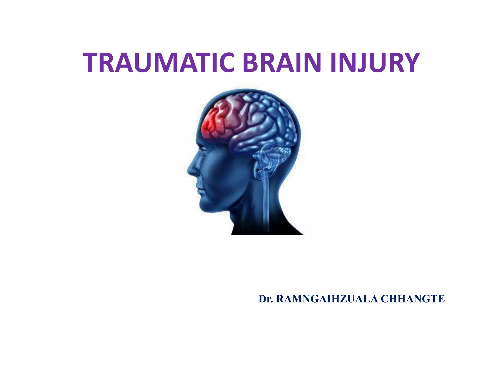
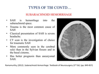
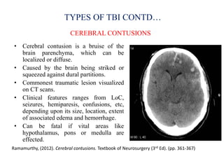
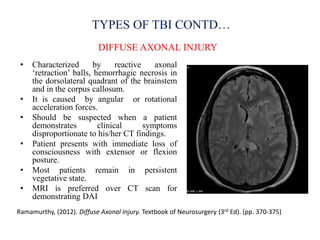
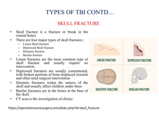
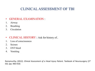
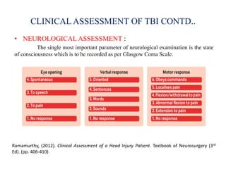
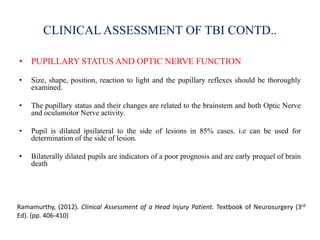
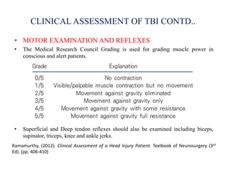
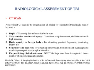
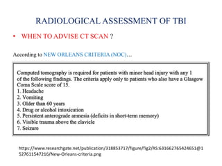
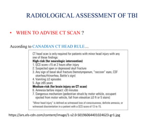
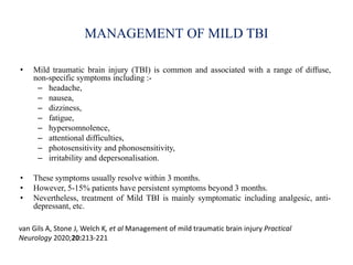
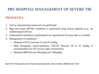
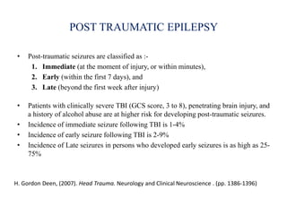
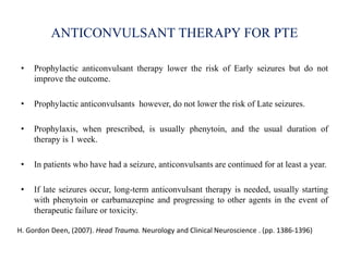
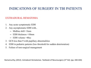
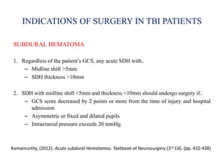
2. INTRODUCTION
• Traumatic Brain Injury (TBI) is defined as an insult to the brain
caused by an external force that may produce diminished or altered
states of consciousness, which results in impaired cognitive abilities
or physical functioning*
• Often referred to as silent epidemic as society in general is largely
unaware of the magnitude of the problem and potential chronicity of
its sequelae**
* National Head Injury Foundation, 1988
** Youmans & Winn, 8th Ed, p.2903
3. EPIDEMIOLOGY OF TRAUMATIC BRAIN INJURY
• Traumatic Brain Injury (TBIs) are a leading cause of morbidity, mortality,
disability and socioeconomic losses in India and other developing
countries.
• It is estimated that nearly 1.5 to 2 million people are injured and 1 million
succumb to death every year in India.
• Road traffic accidents (60%), Falls (20-25%), Violence (10%) are the
leading cause of TBIs in India.
Gururaj G. Epidemiology of traumatic brain injuries: Indian scenario. Neurol Res. 2002 Jan;(1):24-
8.doi:10.1179/016164102101199503. PMID 11783750
4. SEVERITY OF TRAUMATIC BRAIN INJURY
VA/DoD Clinial Practice Guideline for management of Concussion/mTBI (2009)
CRITERIA MILD MODERATE SEVERE
Imaging Normal Normal or
abnormal
Normal or
abnormal
Loss of
Consciousness
0-30 min 30mins to 24 hours > 24 hours
Alteration of mental
state
A moment upto 24
hours
>24 hours, severity based on other
criteria
Post traumatic
Amnesia
0-1 day >1 and < 7 days >7 days
GCS (best score in
the first 24 h)
13-15 9-12 <9
5. CLINICAL FEATURES OF TBI
Vomiting Headache
Confusion ENT bleeed
Seizure Loss of consciousness
Paralysis CSF rhinorrhoea/Otorrhoea
Periorbital echhymosis Dilated pupils/Anisocoria
Bradycardia Aphasia
Inappropriate emotional responses Facial paralysis
Loss of bowel control/bladder control Coma
‘Traumatic Brian Injury’ - A Nitin Agarwal, MD, Rut Thakkar, Khoi Than, MD, FAANS, 2020
6. PATHOPHYSIOLOGY OF TBI
• TBI causes BBB dysfunction which
activates ROS and cytokines/chemokines.
This causes demyelination and axonal
swelling which leads to neurodegeneration.
• Excessive accumulation of
neurotransmitters allow the influx of
calcium ions which results in mitochondrial
dysfunction.
• Mitochondrial dysfunction causes apoptosis
inducing factor (AIF) to be released in
cytosol.
• These cellular and molecular events lead to
caspase dependent and independent
neuronal cell death.
Ray, S. K., Dixon, C. E., and Banik, N. L. (2002). Molecular mechanisms in the pathogenesis of
traumatic brain injury. Histol. Histopathol. 17, 1137–1152. doi: 10.14670/HH-17.1137
7. TYPES OF TRAUMATIC BRAIN INJURY
• PRIMARY TBI : occurs at the moment of initial trauma – skull fractures,
contusions, concussions, diffuse axonal injury, etc.
• Primary TBI can be caused by either penetrating (open) head injury or a
non penetrating (closed) head injury.
• SECONDARY TBI : occurs as an indirect result of the insult. It results
from processes initiated by the initial trauma and typically evolves over
time which include ischemia, hypoxia, cerebral edema, raised ICP,
hypercapnia, meningitis, epilepsy, etc.
Hegde, M.N. (2006). A coursebook on aphasia and other neurogenic language disorders (3rd Ed).
8. TYPES OF TBI CONTD…
CEREBRAL CONCUSSION
• Defined as head injury with a transient loss of brain function and is known
to cause a variety of physical, cognitive and emotional symptoms.
• Most common type of head injury
• Headache and dizziness are the most common symptoms. Amnesia,
confusion, blurring of vision, fatique are also other common symptoms.
• Brain scans are almost always unremarkable.
• Self limiting; needs only symptomatic treatment.
• Caused by acceleration and deceleration forces with minimal axonal
stretch.
Ramamurthy, (2012). Concussion Brain. Textbook of Neurosurgery (3rd Ed). (pp. 257-259)
9. TYPES OF TBI CONTD…
EXTRADURAL HEMATOMA
Ramamurthy, (2012). Extradural Hematomas. Textbook of Neurosurgery (3rd Ed). (pp. 440-444)
• Occurs in aprox 2% of patients with head
injuries.
• Collection of blood between inner table of the
skull and the dura.
• It arises from injury to middle meningeal artery
in over one half, from middle meningeal vein
in on third and from dipolic veins in the
reminder
• Characterized by lucid interval, but not
pathognomic.
• Temporopariental region are most commonly
effected. Commonly associated with overlying
skull fracture.
• Usually occurs in young adults. Rare in
children below 2 years or after the age of 60
years.
• CT scan is the investigation of choice.
• Treatment is mainly surgical.
10. TYPES OF TBI CONTD…
SUBDURAL HEMATOMA
Ramamurthy, (2012). Acute subdural Hematomas. Textbook of Neurosurgery (3rd Ed). (pp. 432-438)
• Accumulation of blood within the
subdural space of the brain.
• Occurs in 5-29% of patients with head
injury.
• It is due to avulsion of bridging veins or
rupture of small coritcal arteries.
• Mechanism of injury is rapid deceleration
of the cranium with a relatively low
magnitude of the shear force applied.
• Convexity of the brain is most commonly
affected
• Usually occurs in the fifth and sixth
decades (mean age 31-47 years).
• 50% of acute SDH is associated with
intracranial injuries
11. TYPES OF TBI CONTD…
SUBARACHNOID HEMORRHAGE
Ramamurthy, (2012). Subarachnoid Hemorrhage. Textbook of Neurosurgery (3rd Ed). (pp. 849-857)
• SAH is hemorrhage into the
subarachnoid space.
• Trauma is the most common cause of
SAH
• Classical presentation of SAH is severe
headache.
• CT scan is the investigation of choice
for traumatic SAH
• More commonly seen in the cerebral
sulci than in the Sylvian fissure and in
the basal cisterns.
• Has better prognosis than aneurysmal
SAH.
12. TYPES OF TBI CONTD…
CEREBRAL CONTUSIONS
Ramamurthy, (2012). Cerebral contusions. Textbook of Neurosurgery (3rd Ed). (pp. 361-367)
• Cerebral contusion is a bruise of the
brain parenchyma, which can be
localized or diffuse.
• Caused by the brain being striked or
squeezed against dural partitions.
• Commonest traumatic lesion visualized
on CT scans.
• Clinical features ranges from LoC,
seizures, hemiparesis, confusions, etc,
depending upon its size, location, extent
of associated edema and hemorrhage.
• Can be fatal if vital areas like
hypothalamus, pons or medulla are
effected.
13. TYPES OF TBI CONTD…
DIFFUSE AXONAL INJURY
Ramamurthy, (2012). Diffuse Axonal Injury. Textbook of Neurosurgery (3rd Ed). (pp. 370-375)
• Characterized by reactive axonal
‘retraction’ balls, hemorrhagic necrosis in
the dorsolateral quadrant of the brainstem
and in the corpus callosum.
• It is caused by angular or rotational
acceleration forces.
• Should be suspected when a patient
demonstrates clinical symptoms
disproportionate to his/her CT findings.
• Patient presents with immediate loss of
consciousness with extensor or flexion
posture.
• Most patients remain in persistent
vegetative state.
• MRI is preferred over CT scan for
demonstrating DAI
14. TYPES OF TBI CONTD…
SKULL FRACTURE
https://operativeneurosurgery.com/doku.php?id=skull_fracture
• Skull fracture is a fracture or break in the
cranial bones
• There are four major types of skull fractures :
– Linear Skull fracture
– Depressed Skull fracture
– Diastatic fracture
– Basilar fracture
• Linear fractures are the most common type of
skull fracture and usually require no
intervention.
• Depressed fractures are usually comminuted,
with broken portions of bone displaced inwards
and often need surgical intervention.
• Diastatic fractures widen the sutures of the
skull and usually affect children under three
• Basilar fractures are in the bones at the base of
the skull
• CT scan is the investigation of choice.
15. CLINICAL ASSESSMENT OF TBI
• GENERAL EXAMINATION :
1. Airway
2. Breathing
3. Circulation
• CLINICAL HISTORY : Ask for history of..
1. Loss of consciousness
2. Seizure
3. ENT bleed
4. Vomiting
Ramamurthy, (2012). Clinical Assessment of a Head Injury Patient. Textbook of Neurosurgery (3rd
Ed). (pp. 406-410)
16. CLINICAL ASSESSMENT OF TBI CONTD..
• NEUROLOGICAL ASSESSMENT :
The single most important parameter of neurological examination is the state
of consciousness which is to be recorded as per Glasgow Coma Scale.
Ramamurthy, (2012). Clinical Assessment of a Head Injury Patient. Textbook of Neurosurgery (3rd
Ed). (pp. 406-410)
17. CLINICAL ASSESSMENT OF TBI CONTD..
• PUPILLARY STATUS AND OPTIC NERVE FUNCTION
• Size, shape, position, reaction to light and the pupillary reflexes should be thoroughly
examined.
• The pupillary status and their changes are related to the brainstem and both Optic Nerve
and oculumotor Nerve activity.
• Pupil is dilated ipsilateral to the side of lesions in 85% cases. i.e can be used for
determination of the side of lesion.
• Bilaterally dilated pupils are indicators of a poor prognosis and are early prequel of brain
death
Ramamurthy, (2012). Clinical Assessment of a Head Injury Patient. Textbook of Neurosurgery (3rd
Ed). (pp. 406-410)
18. CLINICAL ASSESSMENT OF TBI CONTD..
• MOTOR EXAMINATION AND REFLEXES
• The Medical Research Council Grading is used for grading muscle power in
conscious and alert patients.
• Superficial and Deep tendon reflexes should also be examined including biceps,
supinator, triceps, knee and ankle jerks.
Ramamurthy, (2012). Clinical Assessment of a Head Injury Patient. Textbook of Neurosurgery (3rd
Ed). (pp. 406-410)
19. RADIOLOGICAL ASSESSMENT OF TBI
• CT SCAN
Non contrast CT scan is the investigation of choice for Traumatic Brain Injury mainly
because :-
1. Rapid : Takes only few minutes for brain scan
2. Very sensitive to calvarial injury : Can detect scalp hematoma, skull fracture with
high accuracy
3. Radio opacity to foreign body : For detecting gunshot fragments, penetrating
injury, etc
4. Sensitivity and accuracy for detecting hemorrhage, herniation and hydrocephalus
requiring emergent neurosurgical attention.
5. For predicting clinical outcomes : NCCT findings have been incorporated into a
number of outcome prediction rules.
Mutch CA, Talbott JF. Imaging Evaluation of Acute Traumatic Brain Injury. Neurosurg Clin N Am. 2016
Oct;27(4):409-39. doi: 10.1016/j.nec.2016.05.011. Epub 2016 Aug 10. PMID: 27637393; PMCID:
PMC5027071
20. RADIOLOGICAL ASSESSMENT OF TBI
• WHEN TO ADVISE CT SCAN ?
According to NEW ORLEANS CRITERIA (NOC)…
https://www.researchgate.net/publication/318853717/figure/fig2/AS:631662765424651@1
527611547216/New-Orleans-criteria.png
21. RADIOLOGICAL ASSESSMENT OF TBI
• WHEN TO ADVISE CT SCAN ?
According to CANADIAN CT HEAD RULE…
https://ars.els-cdn.com/content/image/1-s2.0-S0196064401024623-gr1.jpg
22. RADIOLOGICAL ASSESSMENT OF TBI
• MRI
• MRI is more sensitive for the detection of certain intracranial injuries especially
axonal injuries
• It is also more sensitive in detecting blood products 24-48 hours after injury.
• Therefore MRI is advised when CT findings are normal and there are persistent
unexplained neurological findings or at subacute or chronic periods.
• Limitations of MRI include.. :-
1. Accessibility
2. Sensitivity to motion
3. Cost
Andrew D. Schweizer, Sumit N. Niogi, Christopher T. Whitlow, A. John Tsiouris. Traumatic Brain
Injury: Imaging Patterns and Complications. 2019 Oct 7. https://doi.org/10.1148/rg.2019190076
23. MANAGEMENT OF MILD TBI
• Mild traumatic brain injury (TBI) is common and associated with a range of diffuse,
non-specific symptoms including :-
– headache,
– nausea,
– dizziness,
– fatigue,
– hypersomnolence,
– attentional difficulties,
– photosensitivity and phonosensitivity,
– irritability and depersonalisation.
• These symptoms usually resolve within 3 months.
• However, 5-15% patients have persistent symptoms beyond 3 months.
• Nevertheless, treatment of Mild TBI is mainly symptomatic including analgesic, anti-
depressant, etc.
van Gils A, Stone J, Welch K, et al Management of mild traumatic brain injury Practical
Neurology 2020;20:213-221
24. PRE HOSPITAL MANAGEMENT OF SEVERE TBI
PROTOCOLS
1. Airway repositioning maneuvers are performed
2. Bag-valve-mask (BVM) ventilation is performed using airway adjuncts (e.g., an
oropharyngeal airway).
3. Endotracheal intubation is performed if an experienced ALS provider is available.
4. Management of ventilation:-
– Maintain ETCO2 between 35 and 45 mmHg.
– Mild therapeutic hyperventilation, ETCO2 between 30 to 35 mmHg is
recommended only for obvious signs of herniation.
– Maintain SBP between 90mmHg and 140mmHg.
Dash HH. Prehospital care of head injured patients. Neurol India. 2008;56:415–419
25. MANAGEMENT PROTOCOL FOR RAISED ICP
Nathens AB, Cryer HG, Fildes J. The American college of surgeons trauma quality improvement
program. Surg Clin North Am. 2012;92:441–454.
26. MANAGEMENT PROTOCOL FOR RAISED ICP
Nathens AB, Cryer HG, Fildes J. The American college of surgeons trauma quality improvement
program. Surg Clin North Am. 2012;92:441–454.
27. POST TRAUMATIC EPILEPSY
• Post-traumatic seizures are classified as :-
1. Immediate (at the moment of injury, or within minutes),
2. Early (within the first 7 days), and
3. Late (beyond the first week after injury)
• Patients with clinically severe TBI (GCS score, 3 to 8), penetrating brain injury, and
a history of alcohol abuse are at higher risk for developing post-traumatic seizures.
• Incidence of immediate seizure following TBI is 1-4%
• Incidence of early seizure following TBI is 2-9%
• Incidence of Late seizures in persons who developed early seizures is as high as 25-
75%
H. Gordon Deen, (2007). Head Trauma. Neurology and Clinical Neuroscience . (pp. 1386-1396)
28. ANTICONVULSANT THERAPY FOR PTE
• Prophylactic anticonvulsant therapy lower the risk of Early seizures but do not
improve the outcome.
• Prophylactic anticonvulsants however, do not lower the risk of Late seizures.
• Prophylaxis, when prescribed, is usually phenytoin, and the usual duration of
therapy is 1 week.
• In patients who have had a seizure, anticonvulsants are continued for at least a year.
• If late seizures occur, long-term anticonvulsant therapy is needed, usually starting
with phenytoin or carbamazepine and progressing to other agents in the event of
therapeutic failure or toxicity.
H. Gordon Deen, (2007). Head Trauma. Neurology and Clinical Neuroscience . (pp. 1386-1396)
29. INDICATIONS OF SURGERY IN TBI PATIENTS
EXTRADURAL HEMATOMA
1. Any acute symptomatic EDH
2. Any asymptomatic EDH with..
– Midline shift >5mm
– EDH thickness >10mm
– EDH volume >40cc
3. GCS less than 9 with pupillary abnormalities
4. EDH in pediatric patients (low threshold for sudden deterioration)
5. Failure of non-surgical management
Ramamurthy, (2012). Extradural Hematomas. Textbook of Neurosurgery (3rd Ed). (pp. 440-444)
30. INDICATIONS OF SURGERY IN TBI PATIENTS
SUBDURAL HEMATOMA
1. Regardless of the patient’s GCS, any acute SDH with..
– Midline shift >5mm
– SDH thickness >10mm
2. SDH with midline shift <5mm and thickness <10mm should undergo surgery if..
– GCS score decreased by 2 points or more from the time of injury and hospital
admission
– Asymmetric or fixed and dilated pupils
– Intracranial pressure exceeds 20 mmHg.
Ramamurthy, (2012). Acute subdural Hematomas. Textbook of Neurosurgery (3rd Ed). (pp. 432-438)
31. INDICATIONS OF SURGERY IN TBI PATIENTS
CONTUSIONS
1. Progressive neurological deterioration
2. Signs of mass effect on brain CT
3. Unresponsive increased ICP
4. Midline shift >5mm
5. Cistern compression evidenced on brain CT
6. Temporal or frontal contusions >20cc
7. Any contusions >50cc
Ramamurthy, (2012). Cerebral contusions. Textbook of Neurosurgery (3rd Ed). (pp. 361-367)
32. INDICATIONS OF SURGERY IN TBI PATIENTS
SKULL FRACTURES
1. Depressed skull fracture > thickness (8-10mm) of the calvaria
2. Dural penetration
3. CSF fistula
4. Intradural Pneumocephalus
5. Significant intracranial hematoma
6. Frontal sinus involvement
7. Wound infection or gross contamination
8. Cosmetic deformity
Stein SC. The Evolution of Modern Treatment for Depressed Skull Fractures. World Neurosurg. 2018
Oct 13. pii: S1878-8750(18)32341-6. doi:10.1016/j.wneu.2018.10.045.
33. SEQUELAE OF TBI
• Post-traumatic amnesia
• Post-concussion syndrome
• Neurobehavioural sequele
• Post-traumatic epilepsy
• Infections – meningitis, osteomyelitis, abscess
• Normal Pressure Hydrocephalus
• Metabolic abnormalities
• Vascular abnormalities
Ramamurthy, (2012). Complications and Sequelae of Head Injuries. Textbook of Neurosurgery (3rd
Ed). (pp. 458-470)