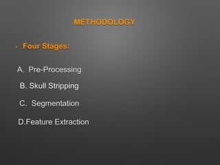
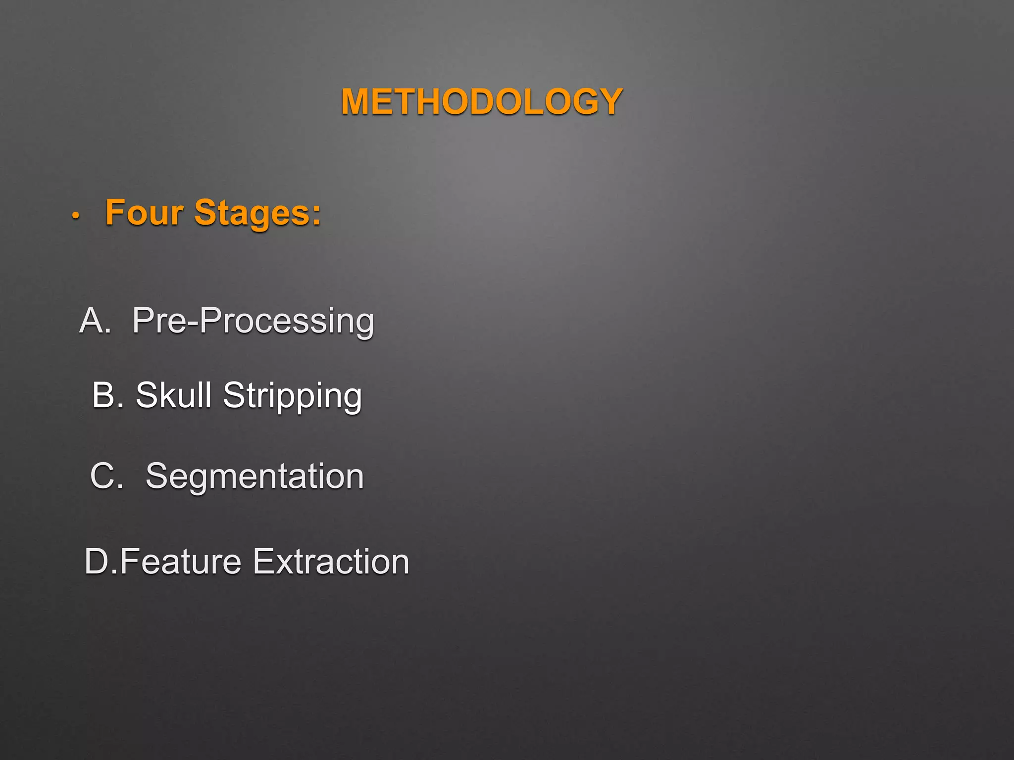
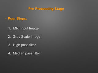
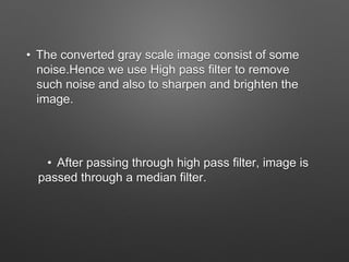
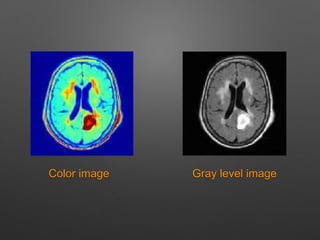
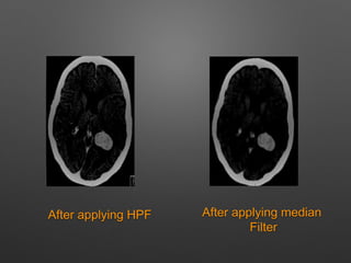
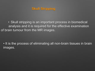
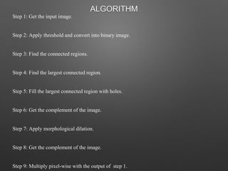
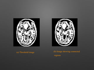
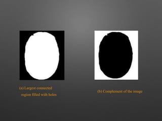
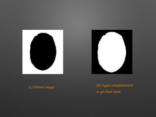
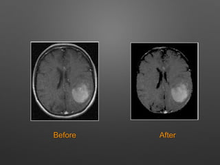
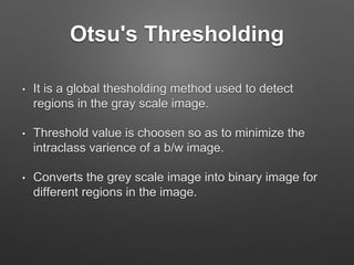
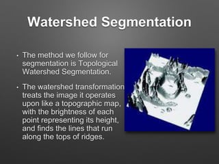
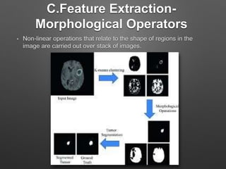
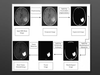
METHODOLOGY
• Four Stages:
A. Pre-Processing
C. Segmentation
D.Feature Extraction
B. Skull Stripping
Pre-Processing Stage
• Four Steps:
1. MRI Input Image
2. Gray Scale Image
3. High pass filter
4. Median pass filter
PROCESS:
• MRI images are magnetic resonance images which
can be acquired on computer when a patient is
scanned by MRI machine.
• This input image is converted to gray scale image
which is the black and white image.
• The converted gray scale image consist of some
noise.Hence we use High pass filter to remove
such noise and also to sharpen and brighten the
image.
• After passing through high pass filter, image is
passed through a median filter.
Gray level imageColor image
After applying HPF After applying median
Filter
Skull Stripping
• Skull stripping is an important process in biomedical
analysis and it is required for the effective examination
of brain tumour from the MR images.
• It is the process of eliminating all non-brain tissues in brain
images.
METHODS:
• Morphology-based
• Intensity-based
Step 1: Get the input image.
Step 2: Apply threshold and convert into binary image.
Step 3: Find the connected regions.
Step 4: Find the largest connected region.
Step 5: Fill the largest connected region with holes.
Step 6: Get the complement of the image.
Step 7: Apply morphological dilation.
Step 8: Get the complement of the image.
Step 9: Multiply pixel-wise with the output of step 1.
ALGORITHM
(a) Threshold image (b) Image showing connected
regions
(a) Largest connected
region filled with holes
(b) Complement of the image
(c) Dilated image
(d) Again complemented
to get final mask
Intensity-Based Methods:
• Region growing
• Edge based
• Histogram based
Before After
B.Segmentation Stage
>Otsu's Thresholding method
>Watershed Segmentation
Otsu's Thresholding
• It is a global thesholding method used to detect
regions in the gray scale image.
• Threshold value is choosen so as to minimize the
intraclass varience of a b/w image.
• Converts the grey scale image into binary image for
different regions in the image.
Watershed Segmentation
• The method we follow for
segmentation is Topological
Watershed Segmentation.
• The watershed transformation
treats the image it operates
upon like a topographic map,
with the brightness of each
point representing its height,
and finds the lines that run
along the tops of ridges.
C.Feature Extraction-
Morphological Operators
• Non-linear operations that relate to the shape of regions in the
image are carried out over stack of images.
From the obtained images the one responsible for
tumor is choosen.
That image is subtracted from the original gray
scale image to get the tumor detected image.
Thank you!!!

Brain tumor detection

  • 1.
    METHODOLOGY • Four Stages: A.Pre-Processing C. Segmentation D.Feature Extraction B. Skull Stripping
  • 2.
    Pre-Processing Stage • FourSteps: 1. MRI Input Image 2. Gray Scale Image 3. High pass filter 4. Median pass filter
  • 3.
    PROCESS: • MRI imagesare magnetic resonance images which can be acquired on computer when a patient is scanned by MRI machine. • This input image is converted to gray scale image which is the black and white image.
  • 4.
    • The convertedgray scale image consist of some noise.Hence we use High pass filter to remove such noise and also to sharpen and brighten the image. • After passing through high pass filter, image is passed through a median filter.
  • 5.
  • 6.
    After applying HPFAfter applying median Filter
  • 7.
    Skull Stripping • Skullstripping is an important process in biomedical analysis and it is required for the effective examination of brain tumour from the MR images. • It is the process of eliminating all non-brain tissues in brain images.
  • 8.
  • 9.
    Step 1: Getthe input image. Step 2: Apply threshold and convert into binary image. Step 3: Find the connected regions. Step 4: Find the largest connected region. Step 5: Fill the largest connected region with holes. Step 6: Get the complement of the image. Step 7: Apply morphological dilation. Step 8: Get the complement of the image. Step 9: Multiply pixel-wise with the output of step 1. ALGORITHM
  • 10.
    (a) Threshold image(b) Image showing connected regions
  • 11.
    (a) Largest connected regionfilled with holes (b) Complement of the image
  • 12.
    (c) Dilated image (d)Again complemented to get final mask
  • 13.
    Intensity-Based Methods: • Regiongrowing • Edge based • Histogram based
  • 14.
  • 15.
    B.Segmentation Stage >Otsu's Thresholdingmethod >Watershed Segmentation
  • 16.
    Otsu's Thresholding • Itis a global thesholding method used to detect regions in the gray scale image. • Threshold value is choosen so as to minimize the intraclass varience of a b/w image. • Converts the grey scale image into binary image for different regions in the image.
  • 18.
    Watershed Segmentation • Themethod we follow for segmentation is Topological Watershed Segmentation. • The watershed transformation treats the image it operates upon like a topographic map, with the brightness of each point representing its height, and finds the lines that run along the tops of ridges.
  • 20.
    C.Feature Extraction- Morphological Operators •Non-linear operations that relate to the shape of regions in the image are carried out over stack of images.
  • 21.
    From the obtainedimages the one responsible for tumor is choosen. That image is subtracted from the original gray scale image to get the tumor detected image.
  • 23.