Downloaded 52 times
![IJRET: International Journal of Research in Engineering and Technology eISSN: 2319-1163 | pISSN: 2321-7308
_______________________________________________________________________________________
Volume: 03 Issue: 03 | Mar-2014, Available @ http://www.ijret.org 367
BRAIN TUMOR DETECTION AND SEGMENTATION USING
WATERSHED SEGMENTATION AND MORPHOLOGICAL
OPERATION
Swe Zin Oo1
, Aung Soe Khaing2
1
Demonstrator, Department of Electronic Engineering, Mandalay Technological University, Myanmar
2
Associate Professor, Department of Electronic Engineering, Mandalay Technological University, Myanmar
Abstract
In the field of medical image processing, detection of brain tumor from magnetic resonance image (MRI) brain scan has become
one of the most active research. Detection of the tumor is the main objective of the system. Detection plays a critical role in
biomedical imaging. In this paper, MRI brain image is used to tumor detection process. This system includes test the brain image
process, image filtering, skull stripping, segmentation, morphological operation, calculation of the tumor area and determination
of the tumor location. In this system, morphological operation of erosion algorithm is applied to detect the tumor. The detailed
procedures are implemented using MATLAB. The proposed method extracts the tumor region accurately from the MRI brain
image. The experimental results indicate that the proposed method efficiently detected the tumor region from the brain image. And
then, the equation of the tumor region in this system is effectively applied in any shape of the tumor region.
Key Words: Magnetic resonance image, skull stripping, segmentation, morphological operation, detection
--------------------------------------------------------------------***----------------------------------------------------------------------
1.INTRODUCTION
In the medical field, magnetic resonance image (MRI) is
widely used in many research [1]. MRI techniques is a
noninvasive method and uses powerful magnet and radio
waves to create the picture of the body. It is suited for
examining soft tissues of the human body such as Ligament
and tendon injury, spinal cord injury and brain tumors, etc.
[2]. The detail information of the human brain can be get
using MRI techniques [1].
Brain, heart and lung etc. are the most important parts of the
human body. And then, all parts of the body are controlled
by the brain cells [3]. Therefore, brain is a vital organ of the
body. Nowadays, brain tumor is a very serious disease
among children and adults. The most deadly and intractable
diseases are brain tumor [4]. Brain tumor’s location and
quickly spreading make a critical problem in treatment of
tumor [5]. Thus, image segmentation and detection are vital
method to solve the medical problem of the various
diseases. Imaging of the brain tumor can be done by
computer tomography (CT) scan, magnetic resonance image
(MRI) scan, Ultrasound, etc. In this research, MRI scan is
used to implement the system [2].
Several works for detection of the brain tumor have been
reported in [1],[5] - [8]. Pratik P et al [1] proposed brain
tumor detection method using connected component
analysis. The method proposed by Manor K Kowari et al [5]
requires to do image cropping so that the exact result of the
tumor region is not obtained. M. Masroor Ahmed et al [6]
proposed the method of the brain tumor detection using
Kmeans Clustering. Nagalkar V J et al [7] proposed brain
tumor detection using soft computing method. This method
can cause false detection in seeing scan. Rajesh C. Patil et al
[8] proposed the method of the brain tumor extraction from
MRI images using MATLAB. Image segmentation can be
done by various techniques: histogram thresholding, region
growing, K-means Clustering and watershed segmentation
[9]. Watershed segmentation is suitable for tumor region
that have higher intensity values [10]. In this paper, marker
controlled watershed segmentation is used to prevent over
segmentation [11]. Preprocessing of the MRI image is the
primary step which removes noise and smooth the image.
To prevent misclassification of brain tissue and non-brain
tissues, skull stripping is done [8]. And, image segmentation
is carried out using marker controlled watershed
segmentation [12]. Then, the tumor region is detected from
the segmented image using morphological operation and
calculated the tumor region [4]. Finally, the location of the
tumor region is determined.](https://image.slidesharecdn.com/braintumordetectionandsegmentationusingwatershedsegmentationandmorphologicaloperation-140812033724-phpapp02/85/Brain-tumor-detection-and-segmentation-using-watershed-segmentation-and-morphological-operation-1-320.jpg)
![IJRET: International Journal of Research in Engineering and Technology eISSN: 2319-1163 | pISSN: 2321-7308
_______________________________________________________________________________________
Volume: 03 Issue: 03 | Mar-2014, Available @ http://www.ijret.org 368
2.MATERIALS AND METHODS
2.1Preprocessing
Preprocessing include the input MRI brain tumor image and
image filtering. In image filtering, several different filters
can be used but the magnetic resonance image (MRI) image
does not contain a lot of noise. So, in this research, average
filter is used to smooth the image. The smoothed image is
used to operate the next step of the system quickly. Average
filter is low pass filter. Average filter is a simple and easy to
implement method of smoothing images. The operation of
average filter is
g (x, y)=1/M (1)
Where, S=neighborhood of pixel (x, y)
M=number of pixels in neighborhood S
2.2 Skull Stripping
Skull stripping is important process in biomedical image
analysis. It is needed to make only in brain image but is not
needed to make in other medical image analysis such as
heart, lung, etc. It must be done before other image
processing step. It is a process of eliminating all non-brain
tissues from brain image. In skull stripping, it is removed
extra cerebral tissues such as skull, fat, skin, etc. Skull
stripping can be done by various methods. They are
automatic skull stripping using image contour, skull
stripping based on region growing and mathematical
morphology, skull stripping based on histogram analysis,
skull stripping based on resonance principle and skull
stripping based on threshold value. Skull stripping based on
threshold value is used to remove the skull tissues in this
paper. In the skull stripping based on threshold value, the
threshold value of the skull tissues and that of normal brain
tissues are manually determined for every image [13].
2.3Watershed Segmentation
Watershed segmentation is a gradient-based segmentation
technique. It considers the gradient map of the image as a
relief map. It segments the image as a dam. The segmented
regions are called catchment basins. Watershed
segmentation solves a variety of image segmentation
problem. It is suitable for the images that have higher
intensity value. Watershed segmentation is caused over
segmentation. To control over segmentation, marker
controlled watershed segmentation is used. Sobel operator is
suitable for edge detection. In marker controlled watershed
segmentation, sobel operator is used to distinct the edge of
the object [10].
The sobel masks in matrix form are as follow:
Mx = , My =
The equation of gradient magnitude used in marker
controlled watershed segmentation is
M= (2)
Angle, ɵ=tan-1
(3)
2.4Morphological Operation
Morphological image processing is a collection of non-
linear operations related to the shape or morphology of
features in an image. A morphological operation on a binary
image creates a new binary image in which the pixel has a
non-zero value. Morphological operations transform the
image. In this paper, erosion is applied to detect the tumor
[6]. The erosion of A by B is given by the expression:
A Ө B = {( i , j) : B( i, j) } (4)
Where, A= the binary image,
B= the structuring element
(i, j)= the center pixel of structuring element
2.5Calculation the tumor region
The area of the tumor region is calculated by the following
equation:
Tumor area=Axtotal number of pixel in the tumor region (5)
A= V x H (6)
Where, A=the area of each pixel
H=horizontal dimension of the image
V=vertical dimension of the image
H=1/horizontal resolution of the image
V=1/vertical resolution of the image
3.IMPLEMENTATION
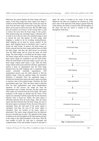
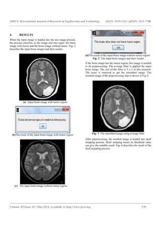
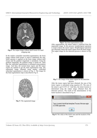
In this paper, the system is implemented as shown in Fig-1.
In this system, the input MRI brain image is used to
implement the algorithms. MRI image gives the detailed
information of the brain tissues than other scan of the brain.
Firstly, the brain image is checked out as the brain image
with tumor region or the brain image without tumor region.
So, the tumor region is extracted from the brain image and
the number of pixel for the tumor region is calculated. If the
number of pixel for the tumor region is equal to zero, the
system displays the brain image without tumor region.](https://image.slidesharecdn.com/braintumordetectionandsegmentationusingwatershedsegmentationandmorphologicaloperation-140812033724-phpapp02/85/Brain-tumor-detection-and-segmentation-using-watershed-segmentation-and-morphological-operation-2-320.jpg)
![IJRET: International Journal of Research in Engineering and Technology eISSN: 2319-1163 | pISSN: 2321-7308
_______________________________________________________________________________________
Volume: 03 Issue: 03 | Mar-2014, Available @ http://www.ijret.org 373
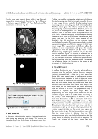
determined based on the pixel value of the tumor region.
The right part of the image and the left part of the image are
defined by manually in this process. Therefore, the location
of the tumor is needed to define automatically. In this paper,
the tumor region is extracted from the single MRI brain
slice. Really, MRI scan take the picture of the brain in many
slices. And the tumor region is not located in the single
slice. Above the reason, the tumor volume is needed to
calculate. The volume of the tumor is calculated to get the
exact result of the tumor region. Therefore, the tumor
volume is needed to calculate. The tumor volume can be
calculated using Frustum model.
ACKNOWLEDGMENT
The author would like to acknowledge the head of
Department of Electronic Engineering, Mandalay
Technological University. The author is highly grateful to
all the teachers, Department of Electronic Engineering,
Mandalay Technological University. The author specially
wants to thank her supervisor Dr. Aung Soe Khaing,
Department of Electronic Engineering, Mandalay
Technological University for his guidance and constant
encouragement throughout the work.
REFERENCES
[1]. Skull Stripping of MRI Head Scans based on Chan-
Vese Active Contour Model, [Online Available],
www.med.harvad.edu/AANLIB/home.html, accessed
on 8 August 2013.
[2]. Magnetic Resonance Image, [Online Available],
www.CEwebsource.com, accessed on 18 June 2013.
[3]. Pratik P. Singhai, Siddharth A. Ladhake, “Brain Tumor
Detection using Marker Based Watershed
Segmentation from Digital MR images”, International
Journal of Innovative Technology and Exploring
Engineering (IJITEE) ISSN: 2278-3075, Volume-2,
Issue-5, April 2013.
[4]. A.Jeeviitha, P. Narendran, “BTS (Brain Tumor
Segmentation) Based on Otus Thresholding,” Indian
Journal of Research, Volume:2, Issue:2, ISSN- 2250-
1991, February 2013.
[5]. Manor K Kowari and Sourabh Yadav, “Brain Tumor
Detection and Segmentation using Histogram
Thresholding”, International Journal of Engineering
and Advanced Technology (IJEAT) ISSN: 2249-898,
Volume-1, Issue-4, Journal, India, April 2012.
[6]. M. Masroor Ahmed, Dzulkifli Bin Mohamad,
“Segmentation of Brain MR Images for Tumor
Extraction by Combining Kmeans Clustering and
Perona-Malik Anisotropic Diffusion model.
[7]. Nagalkaar. V.J and Asole S.S, “Brain Tumor
Detection using Digital Image Processing based on
Soft Computing,” Journal of Signal and Image
Processing, Volume 3, Issue 3, Issn: 0976-8882, 2012.
[8]. Rajesh C.Patil, Dr. A. S.Bhalchandra, “Brain Tumor
Extraction from MRI Images using MATLAB,”
International Journal of Electronics, Communication &
Soft Computing Science and Engineering, Volume 2,
Issue 1, ISSN: 2277-9477.
[9]. S Jayaraman, S Esakkirajan and TVeerakumar, Digital
Image Processing, 3rd
Edition, Tata McGraw Hill,
2010, ISBN (13): 978-0-07-014479-8, ISBN (10): 0-
070114479-6.
[10]. M. C Jobin Christ, R.M.S. Paravathi, “Segmentation of
Medical Image using Clustering and Watershed
Algorithms”, American Journal of Applied Sciences
8(2): 1349-152, 2011 ISSN 1546-9239© 2011 Science
Publication.
[11]. Dibyendu Goshal, Pinaki Pratim Acharjya, “MRI
Image Segmentation using Watershed Transform”,
International Journal of Emerging Technology and
Advanced Engineering, ISSN 2250-2459, Volume 2,
Issue 4, April 2012.
[12]. “Biosignal and Biomedical Image Processing”,
[Online Available], www.dekker.com, accessed on 11
October 2013.
[13]. Rosniza Roslan, Nursuriati Jamil and Rozi Mahmud,
“Skull Stripping Magnetic Resonance Images Brain
Images: region Growing versus Mathematical
Morphology”, International Journal of Computer
Information Systems and Industrial Management
applications, ISSN 2150-7988, Volume (2011).
BIOGRAPHIES
Swe Zin Oo received her
bachelor of engineering in
Electronics from Mandalay
Technological University,
Myanmar in 2007. She is
presently a master student at the
Mandalay Technological
University, Myanmar. Her
research interests include skull
stripping, image segmentation
and tumor detection.](https://image.slidesharecdn.com/braintumordetectionandsegmentationusingwatershedsegmentationandmorphologicaloperation-140812033724-phpapp02/85/Brain-tumor-detection-and-segmentation-using-watershed-segmentation-and-morphological-operation-7-320.jpg)

The paper presents a method for brain tumor detection using magnetic resonance imaging (MRI) that involves preprocessing steps like image filtering, skull stripping, and marker-controlled watershed segmentation. Morphological operations are employed to accurately detect and calculate the tumor area and location within the brain images. The proposed system effectively distinguishes between normal and abnormal brain images, providing reliable results for tumor identification.