Downloaded 12 times
![Multimodal Medical Image Fusion Based On SVD
www.iosrjournals.org 28 | Page
2.2 Principal Component Analysis (PCA) method
Principal Component Analysis is a sub space method, which reduces the multidimensional data sets
into lower dimensions for analysis. This method determines the weights for each source. image using the
eigenvector corresponding to the largest eigen value of the covariance matrix of each source image.
2.3 Discrete Wavelet Transform Method
Wavelet transforms are multi-resolution image decomposition tool that provide a variety of channels
representing the image feature by different frequency subbands at multi-scale. It is a famous technique in
analyzing signals. When decomposition is performed, the approximation and detail component can be separated
2-D Discrete Wavelet Transformation (DWT) converts the image from the spatial domain to frequency domain.
The image is divided by vertical and horizontal lines and represents the first-order of DWT, and the image can
be separated with four parts those are LL1, LH1, HL1 and HH1. Applying Inverse Discrete Wavelet Transform
on fused decomposed level, reconstruct the fused output image.
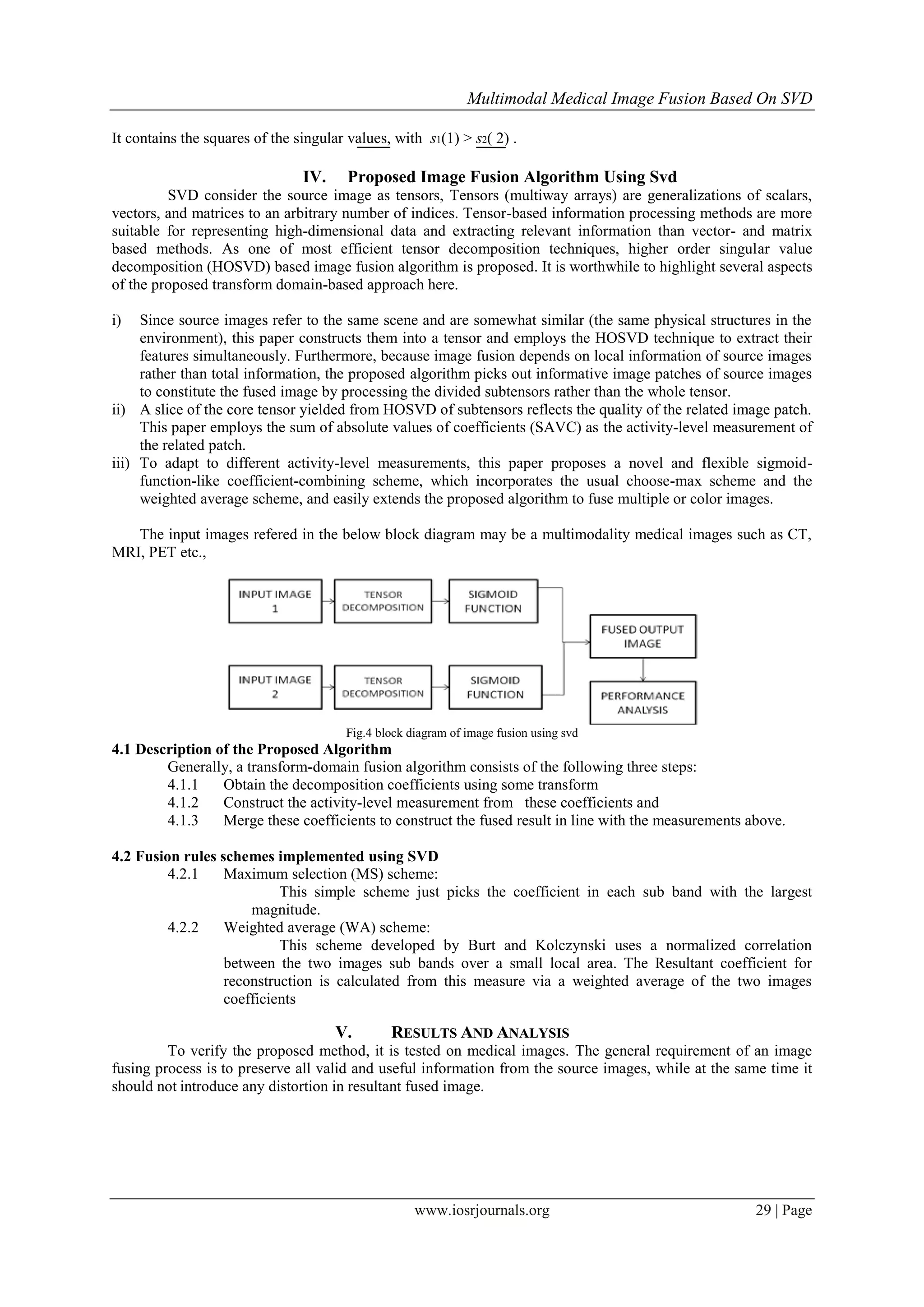
III. IMAGE FUSION TECHNIQUE USING SVD
In this paper, a novel Image fusion technique based on Higher Order singular value decomposition
(HOSVD) is presented to fuse the source images. SVD is a method for identifying and ordering the dimensions
along which data points exhibit the most variations. Using SVD it’s possible to find the best approximation of
the original data points using fewer dimensions. SVD takes a high dimensional, highly variable set of data
points and reducing it to a lower dimensional space that exposes the substructure of the original data more
clearly and orders it from most variation to the least. SVD is based on a theorem from linear algebra which says
that a rectangular matrix A can be broken down into the product of three matrices - an orthogonal matrix U, a
diagonal matrix S, and the transpose of an orthogonal matrix V. The theorem is usually presented like this:
A = USV T
(3.1)
where UTU = I, VTV = I; the columns of U are orthonormal eigenvectors of AAT
, the columns of V are
orthonormal eigenvectors of AT
A, and S is a diagonal matrix containing the square roots of eigenvalues from U
or V in descending order. To compute the SVD of a matrix A with m rows and n columns, with rank r and r ≤ n
≤ m. Then the A can be factorized into three matrices.
Fig.3.1. Illustration of Factoring A to USVT
Where U and V are orthonormal and S is diagonal. Matrix U is an m × m orthogonal matrix
U= [u1, u2,...ur, ur+1 ,..., um ] (3.2)
column vectors ui , for i = 1, 2, …, m, form an orthonormal set:
And matrix V is an n × n orthogonal matrix
V= [ v1, v2,...vr, vr+1 ,..., vn ] (3.3)
Column vectors i v for i = 1, 2, …, n, form an orthonormal set:
Here, S is an m × n diagonal matrix with singular values (SV) on the diagonal. The
matrix S can be showed in following
S1(1)2
0
S12 =
0 S2(2)2
(3.4)](https://image.slidesharecdn.com/f016132731-150505072003-conversion-gate01/75/Multimodal-Medical-Image-Fusion-Based-On-SVD-2-2048.jpg)
![Multimodal Medical Image Fusion Based On SVD
www.iosrjournals.org 31 | Page
c. Entropy (EN)
Entropy is used to calculate the amount of information. Higher value of entropy indicates
that the information increases and the fusion performances are improved.
(5.3)
TABLE 1
Comparison of Different Image Fusion Techniques
S.NO FUSION
TECHNIQUE
DOMAIN MEASURING
PARAMETER
ADVANTAGE DISADVANTAGE
1 Simple
Average
Spatial PSNR= 46.66
NCC= 0.95
EN=7.4122
Simple to
implement
High information
loss
2 PCA Spatial PSNR= 66.66
NCC= 0.96
EN=7.4128
Simple algorithm Presence of
spectral
degradation
3 DWT Transform PSNR= 66.66
NCC= 0.97
EN=7.4244
Better Signal to
noise ratio
Less spatial
Resolution
4 SVD Transform PSNR= 70.44
NCC= 0.98
EN=7.4216
Robust, simple,
fast to implement,
efficiency is more
even in the
presence of noise
Efficiency is still
improved if noise is
reduced
VI. CONCLUSION
Thus a novel image adaptive transform called SVD has been proposed. The proposed algorithm splits
the multiple input images into tensor and can evaluate the quality of image patches using SVD of subtensors.
Finally fused result is obtained by employing sigmoid function. Experimental results and performance analysis
prove that the proposed algorithm is an alternative image fusion approach because it is simple, robust, fast to
implement and it works well in constrained environment.
REFERENCES
[1] Junli Liang, Yang He, Ding Liu and Xianju Zeng (2012) ‘Image Fusion Using Higher Order Singular Value Decomposition’, IEEE
Transactions On Image Processing, Volume 21, No.5, pp 2898-2909.
[2] Amarjot Kaur and Sunil Khullar (2013) ‘Image Fusion using HIS, PCA and Wavelet Technique’, International Journal of
Computer Science and Communication Engineering, Volume 2, No. 2, pp. 92-94.
[3] V.P.S. Naidu (2011) ‘Image Fusion Technique using Multi-resolution Singular Value Decomposition’, Defence Science Journal,
Volume 61, No. 5 pp. 479-484.
[4] G. Bergqvist and E.G Larsson (2010) ‘The higher-order singular value decomposition Theory and application’, IEEE Signal
Processing Magazine, volume 27, No. 3, pp. 151–154.
[5] Li H., S. Manjunath and S. Mitra (1995) ‘Multi sensor image fusion using the wavelet transform’, International journal of
Graphical Models and Image Processing, volume 57, No.3, pp. 235–245.
[6] Cyn Dwith., Vivek Angoth and Amarjoth Singh (2013) ‘Wavelet Based Image Fusion for Detection of Brain Tumor’, International
Journal of Image, graphics and Signal Processing, Volume 1, pp.25-31.
[7] J. Canny (1986) ‘A Computational Approach to Edge Detection’, IEEE Transactions Pattern Analysis Intelligence Volume 8, pp.
679-714.
[8] K.L. Chung, C.H. Shen and L.C. Chang (2001) ‘A novel SVD and VQ based image hiding scheme’, International Journal of
Advance Research Electronics and communication, Volume 22, pp. 105- 108.
[9] Daljit Kaur and Palwinder Singh Mann (2013) ‘Hybrid Transform Domain Algorithm for Medical Image Fusion’, International
Journal for Science and Emerging Technologies with Latest Trends, Volume 8, No.1, pp. 23-27.
[10] R. Costantini, L. Sbaiz and S. Susstrunk (2008) ‘Higher order SVD analysis for dynamic texture synthesis’, IEEE Transactions on
Image Processing, volume 17, No.1, pp.42–52
[11] Bibo Lu. Chunli Miao. (2010) ‘Structure Tensor Based Image Fusion’, Proc.Third International Symposium on Electronic
Commerce and Security Workshops, Volume 11, No.4, pp. 343-346.
[12] Aristeidis Sotiras., Christos Davatzikos and Nikos Paragios (2013) ‘Deformable Medical Image Registration: A Survey’, IEEE
Transactions on Medical imaging, Volume 32, No. 7, pp. 1153-1190.
[13] Q.M. Gaurav Bhatnagar, JonathanWu and Zheng Liu ‘Directive Contrast Based Multimodal Medical Image Fusion in NSCT
Domain’ IEEE TRANSACTIONS ON MULTIMEDIA, VOLUME 15, NO. 5, AUGUST 2013](https://image.slidesharecdn.com/f016132731-150505072003-conversion-gate01/75/Multimodal-Medical-Image-Fusion-Based-On-SVD-5-2048.jpg)

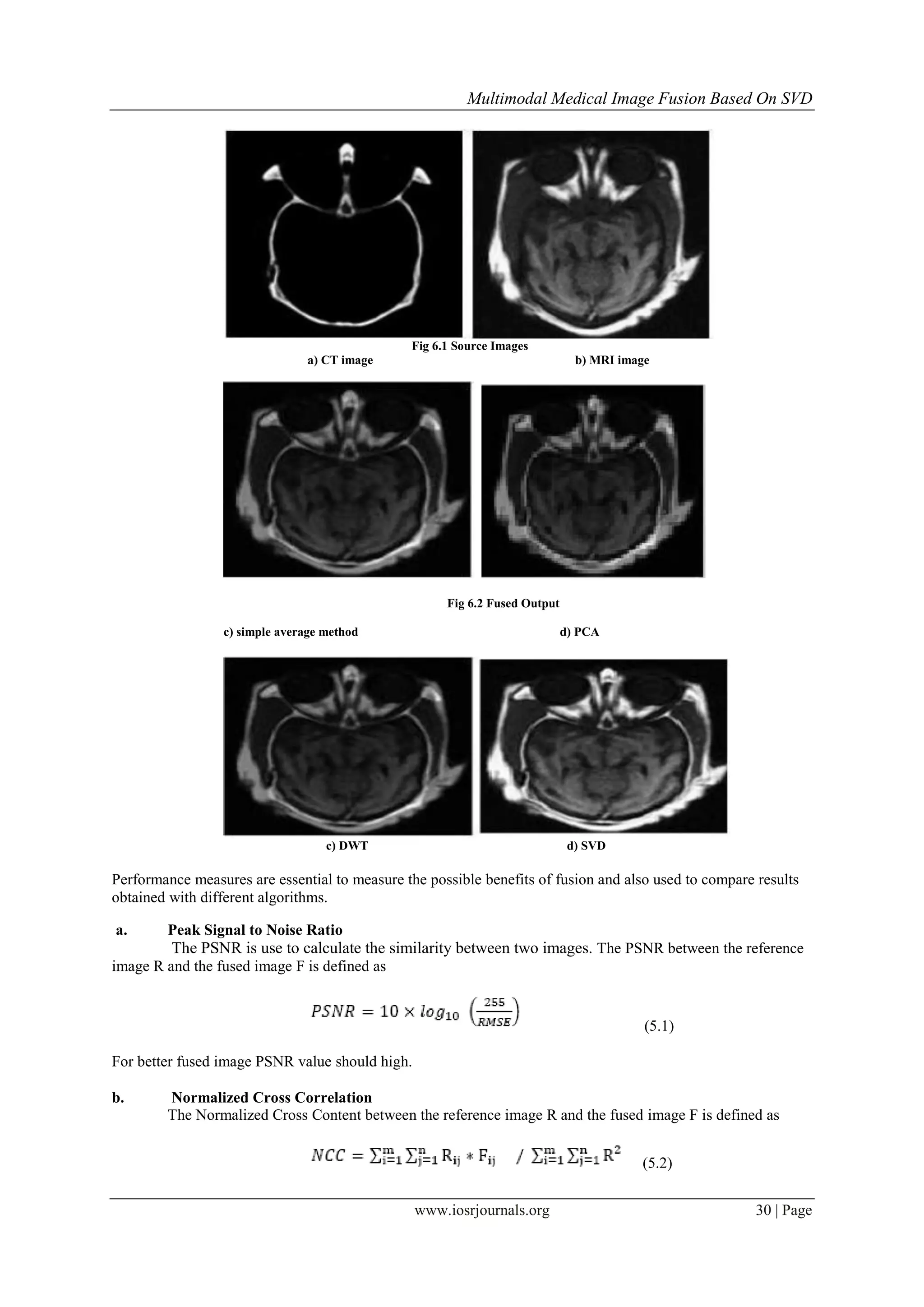
The paper presents a novel multimodal medical image fusion algorithm using Singular Value Decomposition (SVD), which combines informative patches from multiple images to enhance overall image quality. The proposed method utilizes tensor decomposition through Higher Order Singular Value Decomposition (HOSVD) and a sigmoid-function-like coefficient-combining scheme for improved results. Experimental outcomes indicate that the SVD-based approach outperforms traditional methods in terms of image quality and robustness in medical applications.


![Coded Agents – with UiPath SDK + LangGraph [Virtual Hands-on Workshop]](https://cdn.slidesharecdn.com/ss_thumbnails/codedagentsdeck-251215155422-5497c599-thumbnail.jpg?width=640&height=640&fit=bounds)


