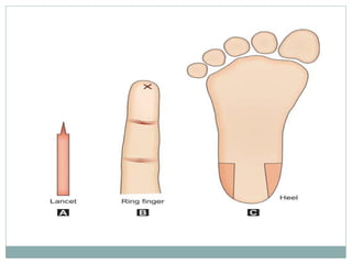
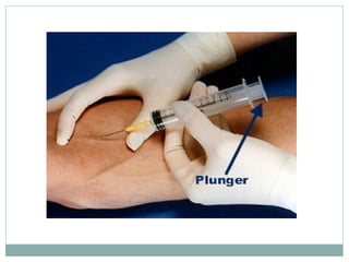
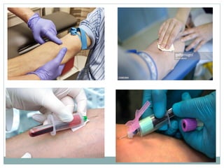
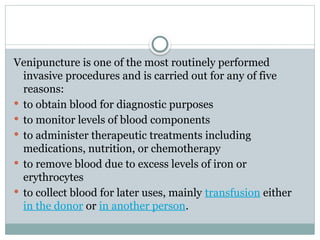
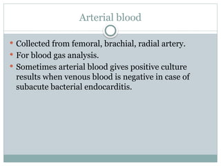
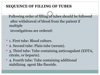
Sample collection methods and
preservation
 It is essential to follow a standard procedure for the
collection of blood specimens to get accurate
laboratory results.
 Any error in collecting the blood or filling the test
tubes may lead to erroneous laboratory results.
 Correct labelling and time to sent to a lab.
Methods of blood collection
Blood sample can be obtained from the skin puncture
or venepuncture.
 Capillary blood
 Venous blood
 Arterial blood
SKIN PUNCTURE
 Commonly used in infants and small children and if
the amount of blood required is small .
 Also called as capillary blood.
 The puncture site is cleansed with 70% ethanol or
other suitable disinfectant.
 Puncture, sufficiently deep to allow free flow of
blood.
 The first drop of blood is wiped away.
 Excessive squeezing should be avoided.
 Should not be collected from cold, cyanosed skin.
VENOUS BLOOD COLLECTION
 When multiple tests are to be done and larger quantity of blood
is needed, anticoagulated venous blood should be obtained.
 Due to the ease of access- veins of the antecubital fossa.
 Rubber tourniquet is tied to the arm.
 Cleansed with 70% ethanol and allowed to dry.
 Needle size should be 19- to 21-gauge in adults and 23-gauge in
children.
 It is performed with the bevel of the needle up and along the
direction of the vein.
 Containers may be glass bottles or disposable plastic tubes with
caps and flat bottom.
 Blood is mixed with the anticoagulant in the
container thoroughly by gently inverting the
container several times.
 The container should not be shaken vigorously as it
can cause frothing and hemolysis.
 After use, disposable needles should be placed in a
puncture-proof container for proper disposal.
 Recapping of needle by hand can cause needle-stick
injury.
Venipuncture is one of the most routinely performed
invasive procedures and is carried out for any of five
reasons:
 to obtain blood for diagnostic purposes
 to monitor levels of blood components
 to administer therapeutic treatments including
medications, nutrition, or chemotherapy
 to remove blood due to excess levels of iron or
erythrocytes
 to collect blood for later uses, mainly transfusion either
in the donor or in another person.
 Proper labelling .
 Precautions.
 Complications.
Arterial blood
 Collected from femoral, brachial, radial artery.
 For blood gas analysis.
 Sometimes arterial blood gives positive culture
results when venous blood is negative in case of
subacute bacterial endocarditis.
Anticoagulants
 Anticoagulants, commonly known as blood thinners,
are chemical substances that prevent or
reduce coagulation of blood, prolonging the clotting
time.
 Anticoagulants used for hematological investigations
are ethylene diamine tetra-acetic acid (EDTA),
heparin,double oxalate, and trisodium citrate.
Ethylene Diamine Tetra-acetic Acid (EDTA)
 This is also called as Sequestrene or Versene.
 This is the recommended anticoagulant for routine
hematological investigations.
 It is used in a concentration of 1.5 mg/ml of blood.
 Dried form of anticoagulant is used as it avoids
dilution of sample.
 Its mechanism of action is chelation of calcium.
 EDTA is used for estimation of hemoglobin,
hematocrit,
cell counts, making blood films, sickling test,
reticulocyte count, and hemoglobin electrophoresis
Changes occurring due to prolonged storage of blood in EDTA
 Blood smear:
– Red cells: Crenation and spherocytic change
– Neutrophils: Separation of nuclear lobes, loss of granules,
vacuoles in cytoplasm
– Monocytes and lymphocytes: Irregular lobulation of
nucleus, vacuoles in cytoplasm
– Platelets: Disintegration
 Hematological tests:
– Packed cell volume and mean cell volume: Increase due to
red cell swelling
– Osmotic fragility: Increases
– Total leucocyte and platelet counts: Progressive decrease.
Double Oxalate (Wintrobe Mixture)
 This consists of ammonium oxalate and potassium
oxalate in 3:2 proportion.
 This combination is used to balance the swelling of red
cells caused by ammonium oxalate and shrinkage caused
by potassium oxalate.
 Mechanism of anticoagulant action is removal of calcium.
 It is used for routine hematological tests and for
estimation of erythrocyte sedimentation rate by Wintrobe
method.
 Place 0.5 ml of this solution in a bottle for 5 ml of blood.
Anticoagulant should be dried in an incubator at 37°C or
on a warm bench before use.
Trisodium Citrate (3.2%)
 This is the anticoagulant of choice for coagulation
studies and for estimation of erythrocyte
sedimentation rate by Westergren method.
 Use 1:9 (anticoagulant: blood) proportion for
coagulation studies; for ESR, 1:4 proportion is
recommended.
Heparin
 Heparin prevents coagulation by enhancing the
activity of antithrombin III (AT III).
 AT III inhibits thrombin and some other coagulation
factors.
 It is used in the proportion of 15-20 IU/ ml of blood.
 Sodium, lithium, or ammonium salt of heparin is
used.
 It is used for osmotic fragility test (since it does not
alter the size of cells) and for immunophenotyping .
 Causes leukocyte clumping and imparts a blue
background to the smear.
SEQUENCE OF FILLING OF TUBES
Following order of filling of tubes should be followed
after withdrawal of blood from the patient if
multiple
investigations are ordered:
 1. First tube: Blood culture.
 2. Second tube: Plain tube (serum).
 3. Third tube: Tube containing anticoagulant (EDTA,
citrate, or heparin).
 4. Fourth tube: Tube containing additional
stabilizing agent like fluoride.

blood collection and method preservation

  • 1.
    Sample collection methodsand preservation
  • 2.
     It isessential to follow a standard procedure for the collection of blood specimens to get accurate laboratory results.  Any error in collecting the blood or filling the test tubes may lead to erroneous laboratory results.  Correct labelling and time to sent to a lab.
  • 3.
    Methods of bloodcollection Blood sample can be obtained from the skin puncture or venepuncture.  Capillary blood  Venous blood  Arterial blood
  • 4.
    SKIN PUNCTURE  Commonlyused in infants and small children and if the amount of blood required is small .  Also called as capillary blood.  The puncture site is cleansed with 70% ethanol or other suitable disinfectant.  Puncture, sufficiently deep to allow free flow of blood.  The first drop of blood is wiped away.  Excessive squeezing should be avoided.  Should not be collected from cold, cyanosed skin.
  • 6.
    VENOUS BLOOD COLLECTION When multiple tests are to be done and larger quantity of blood is needed, anticoagulated venous blood should be obtained.  Due to the ease of access- veins of the antecubital fossa.  Rubber tourniquet is tied to the arm.  Cleansed with 70% ethanol and allowed to dry.  Needle size should be 19- to 21-gauge in adults and 23-gauge in children.  It is performed with the bevel of the needle up and along the direction of the vein.  Containers may be glass bottles or disposable plastic tubes with caps and flat bottom.
  • 7.
     Blood ismixed with the anticoagulant in the container thoroughly by gently inverting the container several times.  The container should not be shaken vigorously as it can cause frothing and hemolysis.  After use, disposable needles should be placed in a puncture-proof container for proper disposal.  Recapping of needle by hand can cause needle-stick injury.
  • 13.
    Venipuncture is oneof the most routinely performed invasive procedures and is carried out for any of five reasons:  to obtain blood for diagnostic purposes  to monitor levels of blood components  to administer therapeutic treatments including medications, nutrition, or chemotherapy  to remove blood due to excess levels of iron or erythrocytes  to collect blood for later uses, mainly transfusion either in the donor or in another person.
  • 14.
     Proper labelling.  Precautions.  Complications.
  • 15.
    Arterial blood  Collectedfrom femoral, brachial, radial artery.  For blood gas analysis.  Sometimes arterial blood gives positive culture results when venous blood is negative in case of subacute bacterial endocarditis.
  • 16.
    Anticoagulants  Anticoagulants, commonlyknown as blood thinners, are chemical substances that prevent or reduce coagulation of blood, prolonging the clotting time.  Anticoagulants used for hematological investigations are ethylene diamine tetra-acetic acid (EDTA), heparin,double oxalate, and trisodium citrate.
  • 17.
    Ethylene Diamine Tetra-aceticAcid (EDTA)  This is also called as Sequestrene or Versene.  This is the recommended anticoagulant for routine hematological investigations.  It is used in a concentration of 1.5 mg/ml of blood.  Dried form of anticoagulant is used as it avoids dilution of sample.  Its mechanism of action is chelation of calcium.  EDTA is used for estimation of hemoglobin, hematocrit, cell counts, making blood films, sickling test, reticulocyte count, and hemoglobin electrophoresis
  • 18.
    Changes occurring dueto prolonged storage of blood in EDTA  Blood smear: – Red cells: Crenation and spherocytic change – Neutrophils: Separation of nuclear lobes, loss of granules, vacuoles in cytoplasm – Monocytes and lymphocytes: Irregular lobulation of nucleus, vacuoles in cytoplasm – Platelets: Disintegration  Hematological tests: – Packed cell volume and mean cell volume: Increase due to red cell swelling – Osmotic fragility: Increases – Total leucocyte and platelet counts: Progressive decrease.
  • 19.
    Double Oxalate (WintrobeMixture)  This consists of ammonium oxalate and potassium oxalate in 3:2 proportion.  This combination is used to balance the swelling of red cells caused by ammonium oxalate and shrinkage caused by potassium oxalate.  Mechanism of anticoagulant action is removal of calcium.  It is used for routine hematological tests and for estimation of erythrocyte sedimentation rate by Wintrobe method.  Place 0.5 ml of this solution in a bottle for 5 ml of blood. Anticoagulant should be dried in an incubator at 37°C or on a warm bench before use.
  • 20.
    Trisodium Citrate (3.2%) This is the anticoagulant of choice for coagulation studies and for estimation of erythrocyte sedimentation rate by Westergren method.  Use 1:9 (anticoagulant: blood) proportion for coagulation studies; for ESR, 1:4 proportion is recommended.
  • 21.
    Heparin  Heparin preventscoagulation by enhancing the activity of antithrombin III (AT III).  AT III inhibits thrombin and some other coagulation factors.  It is used in the proportion of 15-20 IU/ ml of blood.  Sodium, lithium, or ammonium salt of heparin is used.  It is used for osmotic fragility test (since it does not alter the size of cells) and for immunophenotyping .  Causes leukocyte clumping and imparts a blue background to the smear.
  • 22.
    SEQUENCE OF FILLINGOF TUBES Following order of filling of tubes should be followed after withdrawal of blood from the patient if multiple investigations are ordered:  1. First tube: Blood culture.  2. Second tube: Plain tube (serum).  3. Third tube: Tube containing anticoagulant (EDTA, citrate, or heparin).  4. Fourth tube: Tube containing additional stabilizing agent like fluoride.