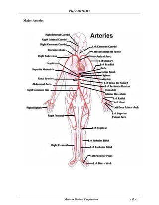
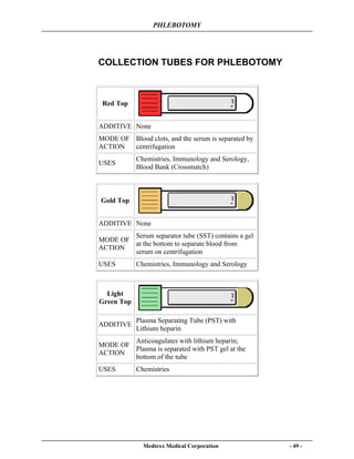
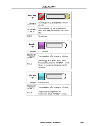
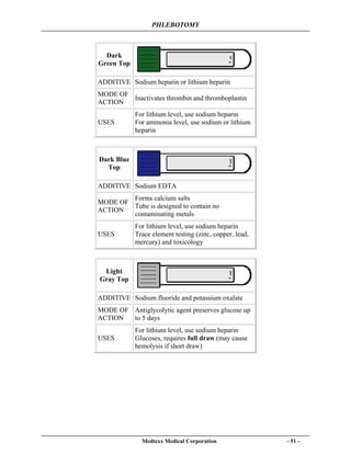
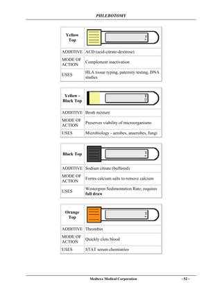
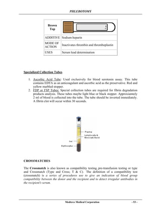
Medtexx Medical Corporation
Fundamentals
Of
Phlebotomy
Second Edition
PHLEBOTOMY
Medtexx Medical Corporation - 1 -
All Medtexx Medical Corporation exams, programs, books, and materials are property of the
Medtexx Corporation and are protected by United States and International copyright laws. All
copyright, logo, trademark, and other intellectual property rights affiliated with the Company are
the property of Medtexx Medical Corporation.
Copying, modifying, publishing, transmitting, or the distribution of the contents of all exams,
books, programs, and materials is not permitted.
The act of infringing on a copyright is a very serious business and is punishable by fines and/or
imprisonment. Medtexx Medical Corporation prosecutes, to the full extent of the law, those who
disregard the rights of this Company for the reproduction and distribution of our material.
Copyright 1999-2007, Medtexx Medical Corporation. All rights reserved.
PHLEBOTOMY
Medtexx Medical Corporation - 2 -
Table of Contents
Phlebotomy: The Historical Perspective 4
History 4
Devices Used for Drawing Blood 5
Universal Precautions & Biosafety Techniques 7
Infection Prevention 8
Human Anatomy & Physiology 10
Skin Layers 10
General Structure: Arteries and Veins 14
The Tourniquet 20
Assembling the Equipment 22
Blood Collection: Routine Venipuncture & Specimen Handling 24
Venipuncture Procedure 24
Order Form/Requisition Form 25
Labeling the Sample 27
Equipment 27
Order of Draw 28
Venipuncture Site Selection 31
Performance of a Venipuncture 31
Troubleshooting 33
Performance of a Fingerstick 39
Blood Collection on Infants 40
Additional Considerations 41
Safety & Infection Control 42
Possible Complications from Phlebotomy 43
Problems Obtaining a Specimen 43
Patient Complications 43
Blood: Samples & Collection 45
Types of Blood Samples 45
Collection Tubes 45
Special Procedures 53
Crossmatches 53
Blood Culture 54
PHLEBOTOMY
Medtexx Medical Corporation - 3 -
Oral Glucose Tolerance Test (GTT) 55
General Collection Requirements 56
Special Collection Techniques 59
Collecting a Blood Sample Using a Butterfly Needle Syringe 60
Drawing Blood Using a Lancet for Microdraw or Infant Heel Stick 62
Measured Bleeding Time 63
Arterial Puncture 65
Glossary 69
Resources 96
Appendix A: Order of Draw for Multiple Tubes i
Appendix B: Sample Requisition Form ii
Appendix C: Hepatitis Overview iii
Appendix D: Proper Glove Removal xi
PHLEBOTOMY
Medtexx Medical Corporation - 4 -
PHLEBOTOMY: THE HISTORICAL PERSPECTIVE
Objective
This lecture will concentrate on the historical perspective of phlebotomy, and show that
man’s initial fascination with his blood and body fluids has had a direct influence on the
study of Biomedical Science today.
Meaning of ‘Phlebotomy’
The term ‘Phlebotomy’ suggests the taking of Blood only. This subject is not only
concerned with “blood letting”, but rather the whole range of skills and knowledge
necessary for the collection of viable specimens for later analysis in a laboratory.
History
‘Phlebotomy’ comes from the Greek word phlebos, meaning veins, and tome, meaning
incision.
Historical evidence suggests the possibility of blood letting for therapeutic reasons may
have begun in Egypt around 1400B.C. Tomb paintings from this time show the
application of a leech to a patient.
Hypocrites (460-377 B.C.), also known as the father of modern science, was responsible
for early medical theory, which believed illness was caused by an “imbalance” in the
body. The removal of this “excess” was thought to restore this balance.
The practice of bloodletting seemed logical when this foundation of all medical treatment
was based on the four body humors: blood, phlegm, yellow bile, and black bile. Health
was thought to be restored by plugging, starving, vomiting, or blood letting.
The art of blood letting was flourishing well before Hypocrites in the fifth century B.C.
By the Middle Ages, both surgeons and barbers were specializing in this bloody practice.
Barbers advertised with a red (for blood) and white (for tourniquet) striped pole. The pole
itself represented the stick squeezed by the patient to dilate the veins.
The practice reached unbelievable heights in the 18th
and early 19th
centuries. The first
U.S. president, George Washington, died from a throat infection in 1799 after being
drained of nine pints of blood within 24 hours. The draining of 16-30 ounces (1-4 pints)
of blood was typical. Blood was often caught in a shallow bowl. When the patient
became faint, the “treatment” was stopped. Bleeding was often encouraged over large
areas of the body by multiple incisions. By the end of the 19th
century (1875-1900),
phlebotomy was declared quackery.
PHLEBOTOMY
Medtexx Medical Corporation - 5 -
Devices Used for Drawing Blood
A variety of devices were used to draw blood,
• Lancet – The lancet was first used before 5th
century B.C. the practitioner
manually perforated the vein. Many shallow cuts were sometimes made.
• Spring-Loaded Lancet – Came into use during the early 18th
century. The device
was cocked and a “trigger” fired the spring-driven blade into the vein.
• The Fleam – Used during the 18th
century. Many varieties existed. Sometimes a
wooden “fleam stick” was used to hit the back of the blade and drive it into the
vein.
• Scarificator – The scarificator, a series of twelve blades, was also in vogue during
the 18th
century. The device was cocked and the trigger released spring-driven
rotary blades that caused many shallow cuts. The scarificator seems more
merciful than other bloodletting instruments. Blood was caught in shallow bowls.
• Flint Cup – During the 17th
to 19th
centuries, blood was also captured in small
flint cups. Heated air inside the cup created a vacuum causing blood to flow into
the cup, a handy technique for drawing blood form a localized area. This practice
was called ‘cupping’.
• Leeches – (Hirudo medicinalis); Leeches were “enticed” by “leeches” to attach
themselves to the skin. The area was chosen and a drop of milk or blood was the
bait. Once the leech was engorged, it was allowed to drop off. In the early 1800’s,
the little animal became quite scarce, and leech farms were having difficulty
keeping up with the demand. Therefore, they became quite expensive.
With this fascination came the need to “see” the components of the body fluids.
“Antony van Leeuwenhoek (1632-1723)” was an unlikely scientist. A tradesman of Delft,
Holland, he came from a family of tradesmen, had no fortune, received no higher
education or university degrees, and knew no languages other than his native Dutch. This
would have been enough to exclude him from the scientific community of his time
completely. Yet with skill, diligence, an endless curiosity, and an open mind free of the
scientific dogma of his day, Leeuwenhoek succeeded in making some of the most
important discoveries in the history of biology. It was he who discovered bacteria, free-
living and parasitic microscopic protists, sperm cells, blood cells, microscopic nematodes
and rotifers, and much more. His researches, which were widely circulated, opened up an
entire world of microscopic life to the awareness of scientists.
Leeuwenhoek is known to have made over 500 "microscopes," of which fewer than ten
have survived to the present day. In basic design, probably all of Leeuwenhoek's
instruments -- certainly all the ones that are known -- were simply powerful magnifying
glasses, not compound microscopes of the type used today. A drawing of one of
Leeuwenhoek's "microscopes" is shown at the left. Compared to modern microscopes, it
PHLEBOTOMY
Medtexx Medical Corporation - 6 -
is an extremely simple device, using only one lens, mounted in a tiny hole in the brass
plate that makes up the body of the instrument. The specimen was mounted on the sharp
point that sticks up in front of the lens, and its position and focus could be adjusted by
turning the two screws. The entire instrument was only 3-4 inches long, and had to be
held up close to the eye; it required good lighting and great patience to use.
It is open to debate when blood first began to be examined for diagnostic purposes. It is
known that other body fluids have been examined since medieval times.
The invention of the microscope in the 17th
century, coupled with advances in
physiologic chemistry and cellular physiology on the 19th
century, paved the way for the
examination of blood as a diagnostic tool.
PHLEBOTOMY
Medtexx Medical Corporation - 7 -
UNIVERSAL PRECAUTIONS AND BIOSAFETY TECHNIQUES
1. Assume ALL human blood, plasma, serum, body fluids (semen, saliva, tears,
cerebrospinal and amniotic fluid, milk and cervical secretions) and tissues to be
contaminated with Human Immunodeficiency Virus (HIV) and/or Hepatitis
Viruses (e.g., HBV). Handle them with appropriate care!
2. Gain knowledge - Be prepared:
• Personnel should understand their risk categorization (per Depts. of Labor and
Health and Human Services) before initiating work: Category I: Personnel
routinely handle blood, body fluids and issues. Category II: Personnel
occasionally handle or work around such materials. Category III: Personnel
never work with or around such materials.
• Be familiar with the CDC/NIH Manual "Biosafety in Microbiological and
Biomedical
• Laboratories’ view biosafety videos and are familiar with the company's
Biosafety Manuals. Ask your supervisor to explain any procedures or
concepts not clear to you before beginning work.
• Category I and II personnel should get the new, safe and effective Hepatitis B
vaccination.
3. Remember: The most susceptible route of laboratory infection for HIV and HBV
is by accidental needle sticks, contamination of the mucous membranes, or
through broken, abraded or irritated skin. Use appropriate caution and maximum
protection to prevent such contact.
4. Avoid spilling, splashing or open aerosolization of human blood or body fluids.
Wear latex gloves, protective lab garments and face and eye shields when
handling human materials.
5. Understand the principles of good microbiological practice before working with
biohazardous materials. Examples include use of aseptic technique, proper
decontamination procedures, emergency biohazard spill management and proper
use of biosafety equipment. Develop proficiency before beginning work.
6. Use Biosafety Level-2 work practices, containment and laboratories when
handling human materials where droplet and aerosol production are likely. Avoid
aerosol-generating activities in handling human materials. When such procedures
are necessary, use biosafety cabinets or other containment and personal protective
equipment.
7. When culturing or manipulating known HIV or HBV, use Biosafety Level-3 (BL-
3) procedures. Any procedure which requires concentration of HIV or HBV or
other human viruses from human materials should be handled under BL-3
containment and handling conditions. Use appropriate biosafety level conditions
(BL-2 or BL-3) when handling non-human primates and other animals inoculated
with human pathogenic materials.
8. Dispose of human and animal biohazardous waste or materials contaminated with
them in accordance with CDC/NIH biosafety and institutional guidelines.
9. Decontaminate laboratory protective garments, gloves and protective equipment
to render them non-infectious.
10. Clean all work areas and equipment used in handling human biohazardous
materials with proven disinfectant (e.g., 1:10 dilution of Clorox) when concluding
work to protect personnel from accidental infection.
PHLEBOTOMY
Medtexx Medical Corporation - 8 -
11. Assume that human serological and biological reagents (e.g., antibody, antigen or
antisera) used in the laboratory are contaminated with HIV or other viruses and
handle them accordingly.
12. Understand your institution's medical surveillance program and be familiar with
the appropriate standard operating procedures for accidental exposure to human
materials. Specific measures must be followed as per CDC/NIH Guidelines in the
Universal Precautions. The specimens involved must be identified and tested for
HIV and HBV and the procedures followed.
13. Report every accident to your supervisor and Occupational Medical Service
personnel.
14. Responsibility for instituting, training and monitoring of biosafety practices in
laboratories handling human materials, HIV or HBV rests with the Laboratory
Director or the designated Principal Investigator (PI). These individuals must
categorize positions; provide facilities, biosafety equipment, biosafety
procedures and training to employees accepting such work assignments to permit
the safe conduct of the work. These responsible individuals must ascertain the
proficiency of the employee in performing the assigned task before permitting the
work to begin.
15. Laboratory personnel have a clear responsibility to fully understand and
consistently adhere to the biosafety practices detailed in the Biosafety and
General Safety Manuals as well as to the biosafety guidelines detailed here and
by the CDC and NIH. Responsibility for conscious or thoughtless non-compliance
with or violation of these guidelines falls on the laboratory worker.
Note: Hand Washing Technique for Skin Exposure to Blood or body Fluids
1. Wash with a good liquid antimicrobial detergent soap.
2. Rinse well with water.
3. Apply solution of 50% isopropyl or ethyl alcohol.
4. Wash again with the liquid soap and rinse well.
INFECTION PREVENTION
Approximately 100% of the infectious agents found in the clinical laboratory are spread
by either airborne (inhalation) or contact transmission. Proper hand washing and strict
adherence to Universal Precautions, body substance isolation and general safety will
minimize the risk of infection. Infectious disease agents are most often transmitted in one
of four methods.
1. Airborne or inhalation transmission. A susceptible individual inhales droplets
of particles of dust containing infectious agents. It is in this manner that
streptococcal sore throat, respiratory viruses, and pulmonary tuberculosis can be
contracted. Example: coughing, sneezing, centrifugation and the “popping” of
specimen container tops are responsible for droplet formation.
2. Contact Transmission. May be direct or indirect. In direct contact, the causative
agent is passed from one individual directly to another. Infections with sexually
PHLEBOTOMY
Medtexx Medical Corporation - 9 -
transmitted diseases, including HIV are examples, of direct contact. Hand contact,
such as shaking hands with an infected person is also thought to be one of the
ways that respiratory virus infections are spread. It is also possible to be infected
by contact with an inanimate object (blood collection tubes etc.). Either accidental
needle sticks or by the sharing of needles by drug users are other examples. A
person may be indirectly exposed by receiving blood infected with Hepatitis C or
HIV.
3. Ingestion Transmission. Food or water containing pathogenic organisms is
ingested. Salmonelosis and Hepatitis A are examples.
4. Vector Borne. A vector or arthropod (mosquitoes, ticks etc.) are capable of
transferring pathogens and resulting in malaria and encephalitis or Lyme disease,
respectively.
PHLEBOTOMY
Medtexx Medical Corporation - 10 -
HUMAN ANATOMY AND PHYSIOLOGY
Skin Layers
PHLEBOTOMY
Medtexx Medical Corporation - 11 -
Major Veins
PHLEBOTOMY
Medtexx Medical Corporation - 12 -
Major Arteries
PHLEBOTOMY
Medtexx Medical Corporation - 13 -
PHLEBOTOMY
Medtexx Medical Corporation - 14 -
General Structure: ARTERIES and VEINS
The arteries and veins are composed pf three coats of tunics and a hollow core, called a
lumen, through which the blood flows.
• Tunica interna (intima), ENDOTHELIUM (simple squamous epithelium) and a
layer of Elastic Tissue called the internal elastic membrane.
• Tunica media, thickest layer Elastic Fibers & Smooth Muscle
• Tunica externa (adventitia) Elastic & Collagenous Fibers
Arteries
• Strong, very Elastic
• Adapted to carry blood under high pressure
• 3 layers
PHLEBOTOMY
Medtexx Medical Corporation - 15 -
o Endothelium
Smooth surface
o Tunica Media
Smooth muscle, Elastic fibers
o Tunica Adventitia
• Sympathetic branch of ans innervate smooth muscle in artery and arteriole
• Hold about 20% total blood volume.
PHLEBOTOMY
Medtexx Medical Corporation - 16 -
Veins
• Carry deoxygenated blood to the heart
• Three layers with middle layer poorly developed
o Less muscle and elastic tissue
• Very distensible
• Carry about 60% of total blood volume
• Functions as blood reservoir
• Vein in limbs have valves like flaps
• Respond to falling BP by vasoconstricting
PHLEBOTOMY
Medtexx Medical Corporation - 17 -
PHLEBOTOMY
Medtexx Medical Corporation - 18 -
Selected Veins of the Upper Limb – Listed Alphabetically
Vein Tributaries Drains Into Regions Drained Notes
Ante brachial,
median
Superficial veins of
the palm and
anterior forearm
Median cubital v. or
basilica
Palm; anterior
forearm
Medium antecubital
v. is variable in size
– it may be large or
absent
Basilic v.
Medial end of the
dorsal venous arch
of the hand;
superficial veins of
the forearm;
median cubital v.
It unites with the
brachial vein(s) to
form the axillary v.
Superficial parts of
the medial side of
the hand and
medial side of the
forearm
Basilica v.
communicated with
deep veins of the
forearm through
perforating veins,
especially in cubital
region.
Brachiocephalic v.
Formed by the
union of the
subclavian v. and
the internal jugular
v.; tributaries;
vertebral v., thymic
v., inferior thyroid
v., internal thoracic
v., 1st
posterior
intercostals v., left
superior intercostals
v. (to the left
brachiocephalic v.)
The left and right
brachiocephalic v.
unite to form the
superior vena cava.
Head; neck; upper
limb; anterior chest
wall
At its origin, the left
brachiocephalic v.
receives the
thoracic duct; at its
origin, the right
brachiocephalic v.
receives the right
lymphatic duct
Cephalic v.
Lateral side of the
dorsal venous arch
of the hand;
superficial veins of
the forearm.
Axillary vein
Superficial parts of
the lateral hand and
lateral forearm
Median cubital vein
usually shunts some
of the blood
collected by the
cephalic v. to the
basilica v.
Dorsal metacarpal
v. of the hand
Dorsal digital vv.
Dorsal venous arch
of the hand
Dorsal aspects of
the digits of the
hand
Dorsal metacarpal
v. drains the
adjacent sides of
the two digits.
Dorsal venous arch
of the hand
Dorsal didgital vv.
And dorsal
metacarpal vv.
Cephalic v. laterally,
basilica v. medially
Dorsal aspects of
the digits and the
superficial
structures of the
dorsal of the hand
Dorsal venous arch
is visible through
the thin skin on the
dorsum of the hand
Median antebrachial
v.
Superficial of the
palm and anterior
forearm
Median cubital v. or
basilica v.
Palm; anterior
forearm
Median antecubital
v. is variable in size-
it may be large or
absent
Median cubital v. Cephalic Basilica
Superficial part of
the hand and
forearm
A median
anterbrachial vein
occurs occasionally
and when present,
it may drain into
the median cubital
vein.
PHLEBOTOMY
Medtexx Medical Corporation - 19 -
Selected Veins of the Lower Limb – Listed Alphabetically
Veins Tributaries Drains into
Regions
Drained
Notes
Dorsal metatarsal
v. of the foot
Dorsal digital vv.
Dorsal venous
arch of the foot
Dorsal aspects of
the digits of the
foot
Dorsal metatarsal
v. drains the
adjacent sides of
two digits
Dorsal venous
arch of the foot
Dorsal digital vv.
And dorsal
metatarsal vv.
Great saphenous
v. medially, small
saphenous v.
laterally
Dorsum of the
digits and the
superficial
structures of the
dorsum of the
foot
Dorsal venous
arch is visible
through the thin
skin on the
dorsum of the
foot
Greater
saphenous v.
Medial end of
dorsal venous
arch of foot,
perforating
communications
with deep veins,
superficial
circumflex iliac
v., superficial
external
pudendal v.
Femoral v.
Skin and
superfiaicl fascia
of the medial
side of the foot
and leg; skin and
superficial fascia
of most of the
thigh; lower
abdominal wall;
perineal region
Greater
saphenous v. is
frequently used
as graft material
in coronary
bypass surgery
Lesser
saphenous v.
Lateral end of
the dorsal
venous arch of
foot
Popliteal v.
Skin and
superficial fascia
of the lateral side
of the foot and
leg
Passes deeply
into the popliteal
fossa
Metatarsal,
dorsal of the foot
Dorsal digital vv.
Dorsal venous
arch of the foot
Dorsal aspects of
the digits of the
foot
Dorsal metatarsal
v. drains the
adjacent sides of
two digits
PHLEBOTOMY
Medtexx Medical Corporation - 20 -
Proper Method for Tying a Tourniquet
PHLEBOTOMY
Medtexx Medical Corporation - 21 -
PHLEBOTOMY
Medtexx Medical Corporation - 22 -
ASSEMBLING THE EQUIPMENT
Equipment Used during a Venipuncture
PHLEBOTOMY
Medtexx Medical Corporation - 23 -
Note: This method of traction is discouraged due to an increased risk of the Phlebotomist
sticking themselves rather than the patient!
Assemble the needle, the barrel, and the first tube you wish to use as in the figures above.
The needle should not be uncovered until ready to perform venipuncture. Place any
additional tubes to be used in a convenient location, keeping some spares handy. The
gauze, alcohol pads, and bandages should be ready. (Note: some phlebotomists may elect
to do this step before applying the tourniquet; this is preferable.)
PHLEBOTOMY
Medtexx Medical Corporation - 24 -
BLOOD COLLECTION: ROUTINE VENIPUNCTURE AND
SPECIMEN HANDLING
Objectives
• Describe and perform the venipuncture process including:
1. Proper patient identification procedures.
2. Proper equipment selection and use.
3. Proper labeling procedures and completion of laboratory requisitions.
4. Order of draw for multiple tube phlebotomy.
5. Preferred venous access sites, and factors to consider in site selection, and
ability to differentiate between the feel of a vein, tendon and artery.
6. Patient care following completion of venipuncture.
7. Safety and infection control procedures.
8. Quality assurance issues.
• Identify the additive, additive function, volume, and specimen considerations to
be followed for each of the various color coded tubes.
• List six areas to be avoided when performing venipuncture and the reasons for the
restrictions.
• Summarize the problems that may be encountered in accessing a vein, including
the procedure to follow when a specimen is not obtained.
• List several effects of exercise, posture, and tourniquet application upon
laboratory values.
VENIPUNCTURE PROCEDURE
The venipuncture procedure requires both knowledge and skill to perform. Each
phlebotomist generally establishes a routine that is comfortable for her or him. Several
essential steps are required for every successful collection procedure:
1. Identify the patient.
2. Assess the patient's physical disposition (i.e. diet, exercise, stress, basal
state).
3. Check the requisition form for requested tests, patient information, and
any special requirements.
4. Select a suitable site for venipuncture.
5. Prepare the equipment, the patient and the puncture site.
PHLEBOTOMY
Medtexx Medical Corporation - 25 -
6. Perform the venipuncture.
7. Collect the sample in the appropriate container.
8. Recognize complications associated with the phlebotomy procedure.
9. Assess the need for sample recollection and/or rejection.
10. Label the collection tubes at the bedside or drawing area.
11. Promptly send the specimens with the requisition to the laboratory.
Equipment
The following are suggested supplies for blood specimen collection.
• Non sterile exam gloves
• Puncture resistant sharps container
• Alcohol wipes
• Tourniquet
• Appropriate specimen collection lab tubes
• 2 x 2 gauze
• Tape
• Evacuated tube holder
• Multisample blood collection needle
ORDER FORM / REQUISITION
A requisition form must accompany each sample submitted to the laboratory. This
requisition form must contain the proper information in order to process the specimen.
The essential elements of the requisition form are:
• Patient's surname, first name, and middle initial.
• Patient's ID number.
• Patient's date of birth and sex.
• Requesting physician's complete name.
• Source of specimen. This information must be given when requesting
microbiology, cytology, fluid analysis, or other testing where analysis and
reporting is site specific.
• Date and time of collection.
• Initials of phlebotomist.
• Indicating the test(s) requested.
PHLEBOTOMY
Medtexx Medical Corporation - 26 -
Figure: An example of a simple requisition form with the essential elements.
PHLEBOTOMY
Medtexx Medical Corporation - 27 -
LABELING THE SAMPLE
• A properly labeled sample is essential so that the results of the test match the
patient. The key elements in labeling are:
o Patient's surname, first and middle.
o Patient's ID number.
o NOTE: Both of the above MUST match the same on the requisition form.
o Date, time and initials of the phlebotomist must be on the label of EACH
tube.
EQUIPMENT
THE FOLLOWING ARE NEEDED FOR ROUTINE VENIPUNCTURE
o Evacuated Collection Tubes - The tubes are designed to fill with a
predetermined volume of blood by vacuum. The rubber stoppers are color
coded according to the additive that the tube contains. Various sizes are
available. Blood should NEVER be poured from one tube to another since
the tubes can have different additives or coatings (see illustrations at end).
o Needles - The gauge number indicates the bore size: the larger the gauge
number, the smaller the needle bore. Needles are available for evacuated
systems and for use with a syringe, single draw or butterfly system.
o Holder/Adapter - use with the evacuated collection system.
o Tourniquet - Wipe off with alcohol and replace frequently.
o Alcohol Wipes - 70% isopropyl alcohol.
o Povidone-iodine wipes/swabs - Used if blood culture is to be drawn.
o Gauze sponges - for application on the site from which the needle is
withdrawn.
o Adhesive bandages / tape - protects the venipuncture site after collection.
o Needle disposal unit - needles should NEVER be broken, bent, or
recapped. Needles should be placed in a proper disposal unit
IMMEDIATELY after their use.
o Gloves - can be made of latex, rubber, vinyl, etc.; worn to protect the
patient and the phlebotomist.
o Syringes - may be used in place of the evacuated collection tube for
special circumstances.
Last Name, First Name
Patient ID: 1234-56
June 5, 2005 8:10 AM Initials: SM
PHLEBOTOMY
Medtexx Medical Corporation - 28 -
ORDER OF DRAW
Blood collection tubes must be drawn in a specific order to avoid cross-contamination of
additives between tubes. The standard order of draw is:
o First - blood culture tube (yellow-black stopper)
o Second - non-additive tube (red stopper or SST)
o Third - coagulation tube (light blue stopper). If just a routine coagulation
assay is the only test ordered, then a single light blue stopper tube may be
drawn. If there is a concern regarding contamination by tissue fluids or
thromboplastins, then one may draw a non-additive tube first, and then the
light blue stopper tube.
o Last draw - additive tubes in this order:
Last draw - additive tubes in this order:
SST (red-gray, or gold, stopper). Contains a gel separator
and clot activator.
Sodium heparin (dark green stopper)
PST (light green stopper). Contains lithium heparin
anticoagulant and a gel separator.
EDTA (lavender stopper)
ACDA or ACDB (pale yellow stopper). Contains acid
citrate dextrose.
Oxalate/fluoride (light gray stopper)
NOTE: Tubes with additives must be thoroughly mixed. Erroneous test results
may be obtained when the blood is not thoroughly mixed with the additive.
NOTE: For plastic tubes, the order of draw for tubes 2 and 3 is reversed.
PHLEBOTOMY
Medtexx Medical Corporation - 29 -
PHLEBOTOMY
Medtexx Medical Corporation - 30 -
PROCEDURAL ISSUES
PATIENT RELATIONS AND IDENTIFICATION
The phlebotomist's role requires a professional, courteous, and understanding manner in
all contacts with the patient. Greet the patient and identify yourself and indicate the
procedure that will take place. Effective communication - both verbal and nonverbal - is
essential.
Proper patient identification is MANDATORY. If an inpatient is able to respond, ask for
a full name and always check the armband for confirmation. DO NOT DRAW BLOOD
IF THE ARMBAND IS MISSING. An outpatient must provide identification other than
the verbal statement of a name. Using the requisition for reference, ask a patient to
provide additional information such as a surname or birth date.
If possible, speak with the patient during the process. The patient who is at ease will be
less focused on the procedure. Always thank the patient and excuse yourself courteously
when finished.
PATIENT'S BILL OF RIGHTS
The Patient's Bill of Rights has been adopted by many hospitals as declared by the Joint
Commission on Accreditation of Healthcare Organizations (JCAHO). The basic patient
rights endorsed by the JCAHO follow in condensed form are given below.
The patient has the right to,
• Impartial access to treatment or accommodations that is available or medically
indicated, regardless of race, creed, sex, national origin, or sources of payment for
care.
• Considerate, respectful care.
• Confidentiality of all communications and other records pertaining to the patient's
care.
• Expect that any discussion or consultation involving the patient's case will be
conducted discreetly and that individuals not directly involved in the case will not
be present without patient permission.
• Expect reasonable safety congruent with the hospital practices and environment.
• Know the identity and professional status of individuals providing service and to
know which physician or other practitioner is primarily responsible for his or her
care.
• Obtain from the practitioner complete and current information about diagnosis,
treatment, and any known prognosis, in terms the patient can reasonably be
expected to understand.
• Reasonable informed participation in decisions involving the patient's health care.
The patient shall be informed if the hospital proposes to engage in or perform
human experimentation or other research/educational profits affecting his or her
care or treatment. The patient has the right to refuse participation in such activity.
• Consult a specialist at the patient's own request and expense.
PHLEBOTOMY
Medtexx Medical Corporation - 31 -
• Refuse treatment to the extent permitted by law.
• Regardless of the source of payment, request and receive an itemized and detailed
explanation of the total bill for services rendered in the hospital.
• Be informed of the hospital rules and regulations regarding patient conduct.
VENIPUNCTURE SITE SELECTION
Although the larger and fuller median cubital and cephalic veins of the arm are used most
frequently, wrist and hand veins are also acceptable for venipuncture.
Certain areas are to be avoided when choosing a site,
• Extensive scars from burns and surgery - it is difficult to puncture the scar tissue
and obtain a specimen.
• The upper extremity on the side of a previous mastectomy - test results may be
affected because of lymphedema.
• Hematoma - may cause erroneous test results. If another site is not available,
collect the specimen distal to the hematoma.
• Intravenous therapy (IV) / blood transfusions - fluid may dilute the specimen, so
collect from the opposite arm if possible. Otherwise, satisfactory samples may be
drawn below the IV by following these procedures:
1. Turn off the IV for at least 2 minutes before venipuncture.
2. Apply the tourniquet below the IV site. Select a vein other than the one
with the IV.
3. Perform the venipuncture. Draw 5 ml of blood and discard before drawing
the specimen tubes for testing.
• Cannula/fistula/heparin lock - hospitals have special policies regarding these
devices. In general, blood should not be drawn from an arm with a fistula or
cannula without consulting the attending physician.
• Edematous extremities - tissue fluid accumulation alters test results.
PROCEDURE FOR VEIN SELECTION
• Palpate and trace the path of veins with the index finger. Arteries pulsate, are
most elastic, and have a thick wall. Thrombosed veins lack resilience, feel cord-
like, and roll easily.
• If superficial veins are not readily apparent, you can force blood into the vein by
massaging the arm from wrist to elbow, tap the site with index and second finger,
apply a warm, damp washcloth to the site for 5 minutes, or lower the extremity
over the bedside to allow the veins to fill.
PERFORMANCE OF A VENIPUNCTURE
Approach the patient in a friendly, calm manner. Provide for their comfort as much as
possible, and gain the patient's cooperation.
Identify the patient correctly.
Properly fill out appropriate requisition forms, indicating the test(s) ordered.
PHLEBOTOMY
Medtexx Medical Corporation - 32 -
Verify the patient's condition. Fasting, dietary restrictions, medications, timing, and
medical treatment are all of concern and should be noted on the lab requisition.
Position the patient. The patient should sit in a chair, lie down, or sit up in bed.
Hyperextend the patient's arm.
Apply the tourniquet 3-4 inches above the selected puncture site. Do not place too
tightly or leave on more than 2 minutes.
The patient should make a fist without pumping the hand.
Select the venipuncture site.
Prepare the patient's arm using alcohol prep. Cleanse in a circular fashion, beginning
at the site and working outward. Allow to air dry.
Grasp the patient's arm firmly using your thumb to draw the skin taut and anchor the
vein. The needle should form a 15 to 30 degree angle with the surface of the arm.
Swiftly insert the needle through the skin and into the lumen of the vein. Avoid
trauma and excessive probing.
When the last tube to be drawn is filling, remove the tourniquet.
Remove the needle from the patient's arm using a swift backward motion.
Press down on the gauze once the needle is out of the arm, applying adequate
pressure to avoid formation of a hematoma.
Dispose of contaminated materials/supplies in designated containers.
Mix and label all appropriate tubes at the patient bedside.
Deliver specimens promptly to the laboratory.
Note: The Bevel
The bevel of a needle is the angled opening at the tip of the needle. The bevel must
always face upward, towards the person holding it, so as to obtain a blood return.
PHLEBOTOMY
Medtexx Medical Corporation - 33 -
It is also very important to remember that if the bevel is exposed during a venipuncture,
before manually withdrawing the needle at the completion of the entire procedure, the
needle is then considered contaminated, and cannot be re-inserted into the patient. The
whole procedure must be stopped, and restarted elsewhere.
Note that when the bevel is exposed, leakage from the site may occur. Never panic,
simply release the tourniquet, remove the needle, and apply pressure with cotton.
Immediately dispose of the needle in the sharps container. This is another reason it is so
important to keep your eyes on the needle, and not on other things around the room!
PROTECT THE PATIENT
• Place blood collection equipment away from patients, especially children and
psychiatric patients.
• Practice hygiene for the patient's protection. When wearing gloves, change them
between each patient and wash your hands frequently. Always wear a clean lab
coat or gown.
TROUBLESHOOTING GUIDELINES
IF AN INCOMPLETE COLLECTION OR NO BLOOD IS OBTAINED
Change the position of the needle. Move it forward (it may not be in the lumen)
Or move it backward (it may have penetrated too far).
PHLEBOTOMY
Medtexx Medical Corporation - 34 -
Adjust the angle (the bevel may be against the vein wall).
Loosen the tourniquet. It may be obstructing blood flow.
Try another tube. There may be no vacuum in the one being used.
Re-anchor the vein. Veins sometimes roll away from the point of the needle and
puncture site.
Corrective Technique
The corrective technique is performed when no there is no blood return. After two
unsuccessful attempts, the rule for Phlebotomists is to call in another Phlebotomist to
perform the venipuncture. However, you must try to successfully obtain the sample
before giving up. The proper procedure to do this is called Corrective Technique.
PHLEBOTOMY
Medtexx Medical Corporation - 35 -
PHLEBOTOMY
Medtexx Medical Corporation - 36 -
PHLEBOTOMY
Medtexx Medical Corporation - 37 -
PHLEBOTOMY
Medtexx Medical Corporation - 38 -
IF BLOOD STOPS FLOWING INTO THE TUBE
The vein may have collapsed; re-secure the tourniquet to increase venous filling.
If this is not successful, remove the needle, take care of the puncture site, and
redraw.
The needle may have pulled out of the vein when switching tubes. Hold
equipment firmly and place fingers against patient's arm, using the flange for
leverage when withdrawing and inserting tubes.
PROBLEMS OTHER THAN AN INCOMPLETE COLLECTION
A hematoma forms under the skin adjacent to the puncture site - release the
tourniquet immediately and withdraw the needle. Apply firm pressure.
The blood is bright red (arterial) rather than venous. Apply firm pressure for more
than 5 minutes.
PHLEBOTOMY
Medtexx Medical Corporation - 39 -
PERFORMANCE OF A FINGERSTICK
Follow the procedure as outlined above for greeting and identifying the patient. As
always, properly fill out appropriate requisition forms, indicating the test(s) ordered.
Verify the patient's condition. Fasting, dietary restrictions, medications, timing, and
medical treatment are all of concern and should be noted on the lab requisition.
Position the patient. The patient should sit in a chair, lie down or sit up in bed.
Hyperextend the patient's arm.
The best locations for finger sticks are the 3rd and 4th fingers of the non-dominant
hand. Do not use the tip of the finger or the center of the finger. Avoid the side of the
finger where there is less soft tissue, where vessels and nerves are located, and where
the bone is closer to the surface. The 2nd (index) finger tends to have thicker,
callused skin. The fifth finger tends to have less soft tissue overlying the bone. Avoid
puncturing a finger that is cold or cyanotic, swollen, scarred, or covered with a rash.
Using a sterile lancet, make a skin puncture just off the center of the finger pad. The
puncture should be made perpendicular to the ridges of the fingerprint so that the drop
of blood does not run down the ridges.
Wipe away the first drop of blood, which tends to contain excess tissue fluid.
Collect drops of blood into the collection device by gently massaging the finger.
Avoid excessive pressure that may squeeze tissue fluid into the drop of blood.
Cap, rotate and invert the collection device to mix the blood collected.
Have the patient hold a small gauze pad over the puncture site for a couple of minutes
to stop the bleeding.
Dispose of contaminated materials/supplies in designated containers.
Label all appropriate tubes at the patient bedside.
Deliver specimens promptly to the laboratory.
PHLEBOTOMY
Medtexx Medical Corporation - 40 -
BLOOD COLLECTION ON INFANTS
The recommended location for blood collection on a newborn baby or infant is the
heel. The diagram below indicates in gray the proper area to use for heel punctures
for blood collection.
Pre-warming the infant's heel (42 C for 3 to 5 minutes) is important to obtain
capillary blood gas samples and warming also greatly increases the flow of blood for
collection of other specimens. However, do not use too high a temperature warmer,
because baby's skin is thin and susceptible to thermal injury.
Clean the site to be punctured with an alcohol sponge. Dry the cleaned area with a dry
cotton sponge. Hold the baby's foot firmly to avoid sudden movement.
Using a sterile blood lancet, puncture the area of the heel that is just “off center” from
the very center of the heel. Do not use the central portion of the heel because you
might injure the underlying bone, which is close to the skin surface. Do not puncture
the outer sides of the heel, either. Do not use a previous puncture site. Make the cut
across the heel print lines so that a drop of blood can well up and not run down along
the lines.
Wipe away the first drop of blood with a piece of clean, dry cotton. Since newborns
do not often bleed immediately, use gentle pressure to produce a rounded drop of
blood. Do not use excessive pressure or heavy massaging because the blood may
become diluted with tissue fluid.
Fill the capillary tube(s) or micro collection device(s) as needed.
When finished, elevate the heel, place a piece of clean, dry cotton on the puncture
site, and hold it in place until the bleeding has stopped.
Be sure to dispose of the lancet in the appropriate sharps container. Dispose of
contaminated materials in appropriate waste receptacles. Remove your gloves and
wash your hands.
PHLEBOTOMY
Medtexx Medical Corporation - 41 -
ADDITIONAL CONSIDERATIONS
To prevent a hematoma
• Puncture only the uppermost wall of the vein
• Remove the tourniquet before removing the needle
• Use the major superficial veins
• Make sure the needle fully penetrates the upper most wall of the vein. (Partial
penetration may allow blood to leak into the soft tissue surrounding the vein by
way of the needle bevel)
• Apply pressure to the venipuncture site
To prevent hemolysis (which can interfere with many tests)
• Mix tubes with anticoagulant additives gently 5-10 times
• Avoid drawing blood from a hematoma
• Avoid drawing the plunger back too forcefully, if using a needle and syringe, and
avoid frothing of the sample
• Make sure the venipuncture site is dry
• Avoid a probing, traumatic venipuncture
Indwelling Lines or Catheters
• Potential source of test error
• Most lines are flushed with a solution of heparin to reduce the risk of thrombosis
• Discard a sample at least three times the volume of the line before a specimen is
obtained for analysis
Hemoconcentration: An increased concentration of larger molecules and formed
elements in the blood may be due to several factors:
• Prolonged tourniquet application (no more than 2 minutes)
• Massaging, squeezing, or probing a site
• Long-term IV therapy
• Sclerosed or occluded veins
Prolonged Tourniquet Application
• Primary effect is hemoconcentration of non-filterable elements (i.e. proteins). The
hydrostatic pressure causes some water and filterable elements to leave the
extracellular space.
• Significant increases can be found in total protein, aspartate aminotransferase
(AST), total lipids, cholesterol, iron
• Affects packed cell volume and other cellular elements
PHLEBOTOMY
Medtexx Medical Corporation - 42 -
Patient Preparation Factors
• Therapeutic Drug Monitoring: different pharmacologic agents have patterns of
administration, body distribution, metabolism, and elimination that affect the drug
concentration as measured in the blood. Many drugs will have "peak" and
"trough" levels that vary according to dosage levels and intervals. Check for
timing instructions for drawing the appropriate samples.
• Effects of Exercise: Muscular activity has both transient and longer lasting
effects. The creatine kinase (CK), aspartate aminotransferase (AST), lactate
dehydrogenase (LDH), and platelet count may increase.
• Stress: May cause transient elevation in white blood cells (WBC's) and elevated
adrenal hormone values (cortisol and catecholamines). Anxiety that results in
hyperventilation may cause acid-base imbalances, and increased lactate.
• Diurnal Rhythms: Diurnal rhythms are body fluid and analyte fluctuations during
the day. For example, serum cortisol levels are highest in early morning but are
decreased in the afternoon. Serum iron levels tend to drop during the day. You
must check the timing of these variations for the desired collection point.
• Posture: Postural changes (supine to sitting etc.) are known to vary lab results of
some analytes. Certain larger molecules are not filterable into the tissue, therefore
they are more concentrated in the blood. Enzymes, proteins, lipids, iron, and
calcium are significantly increased with changes in position.
• Other Factors: Age, gender, and pregnancy have an influence on laboratory
testing. Normal reference ranges are often noted according to age.
SAFETY AND INFECTION CONTROL
Because of contacts with sick patients and their specimens, it is important to follow
safety and infection control procedures.
PROTECT YOURSELF
• Practice universal precautions:
• Wear gloves and a lab coat or gown when handling blood/body fluids.
• Change gloves after each patient or when contaminated.
• Wash hands frequently.
• Dispose of items in appropriate containers.
• Dispose of needles immediately upon removal from the patient's vein. Do not
bend, break, recap, or resheath needles to avoid accidental needle puncture or
splashing of contents.
• Clean up any blood spills with a disinfectant such as freshly made 10%
bleach.
• If you stick yourself with a contaminated needle:
• Remove your gloves and dispose of them properly.
• Squeeze puncture site to promote bleeding.
• Wash the area well with soap and water.
• Record the patient's name and ID number.
• Follow institution's guidelines regarding treatment and follow-up.
PHLEBOTOMY
Medtexx Medical Corporation - 43 -
POSSIBLE COMPLICATIONS FROM PHLEBOTOMY
PROBLEMS OBTAINING A SPECIMEN
Blood Sample That Cannot Be Obtained
Probing is not recommended. Probing is painful to the patient. In most cases another
puncture in a site below the first site, or use of another vein on the other arm, is
advisable.
It is advisable not to attempt a venipuncture more than twice. Notify the patient’s
Registered Nurse
Another person should attempt to draw the specimen
If another person is asked to draw a patient, the new person must re--identify the patient.
If an incomplete collection or no blood is obtained
Ø Change the position of the needle. Move it forward (it may not be in the lumen)
Ø or move it backward (it may have penetrated too far).
Ø Adjust the angle (the bevel may be against the vein wall).
Ø Re-anchor the vein. Veins sometimes roll away from the point of the needle and
puncture site.
If blood stops flowing into the syringe/tube
Ø The vein may have collapsed; resecure the tourniquet to increase venous filling. If this
is not successful, remove the needle, take care of the puncture site, and redraw.
Ø The needle may have pulled out of the vein when switching tubes. Hold equipment
firmly and place fingers against patient's arm, using the flange for leverage when
withdrawing and inserting tubes.
PATIENT COMPLICATION
Problems other than an incomplete collection
Hematoma
A hematoma forms under the skin adjacent to the puncture site - release the tourniquet
immediately and withdraw the needle. Apply firm pressure.
PHLEBOTOMY
Medtexx Medical Corporation - 44 -
To prevent a hematoma
• Puncture only the uppermost wall of the vein (just under the skin)
• Remove the tourniquet before removing the needle
• Use the major superficial veins (the large veins just under the skin)
• Make sure the needle fully penetrates the uppermost wall of the vein. (partial
puncture may allow blood to leak into the tissues just under the skin)
• Apply pressure to puncture site
Petechiae
Little red spots, ranging in size from pinpoint to several millimeters in diameter.
Petechiae consist of extravasated blood. This complication may be a result of a
coagulation abnormality, such as a platelet defect and should be brought to the attention
of the patient’s healthcare provider.
Syncopy (fainting)
Patients may become dizzy and fain at the thought or sight blood, this is the most
common complication phlebotomy. It is caused because of rapid fall in blood pressure.
An automatic nervous system reaction, (psychosomatic trigger), usually based on fear.
Treatment and safe handling of an unconscious patient is a necessity of any qualified
Phlebotomist.
• Abort draw: Remove tourniquet, needle and bend arm
• Call for assistance
• Using good body mechanics, slide patient to floor, keeping hand firmly behind the
cervical spine area. Protect head and neck from injury!
• Elevate feet above heart and monitor blood pressure, breathing, etc.
• Use ammonia only if patient is not responsive within 5 minutes and blood
pressure remains low.
• Assist to upright position in stages (monitor B/P with each change in position)
this is a gradual process. If patient stands up to quickly, he will most likely to
faint again due to drop in B/P.
Scarred Vein
Areas that have been burned or scarred should be avoided during phlebotomy. Burned
area is very sensitive and susceptible to infection, whereas veins under scarred area are
difficult to palpate.
PHLEBOTOMY
Medtexx Medical Corporation - 45 -
BLOOD: Samples & Collection
TYPES OF BLOOD SAMPLES
1. Whole Blood
A blood sample that is drawn and mixed immediately with an anticoagulant to maintain
the integrity of the blood cells and prevent clotting, allowing whole blood analysis to be
accurate. The blood remains in liquid state.
2. Serum
The liquid portion of whole blood that has been allowed to clot. The clotting factors are
bound in the clot. (Blood collected in a tube with no additive will clot within 15-45
minutes. One 10 ml tube of whole blood will yield about 3-4 ml of serum. This is the
only tube that should not be inverted).
3. Plasma
The liquid portion of blood that has not been allowed to clot. Usually, formed when
freshly drawn blood is mixed with anticoagulants. The clotting factors are present in the
plasma. This sample is mixed 6-8 times and immediately centrifuged and plasma
removed.
COLLECTION TUBES
SST- Serum Separator Tube (two types): Gold/Red-Gray Marble
Draw a sufficient amount of whole blood into a plain, red-top tube or serum separator
(SST®) tube. If using an SST® tube, gently invert the tube several times to activate
clotting. Allow blood to clot at ambient temperature for 20-30 minutes. Centrifuge for 10
minutes to separate serum from clot and transfer the serum to a screw-capped, plastic vial
if required; this should be completed within 1 hour of obtaining the specimen.
Additive: Polymer gel and powdered glass clot activator
Stopper Type: HemogardTM
Gold top/Conventional Red-Gray Marble
Tube Type/Size: Plastic tube 13 x 100/16 x 100
Specimen Type: Serum
Draw Amount: 5.0 ml/8.5 ml
Inversions: 5
Laboratory Use: Sterile SST® brand tube for serum clot activator determinations that
require serum in chemistry or infectious disease testing. Gel separates
serum from cells. Tube inversion ensures mixing of clot activator with
blood and clotting within 30 minutes.
PHLEBOTOMY
Medtexx Medical Corporation - 46 -
Mint Green Stopper- PST, Plasma Separator Tube: HOSPITAL PATIENTS ONLY
NOTE: After the tube has been filled with blood, immediately invert the tube several times in order to
prevent coagulation.
Green Stopper Tube (two types): Dark Green/Clear Green
NOTE: After the tube has been filled with blood, immediately invert the tube several times in order to
prevent coagulation.
Lavender Stopper Tube (two types): Dark Lavender/Clear Lavender
NOTE: After the tube has been filled with blood, immediately invert the tube several times in order to
prevent coagulation.
Additive: Lithium Heparin, Polymer gel plasma separator
Stopper Type: HemogardTM
Tube Type/Size: Plastic tube 13 x 75
Specimen Type: Whole Blood, Plasma
Draw Amount: 3.0 ml
Inversions: 8
Laboratory Use: General chemistries and some therapeutic drugs. DO NOT USE FOR
LITHIUM TESTING.
Additive: Sodium Heparin
Stopper Type: HemogardTM
Tube Type/Size: Plastic tube 13 x 75
Specimen Type: Whole Blood, Plasma
Draw Amount: 4.0 ml/2.0 ml (pediatric tube)
Inversions: 8
Laboratory Use: For plasma determinations in chemistry. Tube inversion prevents clotting.
Dark green 4.0 ml Sodium Heparin used for chromosome studies.
Additive: EDTA-K2
Stopper Type: HemogardTM
Tube Type/Size: Plastic tube 13 x 75
Specimen Type: Whole Blood, Plasma
Draw Amount: 4.0 ml/2.0 ml (pediatric tube)
Inversions: 8
Laboratory Use: Dark Lavender 4.0 ml for whole hematology, ammonia, lead, HIV, RNA
quantization determinations and for blood bank testing. Tube inversion
prevents clotting.
PHLEBOTOMY
Medtexx Medical Corporation - 47 -
Light Blue Stopper Tube (two types): Solid Light Blue/Clear Light Blue
NOTE: It is imperative that the tube be completely filled. The ratio of blood to anticoagulant is
critical for valid results. Immediately after draw, invert the tube 6 to 10 times in order to activate the
anticoagulant.
Gray Stopper Tube
NOTE: After the tube has been filled with blood, immediately invert the tube several times in order to
prevent coagulation.
Red Stopper Tube
Additive: Sodium Citrate (3.2%, 0.109M)
Stopper Type: HemogardTM
Tube Type/Size: Glass 13 x 75
Specimen Type: Whole Blood, Plasma
Draw Amount: 2.7 ml/1.8 ml (pediatric tube only)
Inversions: 4 (gently)
Laboratory Use: For coagulation determinations of plasma specimens. Tube inversion
prevents clotting.
Note: Certain tests require chilled specimens. Follow recommended procedures
for collection and transporting of coagulation specimen.
Additive: Sodium fluoride/Potassium oxalate
Stopper Type: HemogardTM
Tube Type/Size: Plastic 13 x 75
Specimen Type: Whole Blood, Plasma
Draw Amount: 4.0 ml
Inversions: 8
Laboratory Use: For glucose, toxicology determinations. Antiglycolytic additives
stabilize glucose values for up to 24 hours at room temperature. Tube
inversion ensures proper mixing of additive and blood.
Additive: Clot Activator (powdered glass)
Stopper Type: HemogardTM
Tube Type/Size: Plastic 13 x 75
Specimen Type: Serum
Draw Amount: 6.0 ml
Inversions: 5
Laboratory Use: For serum determinations in chemistry, serology and blood bank
testing. Can be used as sterile transport tube.
PHLEBOTOMY
Medtexx Medical Corporation - 48 -
Royal Blue Stopper Tube: (two types): No additive/EDTA
There are 2 types of royal blue top Monoject® tubes - one with EDTA anticoagulant and
the other plain. These are used in the collection of whole blood or serum for trace metals
analysis.
Yellow Stopper Tube: (two types)
Additive: None/EDTA
Stopper Type: HemogardTM
Tube Type/Size: Glass 13 x 100
Specimen Type: Whole Blood, Plasma
Draw Amount: 7.0 ml
Inversions: None
Laboratory Use: For trace element, toxicology and nutrition determinations. Special
stopper formulation offers the lowest verified levels of trace elements
available. Refer to specific test for proper tube.
Additive: ACD, solution A
Stopper Type: Conventional
Tube Type/Size: Glass 16 x 100
Specimen Type: Whole blood
Draw Amount: 8.5 ml
Inversions: 8
Laboratory Use: Tissue typing and some Red Cross testing. Refer to specific test for
proper tube.
Additive: ACD, solution B
Stopper Type: Conventional
Tube Type/Size: Glass 16 x 100
Specimen Type: Whole blood
Draw Amount: 6.0 ml
Inversions: 8
Laboratory Use: Tissue typing. Refer to specific test for proper tube.
PHLEBOTOMY
Medtexx Medical Corporation - 49 -
COLLECTION TUBES FOR PHLEBOTOMY
Red Top
ADDITIVE None
MODE OF
ACTION
Blood clots, and the serum is separated by
centrifugation
USES
Chemistries, Immunology and Serology,
Blood Bank (Crossmatch)
Gold Top
ADDITIVE None
MODE OF
ACTION
Serum separator tube (SST) contains a gel
at the bottom to separate blood from
serum on centrifugation
USES Chemistries, Immunology and Serology
Light
Green Top
ADDITIVE
Plasma Separating Tube (PST) with
Lithium heparin
MODE OF
ACTION
Anticoagulates with lithium heparin;
Plasma is separated with PST gel at the
bottom of the tube
USES Chemistries
PHLEBOTOMY
Medtexx Medical Corporation - 50 -
Red-Gray
Top
ADDITIVE
Serum Separating Tube (SST) with clot
activator
MODE OF
ACTION
Forms clot quickly and separates the
serum with SST gel at the bottom of the
tube
USES Chemistries
Purple
Top
ADDITIVE EDTA liquid
MODE OF
ACTION
Forms calcium salts to remove calcium
USES
Hematology (CBC) and Blood Bank
(Crossmatch); requires full draw - invert
8 times to prevent clotting and platelet
clumping
Light Blue
Top
ADDITIVE Sodium citrate
MODE OF
ACTION
Forms calcium salts to remove calcium
USES
Coagulation tests (protime and
prothrombin time), full draw required
PHLEBOTOMY
Medtexx Medical Corporation - 51 -
Dark
Green Top
ADDITIVE Sodium heparin or lithium heparin
MODE OF
ACTION
Inactivates thrombin and thromboplastin
USES
For lithium level, use sodium heparin
For ammonia level, use sodium or lithium
heparin
Dark Blue
Top
ADDITIVE Sodium EDTA
MODE OF
ACTION
Forms calcium salts
Tube is designed to contain no
contaminating metals
USES
For lithium level, use sodium heparin
Trace element testing (zinc, copper, lead,
mercury) and toxicology
Light
Gray Top
ADDITIVE Sodium fluoride and potassium oxalate
MODE OF
ACTION
Antiglycolytic agent preserves glucose up
to 5 days
USES
For lithium level, use sodium heparin
Glucoses, requires full draw (may cause
hemolysis if short draw)
PHLEBOTOMY
Medtexx Medical Corporation - 52 -
Yellow
Top
ADDITIVE ACD (acid-citrate-dextrose)
MODE OF
ACTION
Complement inactivation
USES
HLA tissue typing, paternity testing, DNA
studies
Yellow -
Black Top
ADDITIVE Broth mixture
MODE OF
ACTION
Preserves viability of microorganisms
USES Microbiology - aerobes, anaerobes, fungi
Black Top
ADDITIVE Sodium citrate (buffered)
MODE OF
ACTION
Forms calcium salts to remove calcium
USES
Westergren Sedimentation Rate; requires
full draw
Orange
Top
ADDITIVE Thrombin
MODE OF
ACTION
Quickly clots blood
USES STAT serum chemistries
PHLEBOTOMY
Medtexx Medical Corporation - 53 -
Brown
Top
ADDITIVE Sodium heparin
MODE OF
ACTION
Inactivates thrombin and thromboplastin
USES Serum lead determination
Specialized Collection Tubes
1. Ascorbic Acid Tube: Used exclusively for blood serotonin assay. This tube
contains EDTA as an anticoagulant and ascorbic acid as the preservative. Red and
yellow marbled stopper.
2. FDP or FSP Tubes: Special collection tubes are required for fibrin degradation
products analysis. These tubes maybe light blue or black stopper. Approxiamtely
2 ml of blood is collected into the tube. The tube should be inverted immediately.
A fibrin clot will occur within 30 seconds.
CROSSMATCHES
The Crossmatch is also known as compatibility testing, pre-transfusion testing or type
and Crossmatch (Type and Cross; T & C). The definition of a compatibility test
(crossmatch) is a series of procedures use to give an indication of blood group
compatibility between the donor and the recipient and to detect irregular antibodies in
the recipient's serum.
PHLEBOTOMY
Medtexx Medical Corporation - 54 -
PURPOSE
The main purpose for performing a crossmatch is to promote (not ensure) the safe
transfusion of blood. We are performing testing to the best of our ability that will
demonstrate that the donor blood is compatible with the recipient's blood. Crossmatch
procedures should be designed for speed and accuracy - get the safest blood reasonably
possible available to the patient as soon as possible.
Once donor blood is crossmatched with a potential recipient, the results of the crossmatch
is good only 3 days. If the physician wants the donor blood available longer, we must get
a new recipient sample and repeat tests. This protocol helps detect new antibodies that
may be forming, especially when patient has been transfused within past three months.
PROCEDURE
1. Double check the patient’s identity
2. Draw at least one red top tube (without polymer gel), two if possible. Label the
tubes with information below,
a. Name
b. Date and time collected
c. Hospital number
d. Initials of person collecting
3. Collect specimen carefully to avoid hemolysis of the red cells
4. Most facilities have a type of identification band that is placed on the patient’s
wrist as soon as the specimen has been collected. There is also a label that has a
preprinted ID number, identical to the one on the wrist band. This label must be
affixed to the tube of blood. This same ID number will be placed on the unit of
blood that has been crossmatched. This must be double checked prior to infusion
by the practitioner.
NOTE: Always double or triple check names and ID numbers where there is a
possibility that blood will be transfused. A unit of blood given to the wrong person
could kill.
BLOOD CULTURE
PURPOSE
The detection of septicemia
PREPEARTION OF THE PATIENT
1. Explain that the physician has ordered a series of test and you will have to
perform several sticks.
2. Clean the skin first using alcohol (using concentric rings from the inside out)
3. Follow this with an iodine swab using same technique
PHLEBOTOMY
Medtexx Medical Corporation - 55 -
4. Allow iodine to dry before performing the venipuncture. Once the iodine is dry,
do no palpate the vein again unless you have “sterilized” your own gloved finger
as you did for site puncture.
PROCEDURE
Each laboratory uses its own particular blood culture system. The protocol for the
collection of cultures also varies from hospital to hospital. The following are certain
procedural steps that are common to all blood culture methods.
1. Paint the septum of the blood culture bottle(s) with iodine.
2. For the first culture, if possible, collect a specimen from each arm. The amount of
blood to be drawn depends on the culture system used. (usually 5-10 ml) Draw
the blood in sterile syringes only.
3. After completion of the draw, replace the needle used to make the venipuncture
with a new sterile one. Inject the sample into the blood culture bottle and quickly,
but gently mix to avoid clotting.
4. In subsequent cultures, one venipuncture will be enough; however, each one
should be obtained from alternate arms.
5. After returning to the laboratory, you may be required to “vent” one of the culture
bottles if a two bottle system is used. Check with the lab regarding the proper
procedure to follow.
VALUES: Normal blood cultures should be sterile. The growth of microorganisms in the
blood is a life threatening situation.
ORAL GLUCOSE TOLERANCE TEST (GTT)
PURPOSE
To confirm diabetes mellitus; to aid in diagnosis of malabsorption syndrome and
hypoglycemia.
PREPARATION OF PATIENT
1. The patient should not eat, drink coffee or alcohol, smoke or exercise vigorously
for at least 10 hours prior to or during the testing.
2. If this testing is to be done on an outpatient basis, inform the patient of the time
involved.
PROCEDURE
1. Patient’s height and weight if obtained to determine amount of glucose solution to
give. Several calculators and methods of calculation are available for this purpose.
2. Draw a fasting sample in a gray top tube. Also collect a fasting urine specimen.
3. Give the patient the predetermined amount of glucose solution to drink. Make
sure solution is chilled. NOTE THE TIME. (Patient must drink all the solution in
a 5 minute time limit.)
PHLEBOTOMY
Medtexx Medical Corporation - 56 -
4. Draw a specimen at 30 minutes, 1 hour, 2 hours, and 3 hours. Also collect urine
samples at each blood collection. NOTE TIMES.
Special Considerations: If the patient becomes nauseated or faint, note for vomiting and
should this occur within the first 30 minutes of test, discontinue and notify the physician.
Encourage patient to drink more water during the test to promote adequate urine
excretion.
This test is considered a timed test and therefore the physician can make the most
accurate diagnosis if the testing is followed as closely as possible. If it is impossible to
collect any specimen at the appointment time, notify the lab.
GENERAL COLLECTION REQUIREMENTS
Below is an illustrative list of chemical, hematological, and serologic tests done in many
laboratories. The section labeled “Tube” is the mandatory tube to be drawn for the test
indicated. Any other tubes drawn for that test would be determined by your facility. In
large labs many more tests are performed than those listed below, whereas small labs
may do very few in the list. Good phlebotomy technique includes more than just
collecting a specimen in the proper evacuated tube. It is important that attention be paid
to any special instructions required by the testing laboratory.
CODE
B-Light blue top
Gr-Grey Top
Gn-Green Top
L-Lavender Top
NB-Navy Blue Top
R-Red Top
S-Use Syringe
TEST Tube SPECIAL INSTRUCTIONS
ABO type R At least 7.0 ml clot
Acid Phosphate (PAP) R Freeze if not delivered before 4 hrs.
Albumin R
Alcohol L / Gr
Alkaline Phosphate R
Amylase R
Antinuclear antibodies ANA R Avoid lipemic or hemolyzed serum
B-12, vitamin R Avoid hemolysis and protect from light
Bilirubin, total or direct R Protect from light
Blood urea nitrogen BUN R
Calcium R
Carcineombrynic antigen CEA R / L
PHLEBOTOMY
Medtexx Medical Corporation - 57 -
Cardiac enzymes R
CBC L
Cholesterol, HDL R Collect after 8 – 12 hours fast
Cholesterol, total R
Clot retraction S
Place 3 m in 13x100 mm test tube; place in
37 degree C water bath and allow to clot
Cold agglutinins R
Incubate blood at 37 degree C until clotted,
separate as soon as blood clots.
Complement, C3 R
Complement, C4 R Freeze serum
Complement, total (CH50) R
Let clot in refrigerator, separate
immediately and freeze serum.
Cortisol R/L
CPK enzymes R
C-reactive protein CRP R
Creatinine phosphokinase (CPK
total)
R
Digitoxin R
Dilantin R
Electrolytes (Na, K, CL, CO) R Avoid hemolysis
Febrile agglutination R
Ferratin R
Fibrinogen B Maximum Draw (exact draw)
Folate serum R Avoid hemolysis
Fungal serologies R
Gamma-glutamyl transpepsidase R
GGT R
Gentamicin R
Glucose, fasting R
Glucose, 2hr postprandial R
Glucose tolerance R
3 hr: 5 specimens; 5hr: 7 specimens (urine
sample for each interval)
Glycosylated hemoglobin L
Hematocrit L
Hemoglobin L
Hemoglobin electrophoresis L
Hepatic profiles R
Beta-human chorionic
gonadotropin HCG
R
Human Immunodeficiency virus
HIV
R
Human leukocyte antigen R & Gr
Gn-donor, R-recipient; do not freeze or
refrigerate, record date and time collected
Lactodehydrogenase LDH R
LDH isoenzyme R
Lead R
Lipase L
Lithium R
PHLEBOTOMY
Medtexx Medical Corporation - 58 -
Liver profile R
Magnesium R Avoid hemolysis
Osmalality, serum R
Partial tromboplastin time PTT B Maximum draw
Phenobarbital R
Phosphorus R
Platelet count L
Potassium R Avoid hemolysis
Procainamide R
Prostate specific antigen PSA R Freeze specimen if not to lab in 4 hrs
Prostatic acid phosphate PAP R Freeze specimen if not to lab in 4 hrs
Prothombin time PT B Maximum draw
Quinidine R
Rapid plasma regain RPR R/L
Reticulocyte count L
Rheumatoid arthritis serology RA R
Rubella titer R
Salicylates R
Sedimentation rate L
SGOT (AST) R
SGPT (ALT) R
SMA profiles R
Streptozyme R
T,T R
T4 / t8 ratio L/Gn Blood smear needed, do not freeze
Thioridazine (mellaril) R Protect from light
Thyroid profiles R
Thyroid stimulating hormones
TSH
R
Total iron binding capacity TIBC R
Transferrin R
Triglycerides R
Uric acid R
Viral serologies R
Western blot R
Zinc NB
PHLEBOTOMY
Medtexx Medical Corporation - 59 -
SPECIAL COLLECTION TECHNIQUES
General considerations: Most laboratory testing is routine blood collecting, using the
Evacuated system or in some instances needle and syringe. There are certain test that
require specific handling of the specimen prior to testing if there is any delay in
transporting to the lab.
The following section deals with the test most often encountered in a general care
hospital or health care facility, that require special techniques or handling. This section
does not attempt to cover all lab testing, but is an excellent reference for basic test. The
phlebotomist should refer to the procedure manual provided by their laboratory.
Collecting a Blood Culture
Supplies needed,
• Sterile gloves
• Disposal exam gloves
• Face shield or goggles
• Surgical mask
• Fluid-resistant gown or lab coat
• 2 x 2 gauze pads
• Alcohol sponges or blood culture prep kit
• Tourniquet
• 20 ml syringe
• Sterile needles: 20-22 gauge or 23-25 gauge butterfly
• Blood culture collection tubes
• Permanent black pen for labeling bottles
• Laboratory requisition forms
• Puncture-resistant needle disposal container
• Plastic bag for used supplies
• Bandage
Procedure Guidelines
1. Perform your beginning procedure actions.
2. Check the requisition slip to determine what specimen to collect. Select the proper
supplies.
3. Assemble the needle and syringe. Move the plunger back and forth to break the
seal.
4. Apply a tourniquet and locate a vein. Select the largest, most stable vein in the
area. When palpated, the site should feel firm and rebound slightly.
5. Cleanse the site with alcohol. Wipe in a circular motion. Begin in the center of the
venipuncture site and extend the circle out 3 inches in diameter. Repeat the
cleansing procedure twice. Use a clean swab each time u cleanse the skin.
6. Allow the alcohol or other skin prep to dry thoroughly.
PHLEBOTOMY
Medtexx Medical Corporation - 60 -
7. Remove the needle cover, holding the needle by the wings, with the bevel facing
up, in your dominant hand.
8. Stabilize the vein by holding it with your thumb, approximately 1 inch below the
puncture site.
9. Insert the needle into the patient’s vein at 35-45 degree angle. You will feel a
change of pressure when the needle enters the vein. Advance the needle at least ¼
inches.
10. Rest your dominant hand on patient’s arm. Make sure that the needle does not
move. Blood should begin to flow into the hub of the needle.
11. Holding the syringe with the dominant hand, slowly pull back on the plunger with
the non-dominant hand, withdrawing the required amount of blood.
12. Release the tourniquet when the lat drop of blood is obtained. Hold the collection
device securely, and then pull the upper end of tourniquet downward. Avoid
pulling upward, as his may cause the needle to come out of the patient’s arm.
13. Place the 2 x 2 gauze pad 1 inch above the insertion site. Avoid using cotton balls.
Cotton balls tend to stick to the insertion site, and when removed, remove the
platelet plug, causing bleeding.
14. Quickly withdraw the needle. Immediately bring the gauze pad down over the
site, and apply pressure. Maintain pressure for 3-4 minutes, or until bleeding
stops.
15. Cover the puncture site with a bandage.
16. Using a Kelly or other instrument, carefully remove the needle from the syringe,
and discard the needle in the puncture resistant container. Apply a sterile needle.
17. Apply full protective equipment, including gown, face shield, mask and gloves.
18. Snap the caps off culture bottles.
19. Cleanse the rubber stoppers well, using alcohol. Wipe in a circular motion. Use a
new sponge for each bottle. Allow to dry thoroughly.
20. Remove the cap from the sterile needle and pierce the rubber stopper of the
anaerobic bottle. Slowly depress the plunger, filling the bottle with the
appropriate amount of blood. Withdraw the needleand repeat with the aerobic
bottle. Inject slowly, and exercise slowly, and exercise care to avoid injecting air
into either bottle.
21. Discard the needle and syringe in the puncture-resistant container.
22. Label the bottles according to facility policy.
23. Perform your procedure completion actions.
24. Transport the samples to the laboratory immediately in a plastic transport bag, or
according to facility policy.
Collecting a Blood Sample Using a Butterfly Needle Syringe
Supplies needed,
• 2 pairs of disposable exam gloves
• Face shield or goggles
• Surgical mask
• Fluid-resistant gown or lab coat
• 2 x 2 gauze pads
PHLEBOTOMY
Medtexx Medical Corporation - 61 -
• Alcohol or povidone-iodine sponges
• Tourniquet
• 10 ml syringe
• Sterile 23 gauge butterfly needle
• Sterile 20 gauge needle
• Bandage tape to secure the butterfly in place
• Blood collection tubes
• Labels for collection tubes
• Permanent black pen for labeling tubes
• Laboratory requisition forms
• Puncture- resistant needle disposal container
• Plastic bag for used supplies
• Bandage
Procedure Guidelines
1. Perform your beginning procedure actions.
2. Check the requisition slip to determine what specimen to collect. Select the proper
tubes.
3. Assemble the needle and syringe. Uncoil the butterfly tubing. Move the plunger back
and forth to break the seal.
4. Apply a tourniquet and locate a vein. Select the largest, most stable vein in the area.
When palpated, the site should feel firm and rebound slightly.
5. Cleanse the site with alcohol. Wipe in a circular motion. Begin in the centre if the
venipuncture site and extend the circle out 2 inches in diameter.
6. Allow the alcohol or other skin prep dry thoroughly.
7. Remove the needle cover, holding the needle by the wings, with the bevel facing up,
in your dominant hand.
8. Stabilize the vein by holding it with your non-dominant thumb, approximately 1 inch
below the puncture site.
9. Insert the needle into the patient’s vein. You will feel a change of pressure when the
needle enters the vein. Advance the entire length of the needle.
10. Rest your dominant hand on the patient’s arm. Make sure that the needle does not
move. Blood should begin to flow into the attached tubing.
11. Gently tape the butterfly wings against the skin to hold the needle in place.
12. Holding the syringe with the dominant hand, slowly pull back on the plunger, filling
the syringe with blood.
13. Release the tourniquet when the last drop of blood is obtained. Pull the upper end of
the tourniquet downward. Avoid pulling upward, as this may cause the needle to
come out of patient’s arm.
14. Place a 2x2 gauze pad 1 inch above the insertion site. Avoid using cotton balls.
Cotton balls tend to stick to the insertion site and when removed, remove the platelet
plug, causing bleeding.
15. Quickly withdraw the needle. Immediately bring the gauze pad down over the site,
and apply pressure. Maintain pressure for 3 to 4 minutes, or until bleeding stops.
16. Cover the puncture site with a bandage.
PHLEBOTOMY
Medtexx Medical Corporation - 62 -
17. Remove the butterfly tubing from the syringe. Carefully discard them in the puncture-
resistant container.
18. Open the package for the 20 gauge needle and attach the needle to the syringe.
19. Perform your procedure completion actions.
20. After leaving the room, apply full personal protective equipment.
21. Transfer the blood to the vacuum tubes in a rack by inserting the needle through the
rubber stopper, allowing the tube to fill. Allow the rack to support the tube when the
needle is inserted. Avoid holding it with your hand.
22. Fill the tubes in order of the draw.
23. Gently invert the tubes several times to mix the samples. Avoid shaking.
24. Discard the needle and syringe in the puncture- resistant container.
25. Label the tubes according to the facility policy.
26. Transport the blood to the lab, following facility policy.
Drawing Blood Using a Lancet for Microdraw or Infant Heel Stick
Supplies needed,
• Disposable exam gloves
• Lancets
• Microvette collection devices
• 2 x 2 gauze pads
• Alcohol or povidone-iodine sponges
• Labels for collection
• Permanent black pen for labeling tubes
• Laboratory requisition forms
• Puncture- resistant needle disposal container
• Plastic bag for used supplies
• Bandage or spot adhesive bandages.
Procedure Guidelines
1. Perform your beginning procedure actions. Check the requisition slip to determine
what specimen to collect. Select the proper tubes.
2. Identify the specimen collection site.
3. Cleanse the site with alcohol. Wipe in a circular motion. Begin in the center of the
puncture site and extend the circle out 2 inches in diameter.
4. Allow the alcohol or other skin prep to dry.
5. Hold the plastic end of the lancet in your dominant hand. With your non-dominant
hand, break the plastic cover off the end to expose the needle.
6. Hold the lancet at 45 degree angle. With the sharp of the lancet, pierce the skin. For
an adult fingerstick, make the stick perpendicular to the lines in the fingerprints.
Follow the directions for the type of lancet you are using. If the lancet has a plunger,
depress it to pierce the skin while holding pressure on the site.
7. Remove the lancet. Discard it in the puncture-resistant sharps container.
8. Wipe the first drop of blood away with a sterile 2 x 2 sponge. You will need the rest
to fill the containers.
PHLEBOTOMY
Medtexx Medical Corporation - 63 -
9. Hold the collection tube near the collection site. Position the tube almost horizontally,
with the end slightly down. Squeeze the skin slightly, allowing blood to flow into the
tube. Do not squeeze hard, as this forces tissue fluid into the sample, diluting it. If
blood does not flow freely, create suction by placing your gloved finger over the end
of the capillary tube, or by squeezing the small bulb. Fill the tube approximately 2/3
to ¾ full. Usually, two or three tubes are filled.
10. Apply a gentle pressure with the 2 x 2 gauze to the skin to prevent painful bleeding
inside the tissues. The patient can hold the gauze sponge in place until the bleeding
stops. Avoid using cotton balls. Cotton balls tend to stick to the puncture site, and
when removed, remove the platelet plug, causing bleeding.
11. Wipe any remaining blood from the skin, and cover with an adhesive bandage.
12. Label the sample while in the patient’s room.
13. Perform your procedure completion actions.
Measure Bleeding Time
Supplies needed:
• Disposable exam gloves
• Alcohol or povidone-iodine sponges
• Surgicutt*, template, spring-loaded blade, or similar device.
• Tourniquet
• Blood pressure cuff
• Watch with second hand
• Filter paper
• 2 x 2 gauze pads
• Puncture-resistant sharps container
• Plastic bags for used supplies
• Steri-strips, butterfly bandage, or other bandage
Procedure Guidelines
1. Perform your beginning procedure actions. Double-check the requisition slip.
2. Support the patient’s arm on the bed or other surface, palm up. Make sure the
patient is comfortable and can maintain this position for the duration of the
procedure.
3. Apply the blood pressure cuff to the upper arm. Do not inflate it.
4. Apply gloves.
5. You will perform the test approximately 4 inches below the antecubital space.
Cleanse the site with alcohol or povidone-iodine. Wipe in a circular motion.
Begin in the center of the puncture site and extend the circle 3 inches in diameter.
6. Allow the alcohol or other skin prep to dry.
7. Remove the Surgicutt*, template, or other product from the package. Twist off the
tab on the site, Taking care not to touch the blade or activate the trigger.
8. Inflate the blood pressure cuff until the gauge reads 40mmHg. You must stat the
test within 60 seconds of inflating the cuff.
PHLEBOTOMY
Medtexx Medical Corporation - 64 -
9. Apply the Surgicutt* or other device to the prepared skin, approximately 4 inches
below the antecubital space. Position the device so the blade is parallel to the
bend in the elbow.
10. Depress the trigger while monitoring the second hand on your watch. Remove the
blade from the skin with one second of depressing the trigger. Record the time.
Discard the device containing the blade in the puncture-resistant container.
11. Absorb the blood with the edge of the filter paper. Position the paper near the
incision, without touching the wound directly. Placing the paper directly on the
incision will interfere with the results of the test.
12. With the filter paper, blot the bleeding every 30 seconds. When he blood no
longer stains the paper, stop timing. Discard the filter paper in the plastic bag.
Record the time the test ended.
13. Deflate the blood pressure cuff.
14. Wipe remaining blood from the skin.
15. Apply a Steri-strip, butterfly bandage, or dressing to the incision.
16. Remove the gloves and discard in the plastic bag.
17. Remove the blood pressure cuff.
18. Perform your procedure completion actions.
PHLEBOTOMY
Medtexx Medical Corporation - 65 -
ARTERIAL PUNCTURE
Arterial puncture is a relatively straight forward technique that is easily performed at the
bedside. Pulse oximetry will give a reasonable estimate of the adequacy of oxygenation
in many circumstances but does not assess acid-base status or ventilation and should not
be used alone in cases where these measurements are important.
An arterial blood gas sample reveals how well the lungs are functioning in terms of gas
exchange. It should be clearly explained to the patient that this procedure is more
uncomfortable than a routine venipuncture and more difficult to accomplish.
An arterial blood gas (ABG) will help in the assessment of oxygenation, ventilation, and
acid-base homeostasis. It can also aid in the determination of poisonings
(carboxyhemaglobinemia or methemaoglobinemia) and in the measurement of lactate
concentration.
Who should perform this test?
Paramedics, physicians, nurses (RNs), and respiratory technicians/technologists are the
most trained and experienced at performing arterial blood gas samples. Level Two
Phlebotomists who have undergone specific training involving theory, and under
supervision of a qualified ABG technician.
Technique
Percutaneous puncture of the artery should be performed using standard precautions. The
radial artery is the most common and best site for arterial puncture. The radial artery is
easily compressible, superficial and has good collateral circulation. Except under unusual
circumstances (i.e. severe peripheral vascular disease), it is not necessary to routinely
perform an Allen’s test prior to arterial puncture. In patients that are hypotensive, the
axillary and femoral arteries are potential alternate sites. Contraindications to these
alternate sites include severe coagulaopathy and bypass grafting of that limb.
Complications include pain, vasovagal episodes, hematomas, bleeding, and rarely
aneurysyms.
Site Selection
Several sites can be used; however, the criteria for the selection include the presence of
collateral circulation, how large and accessible the artery is, and the type of tissue
surrounding the puncture site. The site chosen should not be inflamed, irritated,
edematous or close to a wound. Never select a site in an area with an A-V shunt or
fistula.
1. The radial artery – This is the first choice and most common site for ABG
collection. The radial artery is located in the thumb side of the wrist, and is
smaller than arteries at other sites. This artery is easily accessible most of the
time.
PHLEBOTOMY
Medtexx Medical Corporation - 66 -
2. The Brachial Artery – This is the second choice for ABG collection. This artery
is located in the medical anterior aspect of the antecubital fossa near the insertion
of the biceps.
3. The Femoral Artery – Although the largest artery used for arterial blood gas it is
the final artery site to use. It is located superficially in the groin, lateral to the
pubis bone. Due it is location and close proximity to other vital sites, a physician
and/ or ER Trauma Team specialists are most qualified to collect the sample from
this artery.
Necessary Equipment
1) Materials for skin cleansing (Alcohol and cotton)
2) Syringe with 3 to 5 mL of Lidocaine 1% and a 23- to 25-gauge neddle.
3) Preheparinised 3 to 5 mL syringe with 23 to 25 gauge neddle. To heparinize the
syringe, aspirate 0.5 mL of heparin into the syringe, hold the syringe upright, pull the
plunger all the way out to the end, and then return all of the heparin to the original
container. This can be done with butterfly wings.
4) Gloves
5) Ice for transport.
Preparation
Steady State
The patient’s temperature, breathing pattern and concentration of oxygen inhaled affect
the amount of oxygen and carbon dioxide in the blood. Ideally, a patient should have
been in a stable or steady state: meaning no exercise, suctioning or respirator for at least
30 minutes prior to obtaining blood gases.
Anesthetics
These help reduce the painful procedure of having an arterial blood draw. Without this,
the patients may respond with breathing harder, holding the breath, crying or
hyperventilating, which all can affect the blood gas results. Administration of anesthetics
may be omitted for patients who had had the procedure before and are not apprehensive
about it.
Procedure for radial arterial puncture
1. Wear gloves.
2. Consider the use eye protection.
3. Place the patient’s palm upward and gently extend the wrist 10-20 degrees.
4. Clean the site with alcohol.
5. Consider 1-2% lidocaine with a 25 gauge needle to make a wheal over the
puncture site for patient comfort.
6. Enter the skin at a 30-45% angle with a heparinized ABG syringe.
7. Withdraw the needle from the skin and compress the site for 5 minutes.
PHLEBOTOMY
Medtexx Medical Corporation - 67 -
8. Do not recap the needle (except for specially designed hinged caps) and remove
the needle from the syringe and secure a syringe cap.
9. Place in an ice water slurry and transport to the lab expeditiously or
10. Place a drop of blood in an ABG analysis cassette and insert into a bedside testing
device.
Allen's Test
1. Instruct the patient to make a tight fist. If the patient is unresponsive raise the arm
above the heart for several seconds to force blood to leave the hand.
2. Apply direct pressure on the radial and ulner arteries to obstruct blood flow to the hand
as the patient opens and closes his fist rapidly.
3. Instruct the patient to open his hand, with the radial artery remaining compressed. If
the patient is unresponsive, keep the arm above the heart level.
4. Examined the Palmer surface of the hand for an errythematous blush or pallor within
15 seconds.
5. A positive Allen's test is when a blush indicates ulnar patency.
6. A negative Allen's test indicates occlusion of the ulner artery. This radial artery should
not be punctured.
Precautions
1. If a local anesthetic is used check for medication allergies.
2. Alternate sites for serial ABGs.
3. Always do an Allen's Test before puncture.
Complications
• False value
• Discomfort
• Delay in cooling
• Intraluminal clotting
• Hematoma
• Hemorrhage
• Impaired circulation to extremity
• Infection
• Arterial spasm
• Thrombosis
• Nerve injury.
Care after punctured
1. Maintain continuous firm pressure on the countryside for 10 minutes. Make sure all
bleeding stops.
2. The site should be checked for a delayed hematoma and circulation to the extremity
every 15 minutes for the first hour.
3. Assess the results of the arterial blood gases.
PHLEBOTOMY
Medtexx Medical Corporation - 68 -
5. Document decided puncher, ease of puncher, time of applied pressure, site assessment,
and circulatory assessment after arterial puncture.
Specimen Rejection
1. Inadequate volume of specimen for the test
2. Clotted
3. Incorrect or no identification
4. Delay in delivering the sample for analysis
5. Not placed in ice
6. Air bubbles
7. Wrong syringe used
PHLEBOTOMY
Medtexx Medical Corporation - 69 -
GLOSSARY
A
ABG
Arterial Blood Gas.
ABO Blood Group
The major human blood type system which depends on the presence or absence of antigens
known as A and B.
Absorb
To suck up, as through pores.
Acid-citrate-dextrose (ACD)
An anticoagulant containing citric acid, sodium citrate and dextrose. This was formerly used
primarily as a whole blood preservative, but is currently used for plateletpheresis.
Acquired Immunodeficiency Syndrome (AIDS)
An epidemic disease caused by an infection of the human immunodeficiency virus (HIV-1, HIV-
2), a retrovirus that causes immune system failure and debilitation and is often accompanied by
infections such as tuberculosis. AIDS is spread through direct contact with bodily fluids.
Acute
Of short duration. Rapid and abbreviated in onset in reference to a disease process.
Adsorb
to attract and retain other material on the surface.
Aerobic
Having molecular oxygen present. In respect to phlebotomy, blood cultures are often drawn for
the purpose of determining the presence and identification of aerobic microorganisms.
Aerosol canisters
Enclosed containers used to hold specimen tubes for centrifugation.
AHF
Antihemophilic Factor. See: Factor VIII
PHLEBOTOMY
Medtexx Medical Corporation - 70 -
AIDS
See: "Acquired Immunodeficiency Syndrome"
Airborne Precautions
One of a number of newly proposed precautions recommended by the CDC which includes
Standard Precautions plus special precautions for patients known or suspected to be infected with
microorganisms transmitted by airborne droplet nuclei (small-particle residue {5µ or smaller in
size} of evaporated droplets containing microorganisms that remain suspended in the air and that
can be dispersed widely by air currents within a room or over a long distance).
Albumin
Main protein in human blood.
Allergen
An antigenic substance capable of producing an immediate-type hypersensitivity (allergy).
Anaerobic
Growing, living or occurring in the absence of molecular oxygen; pertaining to an anaerobe. As
in phlebotomy, the drawing of blood cultures for the purpose of possible isolation and
identification of anaerobic bacteria.
Anaphylaxis
An acute, generalized life-threatening allergic or hypersensitive reaction in a previously
sensitized person (i.e. a person who has previously been exposed to that particular allergen) who
comes into contact with the same allergen again. Reactions that occur almost immediately tend to
be the most severe. Anaphylaxis can be caused by any allergen. The most common allergens are
medications, insect bites, certain foods, and allergy injections.
Anemia
The condition of having less than the normal number of red blood cells or hemoglobin in the
blood. The oxygen-transporting units are, therefore, insufficient. Patients can feel tired, fatigue
easily, appear pale, develop palpitations, and become short of breath. There are many causes of
anemia, including: bleeding, abnormal hemoglobin formation (such as in sickle cell anemia), iron,
B12 (pernicious anemia), or folate deficiency, rupture of red blood cells (hemolytic anemia), and
bone marrow diseases.
Anesthetic
A drug that causes unconsciousness or a loss of general sensation. A local anesthetic causes loss
of feeling in a part of the body.
PHLEBOTOMY
Medtexx Medical Corporation - 71 -
Antecubital fossa
That part of the arm opposing the elbow.
Anterior
Toward the front or in front of. See ventral.
Antibody
A molecule that has a specific affinity for and reacts with the antigen that was responsible for it's
production or with one which is closely related.
Anticoagulant
Any substance that prevents blood clotting.
• Anticoagulant solutions used for the preservation of stored whole blood and
blood fractions are acid citrate dextrose (ACD), citrate phosphate dextrose
(CPD), citrate phosphate dextrose adenine (cPDA 1) and heparin.
• Anticoagulants used to prevent clotting of blood specimens for laboratory
analysis are heparin and several substances that make calcium ions unavailable to
the clotting process, including EDTA (ethylenediamintetraacetic acid), sodium
citrate and oxalate.
Antigen
A substance that is capable of producing a specific immune response with a specific antibody.
Antihemophilic factor
See: Factor VIII
Anti-platelet agents
Medications that, like aspirin, reduce the tendency of platelets in the blood to clump and clot.
Antiseptic
Something that discourages the growth microorganisms. By contrast, aseptic refers to the absence
of microorganisms. Also, see germicide and disinfectant.
Apheresis
A technique in which blood products are separated from a donor and the desired elements
collected and the rest returned to the donor. This has the advantage of specificity and a good
harvest; for example a good platelet collection may be obtained from two or three donors in
which the conventional method would involve up to ten donors.
PHLEBOTOMY
Medtexx Medical Corporation - 72 -
Arteriole
A small branch of an artery that leads to a capillary. Also, see capillary.
Arteriovenous fistula
The surgical joining of an artery and a vein under the skin for the purpose of hemodialysis. Larger
arteriovenous shunts can place strain on the heart since arterial blood is diverted back to the
venous circulation before it has a chance to deliver nutrients and oxygen to the body tissues.
SYN: arteriovenous shunt.
Artery
Blood vessel carrying blood away from the heart. Arterial blood is normally full of oxygen. The
oxygenated hemoglobin (oxyhemoglobin) makes it look bright red. Arteries are routinely
accessed to retrieve arterial blood samples for blood gas measurements (ABG).
Aseptic
The absence of microorganisms. By contrast, something that just discourages the growth of
microorganisms is antiseptic.
Aseptic technique
A method used by microbiologists and clinicians to keep cultures, sterile instruments and media,
and people free of microbial contamination.
Aspirate
As it relates to blood drawing, the material that is withdrawn with a negative pressure apparatus
(syringe).
Autohemolysis
Hemolysis of red blood cells of a person by his own serum.
B
Bacteremia
The presence of viable bacteria circulating in the bloodstream. Diagnosed with blood cultures.
Basal state
As it pertains to phlebotomy, the basal state is the state of the body early in the morning,
approximately 12 hours after the last ingestion of food or other nutrition. This is the base state of
the body during which fasting blood work is drawn.
PHLEBOTOMY
Medtexx Medical Corporation - 73 -
Basilic vein
Large vein on the inner side of the biceps. Often chosen for intravenous injections and blood
drawing
Basophil
A granular leukocyte with an irregularly shaped nucleus that is partially constricted into two
lobes, and with cytoplasm that contains coarse, bluish-black granules of variable size.
BetadineT
A popular tradename iodine-containing topical antiseptic agent; povidone-iodine.
Bleeding-time
A test which measures the time it takes for small blood vessels to close off and bleeding to stop.
Abnormal results can be seen in those with congenital or acquired platelet function disorders or
thrombocytopenia. Some medications, including aspirin will prolong a bleeding time. For more
information.
Blind stick
Performing a venipuncture with no apparently visible or palpable vein. Though this technique is
discouraged, it is occasionally necessary requiring a skilled phlebotomist who is experienced and
well versed in vascular anatomy.
Blood
The fluid in the body that contains red cells and white cells as well as platelets, proteins, plasma
and other elements. It is transported throughout the body by the Circulatory System. Arterial
blood is the means by which oxygen and nutrients are transported to tissues, venous blood is the
means by which carbon dioxide and metabolic byproducts are transported for excretion. See also:
whole blood; peripheral blood; defibrinated blood.
Blood-borne pathogens
Any disease producing microorganism which is spread through direct contact with contaminated
blood. OSHA defines blood-borne pathogens as "pathogenic microorganisms that are present in
human blood and can cause disease in humans. These pathogens include, but are not limited to,
hepatitis B virus (HBV) and human immunodeficiency virus (HIV)."
Blood cell
There are three main types of cell in the blood stream. The red cell, which carries oxygen, the
white cell, which fights infections and the platelet, which helps prevent bleeding. The correct
balance between each cell type must be maintained for the body to remain healthy.
PHLEBOTOMY
Medtexx Medical Corporation - 74 -
Blood clot
The conversion of blood from a liquid form to solid through the process of coagulation. A
thrombus is a clot which forms inside of a blood vessel. If that clot moves inside the vessel it is
referred to as an embolus (embolism).
Blood clotting factor
Any of a number of different protein factors which, when acting together, can form a blood clot
shortly after platelets have broken at the site of the wound. The factors have Roman numeral
names, like VII, VIII, IX, X, XI, and XIII. Defects in the genes which code for any of these
factors result in genetic diseases like hemophilia, which results from a defect in the gene for
factor VIII or IX.
Blood count
The determination of the proper number of red blood cells, white blood cells and platelets are
present in the patient’s blood. Also known as complete blood count (CBC).
Blood culture
A test which involves the incubation of a blood specimen overnight to determine if bacteria are
present. Blood is collected in a special media which enhances the growth of both aerobic and
anaerobic microorganisms.
Blood film
See: "blood smear"
Blood group
An inherited feature on the surface of the red blood cell. A series of related blood groups make up
a blood group system such as the ABO system or the Rh system. See also: ABO blood group.
Blood letting
The act or process of letting blood or bleeding, as by opening a vein or artery, or by cupping or
leeches; especially applied to venesection.
Blood smear
A sample of blood is applied to a microscope slide and then studied under the microscope. SYN:
blood film
Blood transfer device
A safety device designed to transfer blood from one container into another. In phlebotomy, these
devices are most often used in the transfer of blood from a syringe into a blood culture bottle or
evacuated sample tube.
PHLEBOTOMY
Medtexx Medical Corporation - 75 -
Blood vessel
All the vessels lined with endothelium through which blood circulates.
Bruise
A bruise or "contusion" is an traumatic injury of the soft tissues which results in breakage of the
local capillaries and leakage of red blood cells. In the skin it can be seen as a reddish-purple
discoloration which does not blanch when pressed upon. When it fades it becomes green and
brown as the body metabolizes the blood cells in the skin. It is best treated with local application
of a cold pack immediately after injury. Also see hematoma.
Butterfly
A small needle with two plastic wings attached which are squeezed together to form a tab that is
used to manipulate the needle. A long 6-12" plastic tubing is attached which again offers better
manipulation. This assembly is then attached to a syringe or Evacuated tube holder for the
purpose of drawing a blood sample.
C
Cannula
A tube for insertion into a duct or cavity.
Capillary
Any one of the minute vessels that connect the arterioles and venules, forming a network in
nearly all parts of the body. Their walls act as semipermeable membranes for the interchange of
various substances, including fluids, between the blood and tissue fluid
Carbamate hemoglobin
The hemoglobin compound bound with carbon dioxide in the red blood cells. The carbon dioxide
is transported from body cells, through the venous blood system, to the lungs for exchange with
oxygen. (see oxyhemoglobin)
Carboxyhemoglobin
Hemoglobin which has been bound with carbon monoxide, which has an affinity for hemoglobin
200 times greater than oxygen. Carbon monoxide poisoning.
Catheter
A thin, flexible tube. When a catheter is placed in a vein, it provides a pathway for giving drugs,
nutrients, fluids, or blood products. Also, blood samples can be withdrawn through the catheter.
CBC
See: "complete cell count"
PHLEBOTOMY
Medtexx Medical Corporation - 76 -
Central venous catheter
Small, flexible plastic tube inserted into the large vein above the heart, through which drugs and
blood products can be given and blood samples withdrawn painlessly. SYN: Hickman catheter.
Centrifuge
A laboratory apparatus that separates mixed samples into homogenous component layers by
spinning them at high speed. Different constituents of body fluids can be separated on the basis of
their density by artificially increasing gravity in a centrifuge.
Chelate
Combining with a metallic ion into a ring complex.
Chlorhexidine gluconate
Antiseptic used in bleeding times, blood cultures and surgical procedures. Preparation contains
chlorhexidine gluconate 2% w/v and isopropyl alcohol 70% v/v.
Chromatin
The more readily stainable portion of the cell nucleus. It is a DNA attached to a protein structure
and is the carrier of genes in inheritance.
Circulation
The movement of fluid in a regular or circuitous course. Although the noun "circulation" does not
necessarily refer to the circulation of the blood, for all practical purposes today it does. Heart
failure is an example of a problem with the circulation.
Circulatory System
The circulatory system is composed of the heart, arteries, capillaries and veins. It serves to
transport blood low in oxygen from the body to the lungs and heart (veins) and oxygenated blood
from the lungs and heart throughout the body (arteries).
Citrate
A compound that is an intermediate in the citric acid cycle (Krebs cycle or glycolysis). Citrate
chelates (binds) calcium ions, preventing blood clotting and, thus, is an effective anticoagulant.
Citrate phosphate dextrose (CPD)
An anticoagulant
Citrate phosphate dextrose adenine (CPDA-1)
An anticoagulant used for the preservation of whole blood and red cells for up to 35 days
PHLEBOTOMY
Medtexx Medical Corporation - 77 -
Citric Acid Cycle
A group or series of enzymatic reactions in living aerobic organisms that results in the production
of energy. Also known as the tricarboxylic acid cycle and the Krebs cycle. For a much more
detailed and interactive explanation.
Clot
A semisolid mass of blood found inside or outside the body.
Coagulate
The process of clot formation. Part of an important host defense mechanism call homeostasis.
Coagulation factors
Group of plasma protein substances (Factor I thru XIII) contained in the plasma, which act
together to bring about blood coagulation. For an in-depth explanation of blood coagulation.
Cohorting
In epidemiology, a group of individuals who share common characteristics; for example, patients
in isolation may share the same airspace if the infectious agent is the same.
Collateral circulation
Blood which infuses an area through a secondary or accessory route. Blood which is carried
through secondary channels after the primary vessels of that part have been obstructed or
removed.
Complete blood count (CBC)
The number of red blood cells, white blood cells and platelets (per cubic millimeter) that are
present in the patients sample of blood is determined. Also included is the hematocrit (%),
hemoglobin concentration (gm%) and the differential. Most common test done on the blood.
Contagious
Infectious. May be transmitted from person to person.
Contamination
The soiling by inferior material, as by the introduction of organisms into a wound.
Contusion
A bruise or injury without a break in the skin.
PHLEBOTOMY
Medtexx Medical Corporation - 78 -
Coumadin™
Trademark for the preparation of warfarin sodium.
Cytoplasm
The liquid portion of a cell including organelles and inclusions suspended in it. It is the site of
most chemical activities of the cell.
D
Defibrinated blood
Blood which has been deprived of fibrin.
Dialysis
The process of cleansing the blood by passing it through a special machine. Dialysis is necessary
when the kidneys are not able to filter the blood. Dialysis allows patients with kidney failure a
chance to live productive lives. There are two types of dialysis: hemodialysis and peritoneal
dialysis. Each type of dialysis has advantages and disadvantages. Patients can often choose the
type of long term dialysis that best matches their needs. For more information on dialysis.
Diaphoretic
Formation of profuse perspiration (sweat). A symptom of syncope or vasovagal response
Differential
A count made on a stained blood smear of the proportion of the different leukocytes (WBC's) and
expressed as a percentage. A differential is a normal part of a complete blood count (CBC).
Disinfectant
An agent that disinfects, applied particularly to agents used on inanimate objects.
Distal
Remote, farther from any point of reference, opposed to proximal.
Dorsal
Denoting a position more toward the back surface than some other object of reference; same as
posterior in human anatomy.
E
Ecchymosis
The skin discoloration caused by a bruise (contusion).
PHLEBOTOMY
Medtexx Medical Corporation - 79 -
Edema
The swelling of soft tissues as a result of excess fluid accumulation. Edema may be localized, due
to venous or lymphatic obstruction or to leakage of fluids from the vascular system into the
intercellular tissue spaces. It can also be systemic and generalized due to heart or renal disease.
Development of collateral circulation will result in a reduction of water accumulation.
EDTA
Ethylenediaminetetraacetate. A calcium chelating (binding) agent that is used as an anticoagulant
for laboratory blood specimens. Also used in treatment of lead poisoning.
Efferent
Carrying away. An artery is an efferent vessel carrying blood away from the heart.
Effluent
An outflow, usually of fluid.
Electrolyte
A substance that will acquire the capacity to conduct electricity when put into solution.
Electrolytes include sodium, potassium, chloride, calcium and phosphate. Informally called
"lytes".
Embolus
A sudden blockage of a blood vessel by a blood clot or some other obstruction which has been
transported through blood vessels and lodged at a site too small for passage. Examples of emboli
are a detached blood clot, a clump of bacteria, or other foreign material, such as air. Contrast to
thrombus.
EMLA cream
Also "Eutectic Mixture of Local Anesthetics". A cream mixture of lidocaine and prilocaine, this
topical anesthetic is often used locally on children for mildly invasive procedures such as
venipunctures and intramuscular injections. The cream is placed on the skin in the area where the
procedure is to be performed. After 30-60 minutes, the cream is removed and the procedure
completed.
Endothelium
The layer of cells lining the closed internal spaces of the body such as the blood vessels and
lymphatic vessels.
Engineering control
controls (e.g., sharps disposal containers, self-sheathing needles) that isolate or remove the
bloodborne pathogens hazard from the workplace.
PHLEBOTOMY
Medtexx Medical Corporation - 80 -
Eosinophil
An eosin (red) staining leukocyte with a nucleus that usually has two lobes connected by a
slender thread of chromatin, and cytoplasm containing coarse, round granules that are uniform in
size. See image
Epidemiology
The science concerned with the study of factors influencing the distribution of disease and their
causes in a defined population to establish programs to prevent and control their development and
spread.
Epidermis
The upper or outer layer of the two main layers of cells that make up the skin.
Epithelium
The outside layer of cells that covers all the free, open surfaces of the body including the skin,
and mucous membranes that communicate with the outside of the body.
Erythrocyte
Cells that carry oxygen to all parts of the body. See: red blood cells.
Etiology
The cause or origin of a disease or disorder.
Evacuated tube
An often generic term used to describe equipment used to automatically aspirate blood from a
vessel by venipuncture. The concept was first devised and produced by Becton Dickinson under
the trademark, Evacuated tube.
Evacuated Tube Holder
A cylindrical shaped holder that accepts an Evacuated tube on one end and a Evacuated tube
needle on the other. The holder, tube and needle comprise the Evacuated tube System (see
illustration), used to draw multiple tubes of blood with one venipuncture.
Evacuated Tube Needle
The needle used to attach to a Evacuated tube holder. The needle has a male thread on one end
which screws into the holder. The threaded end also has a large gauge needle, enclosed by a
rubber sheath. This needle will puncture the stopper of a Evacuated tube tube allowing blood to
enter the tube. Upon withdrawal of this needle from the tube, the rubber sheath covers the needle
bevel, stopping the flow of blood. Thus, any number of tubes may be drawn with only a single
venipuncture.
PHLEBOTOMY
Medtexx Medical Corporation - 81 -
Evacuated Tube System
The combination of a Evacuated tube holder, needle and sample tube which allows for a more
automated method of drawing blood. When a multi-sample needle is used the system will allow
for the aspiration of any number of sample tubes with only one venipuncture. (see Illustration)
Evacuated tube
Blood sample tubes containing a vacuum. When the tube stopper is pierced by a Evacuated tube
needle which has been properly positioned in a vein, the vacuum draws blood into the tube.
F
Factor VIII
One of a number of coagulation (clotting) factors. Classic hemophilia (hemophilia A) is due to a
congenital deficiency in the amount (or activity) of factor VIII. Factor VIII is also known as
antihemophilic factor (AHF) or antihemophilic globulin (AHG). The gene for factor VIII (that for
classic hemophilia) is on the X chromosome so females can be silent carriers without symptoms
and males can be hemophiliacs.
Faint
See: syncope
Fasting
Without eating. A number of laboratory tests are performed on "fasting" blood specimens such as
sugar (glucose) levels and tolerance tests such as glucose, lactose and dextrose. Specimens are
usually taken after overnight fasting.
Fibrin
The protein formed during normal blood clotting that is the essence of the clot.
Fibrinogen
The protein from which fibrin is formed/generated in normal blood clotting.
Fistula
An abnormal passageway usually between two internal organs. Such passages may be created
experimentally for the purpose of obtaining body secretions for study. For example, see
arteriovenous fistula.
Flash-back
Relative to venipunctures, the appearance of a small amount of blood in the neck of a syringe or
the tubing of a butterfly. This is a sign that the vein has been properly accessed.
PHLEBOTOMY
Medtexx Medical Corporation - 82 -
Flexion
The process of bending or the state of being bent. Flexion of the fingers results in a clenched fist.
G
Gauge
Needle diameter is measured by gauge; the larger the needle diameter, the smaller the gauge. For
example, a very large diameter needle (16 ga.) may be used for hemodialysis, whereas a much
smaller needle (23 ga.) would be used to draw blood for laboratory testing.
Germicide
An agent that kills pathogenic microorganisms
Glucose
The sugar measured in blood and urine specimens to determine the presence or absence of
diabetes. Glucose is the end product of carbohydrate metabolism and is the chief source of energy
for all living organisms.
Graft
An implant or transplant of any tissue or organ.
H
Harvesting
The collection and preservation of tissues or cells from a donor for the purpose of transplantation.
Hematocrit
The ratio of the total red blood cell volume to the total blood volume and expressed as a
percentage.
Hematoma
A localized collection of blood within tissue due to leakage from the wall of a blood vessel,
producing a bluish discoloration (ecchymosis)and pain.
Hemoconcentration
A decrease in the fluid content of the blood (plasma), resulting in an increase in concentration.
This is determined by an increase in the hematocrit. Caused by a filtration of plasma into body
tissues and often created by dehydration.
PHLEBOTOMY
Medtexx Medical Corporation - 83 -
Hemodialysis
The removal of certain components of the blood by virtue of the difference in their rates of
diffusion through a semipermeable membrane. A method often used for removing undesirable
elements from the blood in kidney patients.
Hemoglobin
The oxygen carrying pigment of the red blood cells.
Hemolysis
The breaking of the red blood cells membrane releasing free hemoglobin into the circulating
blood. In phlebotomy, this is usually the result of mechanical damage due to poor technique.
Hemostasis
The cessation of bleeding, either by vasoconstriction and coagulation or by surgical means.
Heparin
An anticoagulant that acts to inhibit a number of coagulation factors, especially factor Xa.
Heparin is formed in the liver.
Hepatitis
Inflammation of the liver.
• hepatitis A: usually a self limited viral disease caused by the hepatitis A virus.
Transmission is usually the result of poor hygiene and most often through the fecal-oral
route. Most recently implicated in numerous outbreaks at restaurants where employee
hygiene is suspect. Usual symptoms include mild flu-like distress and possible mild
jaundice.
• hepatitis B: An acute form of hepatitis caused by hepatitis B virus. The virus is shed in
body fluids of chronic and acute patients as well as asymptomatic carriers. Transmission
is primarily by blood transfusions, needlestick injuries by health care workers and sharing
of needles by drug abusers. It has also been known to be transferred from mother to
neonate and by intimate sexual contact. Symptoms include fever, nausea, vomiting and
jaundice. This is usually self-limiting but the range varies extensively.
• hepatitis C: Caused by hepatitis C virus, this is the most common for of hepatitis after
blood transfusion. It is also the most prevalent form resulting from needle sharing by
drug abusers and is occasionally implicated in health care worker involving parenteral
transfer through needlesticks or scalpel injuries. Symptoms are generally mild and the
disease may revert from acute to chronic in a large percentage of patients. Cirrhosis may
occur.
Hickman catheter
A hollow silicone (soft, rubber-like material) tube inserted and secured into a large vein in the
chest for long-term use to administer drugs or nutrients. The catheter is inserted through a small
PHLEBOTOMY
Medtexx Medical Corporation - 84 -
incision made near the collarbone. Medication, blood products, nutritional support, and new bone
marrow can be delivered through the catheter.
HIV
See: Human Immunodeficiency Virus
Human Immunodeficiency Virus
The virus known to be responsible for producing Acquired Immunodeficiency Syndrome (AIDS).
Humoral
Pertaining to elements dissolved in blood or body fluids, e.g., humoral immunity from antibodies
in the blood as opposed to cellular immunity.
Hyperglycemia
An abnormally high glucose in the blood.
Hypersensitivity
A state in which the body reacts with an exaggerated immune response to a foreign substance.
Reactions are classified as delayed or immediate types.
Hypodermic needle
A needle that attaches to a syringe for the purpose of injections or withdrawal of fluids such as
blood.
Hypoglycemia
An abnormally low glucose level in the blood.
I
ICD9 code
ICD9 codes describe medical or psychiatric procedures performed by physicians and other health
providers. The ICD9 codes were developed by the Health Care Financing Administration (now
CMS) to assist in the assignment of reimbursement amounts to providers by Medicare carriers. A
growing number of managed care and other insurance companies, however, base their
reimbursements on the ICD9 codes.
Implant
An object or material, such as tissue, partially or totally inserted or grafted into the body of a
recipient.
PHLEBOTOMY
Medtexx Medical Corporation - 85 -
Invitro
Outside the living body; inside a glass; observable in a test tube
Invivo
Inside the living body.
K
Krebs Cycle
See: Citric Acid Cycle
L
Laminar flow hood
Safety cabinets with air flow in such a direction as to carry any harmful materials or fumes away
from the worker. A discussion of biological safety cabinets is provided in the CDC publication,
"Biosafety in Microbiological and Biomedical Laboratories".
Lancet
A small pointed blade usually with two edges used for incising or puncturing.
Lateral
A position farther from the midline of the body or another reference structure.
Leukocyte
See: "white blood cells".
Lymph
Fluid found in lymphatic vessels and nodes derived from tissue fluids. Lymph is collected from
all parts of the body and returned to the blood by the lymphatic system.
Lymphedema
Lymphedema is a type of swelling which occurs in lymphatic tissue when excess fluid collects in
the arms or legs because the lymph nodes or vessels are blocked or removed. Regarding
phlebotomy, this can be a major complication of mastectomies.
Lymphocyte
Any of the mononuclear, nonphagocytic leukocytes, found in the blood and lymph, which are the
body's immunologically competent cells. Most are small, 7-10µ in diameter with a round or
PHLEBOTOMY
Medtexx Medical Corporation - 86 -
slightly indented nucleus that almost fills the cell with a thin rim of cytoplasm that may contain a
few granules.
Lysosome
One of the minute particles seen with the electron microscope in many types of cells, containing
various hydrolytic enzymes and normally involved in the process of localized digestion inside the
cell.
Lytes
Short for "electrolytes".
M
Macrophage
Any of the many forms of mononuclear phagocytes found in tissues and originating from stem
cells in the bone marrow. In normal circulation, the monocyte may be categorized as a
macrophage.
MCH - Mean Corpuscular Hemoglobin
The average hemoglobin content in a red blood cell (erythrocyte), expressed in picograms/RBC.
This is the average amount of hemoglobin per RBC. Calculation:
MCH = (Hgb x 10) ÷ RBC
Where: Hgb = blood hemoglobin concentration (g/dL)
RBC = Red cell count (millions/mL)
MCHC - Mean Corpuscular Hemoglobin Concentration
The average hemoglobin concentration in red blood cells (erythrocytes), expressed in "percent"
(g/dL). This is the amount of hemoglobin relative to the size of the cell per RBC. Calculation:
MCHC = Hgb ÷ Hct
Where: Hgb = blood hemoglobin concentration (g/dL)
Hct = hematocrit (%)
MCV - Mean Corpuscular Volume
Average volume of red blood cells (erythrocytes), expressed in cubic micrometers (µm3
) or
femtoliters. This is the average RBC size. Calculation:
PHLEBOTOMY
Medtexx Medical Corporation - 87 -
MCV = (Hct ÷ RBC) x 10
Where: Hct = hematocrit (%)
RBC = Red cell count (millions/mL)
Medial
Pertaining to the middle aspect; closer to the midline of the body or structure.
Microcapillary
Referring to collection of blood specimens by puncturing capillaries, usually in the heel of infants
or the fingers of children and adults. This procedure is limited to collection of very small
quantities of sample.
Monocyte
A mononuclear, phagocytic leukocyte, 13-25µ in diameter, with an oval to kidney shaped
nucleus, lacey chromatin and abundant gray-blue cytoplasm, sometimes containing fine reddish
granule. See image.
Mononuclear
A cell containing but one nucleus. In blood circulation, monocyte and lymphocyte.
Multi-sample adapter
A device used with a butterfly and evacuated tube holder (see illustration) to allow for the
withdrawal of multiple tubes of blood during a venipuncture.
N
Negative air pressure
Pressure less than that of atmosphere. For example, considering an isolation unit where the room
air is under negative pressure, when the rooms door is open, air from outside the room is brought
into the room which restricts any contaminated air from exiting.
Neutrophil
A polymorphonuclear granular leukocyte having a nucleus with 3-5 lobes connected by slender
threads of chromatin, and cytoplasm containing fine inconspicuous granules. Neutrophils are the
major phagocytes in the circulation.
Nosocomial infection
A hospital-borne infection. An infection whose origin is from within the hospital environment.
PHLEBOTOMY
Medtexx Medical Corporation - 88 -
O
Order of Draw
Terminology used to define the order in which blood sample tubes should be drawn using a multi-
sample technique such as the Evacuated tube System. Evacuated tube is a trademark of Becton
Dickinson. For excellent educational materials provided by BD, go here.
Other Potentially Infectious Material (OPIM)
OPIM, as defined by the OSHA Bloodborne Pathogens Standards, means (1) The following
human body fluids: semen, vaginal secretions, cerebrospinal fluid, synovial fluid, pleural fluid,
pericardial fluid, peritoneal fluid, amniotic fluid, saliva in dental procedures, any body fluid that
is visibly contaminated with blood, and all body fluids in situations where it is difficult or
impossible to differentiate between body fluids; (2) Any unfixed tissue or organ (other than intact
skin) from a human (living or dead); and (3) HIV-containing cell or tissue cultures, organ
cultures, and HIV- or HBV-containing culture medium or other solutions; and blood, organs, or
other tissues from experimental animals infected with HIV or HBV.
Oxyhemoglobin
Hemoglobin that has been bound with oxygen in the lungs for the purpose of transport of oxygen
to cells of the body. In the cells oxygen is exchanged for carbon dioxide (see carbamate
hemoglobin).
P
Pallor
Paleness; decrease of absence of skin color.
Palmar
Referring to the palm surface or side of the hand
Palpate
To examine or feel by the hand. In relation to venipunctures, this technique is used to "feel" a
vein which will tend to rebound when slight pressure is applied with the finger. The technique is
used to help determine the size, depth and direction of a vein. In relation to arterial punctures, this
technique is used to determine the position and depth of an artery.
ParafilmT
A thin film of paraffin used primarily in the laboratory to seal open containers such as test tubes.
Pathogen
Any microorganism that produces disease.
PHLEBOTOMY
Medtexx Medical Corporation - 89 -
Pathogenic
Having the capability of producing disease.
Peripheral blood
Blood obtained from the circulation away from the heart, such as from the fingertip, heel pad,
earlobe or from an antecubital vein.
Peritoneal dialysis
Dialysis through the peritoneum.
Peritoneum
The membrane lining the abdominal and pelvic wall.
pH
The symbol used to depict the hydrogen ion concentration of a solution, i.e. acidity. pH 7.0 is
neutral; above 7.0 is alkaline, below is acid.
Phagocytosis
A phagocyte is any cell capable of ingesting particulate matter. The term usually refers to WBC's,
specifically polymorphonuclear leukocytes , monocytes and macrophages in tissues. The
particulate is taken into the cell in a membrane-bound vacuole called a phagosome. The
phagosome combines with lysosomes within the cell cytoplasm forming phagolysosomes which
then digest and destroy the particulate. (See illustration)
Phlebitis
Inflammation of a vein. The condition is marked by infiltration of the layers of the vein and the
formation of a clot. It produces edema, stiffness and pain in the affected area.
Phlebotomist
One who practices phlebotomy
Phlebotomy
The incision of a vein as for blood letting (venesection); needle puncture of a vein for the purpose
of drawing blood (venipuncture).
Pipet
A glass or transparent plastic tube used to accurately measure small amounts of liquid.
PHLEBOTOMY
Medtexx Medical Corporation - 90 -
Plasma
The fluid portion of the blood in which the cellular components are suspended. Plasma contains
coagulation factors used in the clotting of blood as opposed to serum.
Platelet
Also known as a thrombocyte, this is a particulate component of the blood, approximately 2-4
microns in diameter and known for its involvement in blood coagulation. This structure, which
has no nucleus or DNA, is formed by breaking off from the cytoplasm of the parent cell, known
as a megakaryocyte in the bone marrow. Under normal conditions, platelets will aggregate at the
site of a break in vascular integrity, forming the beginning stages of a clot. Normal platelet counts
range from 150,000-450,000/cm³.
Plateletpheresis
The selective separation and removal of platelets from withdrawn blood. The remainder of the
blood is re-transfused back into the donor. Also: thrombapheresis and thrombocytapheresis.
Polymorphonuclear
A white blood cell with a nucleus so deeply lobed so as to appear to have multiple nuclei.
Leukocytes so categorized include neutrophils, and to a lesser degree, eosinophils and basophils.
Posterior
Situated at the back (dorsal) part of a structure.
Povidone-iodine
Used as a topical antiseptic, this is a compound made by reacting iodine with povidone which
slowly releases iodine. As related to phlebotomy, povidone-iodine is routinely used as the
antiseptic of choice for blood cultures, bleeding times and for patients with allergies to alcohol.
BetadineT.
Prone
Lying face down; opposed to supine.
Prophylaxis
A preventative treatment.
Protoplasm
The viscid, translucent fluid that makes up the essential material of all plant and animal cells. The
protoplasm surrounding the nucleus is called cytoplasm and that composing the nucleus is
nucleoplasm.
PHLEBOTOMY
Medtexx Medical Corporation - 91 -
Proximal
Nearest to any other point of reference.
Q
QNS
"Quantity Not Sufficient"
R
Red blood cell (RBC)
One of the solid components of the blood which is normally a biconcave disc with no nucleus.
This is the component of the blood that contains hemoglobin which is responsible for oxygen and
carbon dioxide exchange. A red cell count is performed as part of a complete blood count and
ranges from 4,500,000-5,000,000 RBC's per cubic millimeter.
Red Blood Cell Indices
See:
• mean corpuscular hemoglobin (MCH)
• mean corpuscular volume (MCV)
• mean corpuscular hemoglobin concentration (MCHC)
Reverse isolation
An isolation procedure designed to protect the patient from contracting disease. Frequently used
for transplant patients or for patients whose immune response has been greatly reduced.
Rh System
The most complex of all human blood groups and is responsible for serious hemolytic disease of
the newborn.
S
Sclerosis
A hardening, especially from inflammation and certain disease states. Though sclerosis may
occur in many areas of the body, the term is most often associated with blood vessels.
Semipermeable
Permitting the passage of certain molecules and hindering others.
PHLEBOTOMY
Medtexx Medical Corporation - 92 -
Serum
Referring to blood, the clear liquid portion of blood that separates out after clotting has taken
place. Since clotting has occurred, serum is fibrinogen deficient. Contrast to plasma.
Standard Precautions
The most important of two categories of precautions under new CDC recommendations to replace
the current "Universal Precautions" guidelines. These precautions are designed for the care of all
patients in hospitals regardless of their diagnosis or presumed infection status and is the primary
strategy for successful nosocomial infection control. Compare to "Transmission-Based
Precautions". Go directly to the CDC for the complete recommendation.
Stat
Abbreviation for the Latin word statim, meaning immediately.
Supine
Lying down with the face up; opposed to prone.
Syncope (vasovagal syncope)
Fainting; a temporary loss of consciousness due to a reduction of blood to the brain. For a much
more in-depth explanationgo here.
Syringe
An instrument used to inject fluids into or aspirate fluids from any vessel or cavity. A syringe
generally consists of two parts, the barrel and the plunger and works much as the piston of an
automobile. As the plunger is pulled up a negative pressure is created which draws fluids up into
the barrel; if the plunger is pushed down a positive pressure is exerted and any fluid in the barrel
is expelled. A hypodermic needle is normally affixed to the end of the syringe for injections and a
butterfly for a venipuncture. The use of a syringe and straight hypodermic needle for phlebotomy
is no longer considered an acceptable procedure.
T
Therapeutic
Pertaining to results obtained through treatment; having medicinal or healing properties; a healing
agent.
Thrombocyte
See: Platelet
Thrombocytopenia
Decrease in the number of blood platelets below normal values.
PHLEBOTOMY
Medtexx Medical Corporation - 93 -
Thrombosis
The formation of a blood clot (thrombus) within a vessel.
Thrombus
A blood clot obstructing a blood vessel or a cavity of the heart. Heparin and Coumadin™ are
being used to assist in dissolving or preventing clot formations.
Tourniquet
In regards to venipuncture, a constrictive band, placed over an extremity to distend veins for the
purpose of blood aspiration or intravenous injections. Materials used may be rubber, latex or
other synthetic elastic material. A blood pressure cuff may also be used.
Transmission-Based Precautions
A new category of precautions as proposed by the CDC to replace the current "Universal
Precautions". This category is used for patients known or suspected to be infected or colonized
with epidemiologically important pathogens that can be transmitted by airborne or droplet
transmission or by contact with dry skin or contaminated surfaces. Compare to "Standard
Precautions".
Transplant
An organ or tissue taken from the body for grafting into another part of the same body or into
another individual. SYN: graft
U
Universal (Standard) Precautions
A set of procedures and protocols designed to protect the healthcare worker which uses the basic
concept that each patient must be treated as though they were infected with an infectious disease
such as AIDS or hepatitis. See the section "Infection Control" in this site for further details.
V
Vacuole
Any small space of cavity formed in the protoplasm of a cell.
Vascular
Pertaining to or composed of blood vessels. The vascular system is composed of the heart, blood
vessels, lymphatics and their parts considered collectively.
PHLEBOTOMY
Medtexx Medical Corporation - 94 -
Vascular graft
Type of an arteriovenous fistula consisting of either a venous autograft or synthetic tube which is
grafted to the artery and vein.
Vasoconstriction
Decrease in the inside diameter of especially arterioles leading to a decrease in blood flow to a
part.
Vasovagal response
a transient vascular and neurogenic reaction marked by pallor, nausea, sweating, slowing heart
rate and a rapid fall in arterial blood pressure which may result in loss of consciousness. It is most
often the result of emotional stress associated with pain or fear. SYN: vasovagal syncope,
vasovagal attack, vasodepressor syncope.
Vein
Blood vessels carrying blood to the heart. Blood contained within these vessels is generally
bound with carbon dioxide which will be exchanged for oxygen in the lungs. The presence of
carbon dioxide and the absence of oxygen accounts for the dark red appearance of the blood in
venous circulation. The only exception to this is the pulmonary vein which is the vein returning to
the heart from the lungs, this time with oxygenated blood (no carbon dioxide).
Venesection
Opening of a vein for the purpose of collecting blood. SYN: Blood letting
Venipuncture
The puncture of a vein for any purpose
Venous
Pertaining to the veins, or blood passing through them.
Ventral
Pertaining to the front side of the body. SYN: anterior
Venule
A very tiny vein, continuous with the capillaries. Compare with arteriole.
Volar
Pertaining to the palm or sole; indicating the flexor portion of the forearm, wrist or hand.
W
PHLEBOTOMY
Medtexx Medical Corporation - 95 -
Warfarin sodium
The sodium salt of warfarin, one of the synthetic anticoagulants. Coumadin™.
White blood cell
Also leukocyte. A variety of cells within the blood and bone marrow whose general purpose is to
help in fighting infection. Each type is differentiated by use of a stained preparation (see
differential) and is separated based on how the cells and their components take up the stain. The
five general cells thus distinguished are neutrophils, lymphocytes , monocytes , basophils and
eosinophils all of which are nucleated cells.
Whole blood
Blood from which none of the elements have been removed. It is usually referred to as that blood,
collected from a donor and anticoagulated for the purpose of blood replenishment for a recipient.
White blood cell count
The number of white blood cells (leukocytes) found in the peripheral blood and measured per
cubic millimeter. See also complete blood count.
PHLEBOTOMY
Medtexx Medical Corporation - 96 -
RESOURCES
• The Internet Pathology Laboratory for Medical Education (1994-2005)
• Morbidity and Mortality Weekly Reports 37: No.S-4,1-19. April 1, 1988
• AllRefer Health, a medical and health information resource containing outstanding
database of health articles and reference materials.
• Discovery Health
• Heal Central
• Blood Matters, Improving patient outcomes from better transfusion practice.
• AABB advancing transfusion and cellular therapies
• MoonDragon's Health Information
• UHS and UTHSCSA Department of Pathology
• Capital Health
• Arterial Puncture, Dr. David Chong
• Legacy Health System
• Phlebotomy, the art of drawing blood by Jack Gray
• Radial Artery Puncture by Carlos Eduardo Reis, MD
• Deaconess Billings Clinic, Health care and Laboratory Services (2000)
• Becton, Dickinson and Company
Appendix A—Order of Draw: Multiple Tube Collections
The following chart reflects the most current standard for the order in which blood samples are
collected in tubes. This includes the change in which CLSI (formerly NCCLS) recommended.
{Order of Draw (NCCLS H3-A5, Vol 23, No 32, 8.10.2)}
Color Type
Yellow
Light Blue
Gold
Red
Dark Green
Light Gray
Purple
(Lavender)
Gray
or
Lt. Purple
(Lt. Lavender)or
or
Blood Cultures—SPS
Citrate Tube
SST Gel Separator Tube
Serum Tube
Heparin Tube
PST Gel Separator Tube
EDTA Tube
Fluoride (Glucose) Tube
Red/Gray
Lt. Gr/Dk.
Gr.
Note: Always follow your facility’s protocol for order of draw!
* When using a winged blood collection set (a.k.a. Butterfly) for venipuncture and a coagula-
tion (citrate) tube is the first specimen to be drawn, a discard tube should be drawn first. The
discard tube must be used to fill the blood collection set tubing’s “dead space” with blood but
the discard tube does not need to be completely filled. This important step will ensure mainte-
nance of the proper blood-to-additive ratio of the blood specimen. The discard tube should be a
nonadditive or coagulation tube.
~i~
Patient Information
Medical Record #_______________________d.o.b.____________
Patient Name___________________________________________
Address _______________________________________________
City______________________State_______Zip_______________
Home (______)______________ Work (______)_____________
Medtexx Medical Corporation
P.O. Box 845
Gotha, FL 34734
Phone: 407-905-0203
Fax: 321-221-9423
Phlebotomy Requisition Form
Ordering / Referring Physician Name:___________________________________________________________
Ordering / Referring Physician Signature:________________________________________________________
Signs or Symptoms (Please Note: Diagnosis Codes Necessitating Reason For Visit Must Be Provided Prior To Rendering Lab
Service. Do Not Include A “Rule Out” Diagnosis. When Ordering Multiple Tests On The Same Order Form, Please Indicate A
Sign Or Symptom/Diagnosis For Each Test/Treatment).
Routine □ STAT □
Call Results to:________________________________
Fax Results to:________________________________
ICD-9 CODE(S)/Reason for Tests:
_______________________________
_______________________________
Test Name CPT
ANA 86038
Alkaline 84075
Amylase 82150
Basic Metabolic Prof. 80048
Bilirubin 82247
Bili, Adult Total 82247
Bili, Neo.Direct/Total 82248
Blood Culture X1 X2 87040
BNP 83519
BUN 84520
Cardio CRP 86140
CRP (C-Reactive prot.) 86140
Calcium 82310
Cardiolipin Antibody 86147
Carbamazepine 80156
CBC w/plts, auto diff 85025
CBC Manual diff 85007
Hemagram 85021
CEA 82378
Cholesterol, Total 82465
CK, Total 82550
Comprehensive Meta. 80053
Coronary Risk Lipids 80061
Creatinine 82565
Digoxin 82565
Test Name CPT
Dilantin 80185
Electrolytes, Serum 80051
Ferritin, Serum 82728
Flu AB A&B, swab 87400
Folate, Serum 82746
GGTP 82977
Culture, Throat, RAPID
Culture, Throat, Routine
Culture, Stool 87045
Stool, Clostridium difficile 87324
Culture, Stool, Hem.E.coli 87045
Stool, Occult Blood 82270
Stool, Ova & Parasite 87177
Stool, Rotavirus 87425
Culture, Wound 87070
Glucose, Serum 82947
2 Hr. GTT (Pregnancy) 82947
3 Hr. GTT (Standard) 82947
Glycohemoglobin, A1C 83036
HCG Beta Quant 84702
Hepatitis B surface AB 86706
Hepatitis B surface AG 87340
Hepatitis C AB 86803
Hepatic Function 80076
Hepatitis Profile (A, B, C) 80074
Test Name CPT
HIV 87536
Homocysteine 82131
Immunoglobulin
(A, G, M)
82784
83883
Immunoelectrophoresis
Iron 83540
Iron & TIBC 83550
LDH 83615
Lipase 83690
Magnesium 83735
Mono Screen 86308
Phosphorus 84100
Protein Electrophor. 84165
Potassium 84132
PSA Total & Free 84154
PSA 84153
PT 85610
PTT 85730
Reticulocyte Count 85045
Sed Rate, ESR 85651
SGPT (ALT) 84460
SGOT (AST) 84450
T3 Total 84480
T4, Free 84439
TSH 84443
Theophylline 80198
Appendix B—Sample Requisition Form
~ii~
~iii~
Appendix C: Hepatitis
Hepatitis is a disease or condition marked by inflammation of the liver. There are several
variations of the virus, but the most common forms of viral hepatitis are Hepatitis A,
Hepatitis B, Hepatitis C, and Hepatitis D. Three of the variations are major concerns for
healthcare workers:
• Hepatitis A
• Hepatitis B
• Hepatitis C
Hepatitis A
Hepatitis A virus, also known as HAV, can affect anyone. It is a liver disease
caused by the hepatitis A virus. It is still considered a common disease in the
United States. Young children can be infected with the virus but not show the
symptoms. These children often spread the virus to older children and adults.
HAV is found in the stool (feces) of persons with hepatitis A. It is spread from
person to person by putting anything in the mouth that has been contaminated
with the stool of a person with hepatitis A. The virus can easily spread in areas
where there is poor sanitation or poor personal hygiene.
In addition to getting HAV directly from infected people, you can get it by:
o Eating fruits, vegetables, or other food that may have become
contaminated during handling
o Eating raw shellfish harvested from sewage-contaminated water
o Swallowing contaminated water or ice
Persons with HAV can spread the virus to household members or to sexual
partners. Casual contact as in the usual office, factory or school setting, does not
spread the virus.
Who is more likely to get Hepatitis A?
Persons who share a household or have sexual contact with someone who has
HAV
Men who have sexual intercourse with men
Persons who use street drugs
Children and employees in child care centers (especially centers that have
children in diapers) where a child or an employee has HAV
Travelers to countries where HAV is common
Persons with clotting factor disorders who receive factor concentrates
~iv~
Residents and staff of institutions for developmentally disabled persons when a
resident or employee has HAV
Workers who handle HAV-infected animals or work with HAV in a research
laboratory setting
Symptoms of HAV
Children who are infected often have no symptoms. Three of every four adults
who get HAV have symptoms. Symptoms usually develop over a period of
several days.
Symptoms may include:
Yellow eyes
Dark urine
Nausea
Fever
Tiredness
Loss of appetite
Stomach ache
Vomiting
A person can spread HAV about one week before symptoms appear and during
the first week of symptoms. Persons with no symptoms can still spread the virus.
This often happens with young children who unknowingly spread HAV to older
children and adults.
HAV usually does not cause death. There is no chronic (long-lasting) infection
with HAV. Recovering from the disease produces lifelong immunity from future
HAV infection. Once a person recovers from Hepatitis A, they will never get it
again.
How to Prevent HAV
Good personal hygiene and proper sanitation can help prevent HAV. Always
wash your hands after using the bathroom, changing a diaper, or before preparing
or eating food.
Vaccines are also available for long-term prevention of Hepatitis A virus infection
in person 2 years of age and older. You will either need two shots of Hepatitis A
vaccine or three shots of the combination Hepatitis A and Hepatitis B vaccine.
After getting your first shot, your doctor or nurse will tell you when to return for
the second shot. Immune globulin (IG) is available for short-term prevention of
HAV infection in all ages. IG might be used for short-term protection in two
situations:
~v~
o For travelers instead of, or in addition to Hepatitis A vaccine
o For unvaccinated persons, who have recently been exposed to HAV
IG must be given within two weeks of exposure to HAV in order to work.
Who Should Receive Hepatitis A Vaccine?
Children in states and countries with consistently increased rates of HAV (county
and state health departments can tell you whether your areas have these higher
HAV rates)
Men who have sexual intercourse with men
Persons use street drugs
Persons who work in or travel to countries where infection with HAV is common
(for the most protection, first dose should be given at least 4 weeks before travel)
Persons with chronic liver disease
Persons with clotting factor disorders, such as hemophilia
Persons who work with HAV-infected animals or work with HAV in a research
setting
Hepatitis B
Hepatitis B, also known as HBV, is a serious disease caused by the Hepatitis B
virus. It can cause lifelong infection, cirrhosis (scarring) of the liver, liver cancer,
liver failure, and death.
HBV is an infection of the liver. It cannot be cured. However, there is a Hepatitis
B vaccine available for all age groups to prevent HBV infection. There are also
promising new treatments available for those who have developed chronic
Hepatitis B infections.
Hepatitis B is the most common serious liver infection in the world. It is
transmitted through blood and infected bodily fluids. HBV is 100 times more
infectious than the AIDS virus, yet it can be prevented with a safe and effective
vaccine. For the 400 million people worldwide who are already chronically
infected with HBV, the vaccine is of no use.
Currently, 2 billion people have been infected (1 out of 3 people) worldwide. 400
million people are chronically infected, and 10 – 30 million people will become
infected each year. An estimated 1 million people die each year from HBV and
its complications. Approximately 2 people die each minute from Hepatitis B. As
for those in the healthcare field, approximately 1 healthcare worker, in America,
dies each day from HBV.
A simple blood test can determine whether a person has been infected with the
virus or not.
~vi~
How HBV is Spread
HBV is transmitted through blood and infected bodily fluids. This can occur through:
Having unprotected sexual intercourse with someone who has the virus
Sharing needles or drugs
Needle sticks or sharps exposures on the job
Sharing earrings, razors, nail clippers, or toothbrushes
By piercing your body or getting a tattoo or through acupuncture when infected
tools are used
Touching infected blood or bodily fluids
From an infected mother to her infant during the delivery process
HBV is not transmitted casually. It cannot be spread through sneezing, coughing,
hugging or eating food prepared by someone who is infected with HBV. Everyone is
at some risk for Hepatitis B infection, but some groups are at higher risk because of
their occupation or life choices.
Who is more likely to get Hepatitis B?
Healthcare workers and emergency personnel
Infants born to mothers who are infected at the time of delivery
Partners or individuals living in close household contact with an infected person
Individuals with multiple sex partners, past or present
Individuals who have been diagnosed with a sexually transmitted disease
Illicit drug users (injecting, inhaling, snorting, popping pills)
Men who have sexual intercourse with men
Individuals who received a blood transfusion prior to 1992
Individuals who get tattoos or body piercing
Individuals who travel to countries where HBV is common (Asia, Africa, South
America, the Pacific Islands, Eastern Europe, and the Middle East)
Individuals emigrating from countries where HBV is common, or born to parents
who emigrated from these countries (see above)
Families adopting children from countries where HBV is common (see above)
Individuals with early kidney disease or undergoing kidney dialysis
Individuals who use blood products for medical conditions (i.e. hemophilia)
Residents and staff of correctional facilities and group homes
Symptoms of HBV
People can have HBV without experiencing any symptoms. This is why it is
called a “silent infection”. About 69% of infected people do not have noticeable
symptoms when they are first infected. They may feel fine, or they may just feel
like they have the flu. Even if there are no signs, HBV can be spread to others.
~vii~
Symptoms include:
Yellow skin or eyes
Loss of appetite
Tiredness
Dark urine
Light or gray stool
Fever
Mild nausea
Vomiting
May experience pain in the stomach or abdomen, muscles, and joints
Bloated or swollen stomach
Long Term Effects
Though there is no cure for HBV, it can go away on its own, in some people.
There is also medicine available that can help the liver of people who have
chronic hepatitis.
Long term effects include:
o The virus can be spread
o A higher chance of getting HIV (the virus that causes AIDS)
o Chronic hepatitis can badly damage the liver. It can lead to cancer and
even death.
If a woman has HBV while she is pregnant, she should tell her doctor
immediately. The virus can be spread to the infant. If this is the case, then the
baby will need special treatment immediately after birth.
Most healthy adults (90%) who are infected with HBV will recover and develop
protective antibodies against future Hepatitis B infections. A small number (5 –
10%) will be unable to get rid of the virus and will develop chronic infections.
Unfortunately, this is not true for infants and young children – 90% of infants and
up to 50% of young children infected with Hepatitis B will develop chronic
infections. Therefore, vaccination is essential to protect infants and children.
Treating HBV
Treatment for HBV is customized to the infected individual by their physician.
The infected person should inform their partner(s) and anyone they live with that
they have HBV. Their partner(s) and/or people living with them will need to get
the vaccine.
~viii~
Acute vs. Chronic Hepatitis B
When a person is first infected with HBV, it is called an “acute infection”. A
person may not have any symptoms or they could become seriously ill. Most
adults will recover and get rid of the virus without any problems. If the virus
remains in the blood for more than six months, then a person is diagnosed as
having a “chronic infection”.
FYI: What is Hepatitis D?
Hepatitis D, also known as HDV Co-infection, is a type of viral hepatitis caused
by the Hepatitis D virus (HDV), which needs the Hepatitis B virus to exist. Only
people who are already infected with HBV can be infected with HDV.
HDV Co-infection occurs simultaneously when first infected with the Hepatitis B
virus.
HDV Super-infection occurs in persons with an existing chronic Hepatitis B
infection.
A co-infection may result in a more severe acute disease and a higher risk (2% -
20%) of developing acute liver failure compared with those infected with HBV
alone.
HDV – HBV Super-infection
Chronic HBV carriers who acquire HDV super-infections usually develop chronic
HDV infection, as well. Progression to cirrhosis is believed to be more common
with HDV – HBV chronic infections. Transmission occurs in the same way as
HBV. The only way to prevent HDV is to prevent HBV. There is really no
effective treatment for HDV. For an acute HDV infection, only supportive care
for symptoms can be provided. For a chronic HDV infection, some doctors may
try interferon-alpha, but this may only slow disease progression. Ultimately, a
liver transplant may be required.
Hepatitis E
Hepatitis E is transmitted in much the same way as Hepatitis A, primarily through
contaminated water. However, HEV does not occur in the United States
frequently. Signs and symptoms are the same as HAV. There is no vaccine.
There is no chronic (long-term) infection.
~ix~
Hepatitis C
Hepatitis C (HCV) is another hepatitis virus. Like all forms of hepatitis, it attacks
the liver. 80% of infected persons have no signs or symptoms. The number of
new infections per year has declined from an average of 240,000 in the 1980’s to
about 30,000 in 2003. Most infections are due to illegal injection drug use. An
estimated 3.9 million Americans have been infected with HCV, of whom 2.7
million are chronically infected.
Symptoms Include:
Jaundice (Yellow skin)
Fatigue (Feeling tired)
Dark urine
Abdominal pain
Loss of appetite
Nausea
Long Term Effects
Chronic infection occurs in 55% - 85% of infected persons. 70% of chronically
infected persons experience chronic liver disease. 1% - 5% of infected persons
may die from chronic liver disease. HCV is the nation’s leading indication for
liver transplant.
How HCV is spread
Hepatitis C virus is a blood borne pathogen, much like Hepatitis B. This means
the virus is spread when blood from an infected person enters the body of a
person who is not infected. This can occur through:
Having unprotected sexual intercourse with someone who has the virus
Sharing needles or drugs
Needle sticks or sharps exposures on the job
Sharing earrings, razors, nail clippers, or toothbrushes
By piercing your body or getting a tattoo or through acupuncture when
infected tools are used
Touching infected blood or bodily fluids
From an infected mother to her infant during the delivery process
Who is more likely to get Hepatitis C?
Injecting drug users
Recipients of clotting factors made before 1987
~x~
Hemodialysis patients
Recipients of blood and/or solid organs before 1992
People with undiagnosed liver problems
Infants born to infected mothers
Healthcare/public safety workers
People having sex with multiple partners
People having sex with an infected steady partner
People at risk for HCV infection might also be at risk for infection with HBV
or HIV
How to Prevent HCV
Currently, there is no vaccine to prevent or to cure Hepatitis C. The best way to
avoid contracting HCV and to prevent the spread of the virus is to:
Do not shoot drugs; if you do shoot drugs, stop and get into a treatment program;
if you can’t stop, never share needles, syringes, water, or “works”, and get
vaccinated against Hepatitis A & B.
Do not share personal care items that might have blood on them (razors,
toothbrushes, etc.).
If you are a healthcare or public safety worker, always follow routine barrier
precautions and safely handle needles and other sharps; get vaccinated against
Hepatitis B.
Consider the risks if you are thinking about getting a tattoo or body piercing. You
might get infected if the tools have someone else’s blood on them or the artist or
piercer does not follow good health practices.
HCV can be spread by sexual intercourse, but this is rare. If you are having sex
with more than one steady partner, use condoms correctly and every time to
prevent the spread of sexually transmitted diseases. You should also get
vaccinated against Hepatitis B.
If you are HCV positive, do not donate blood, organs, or tissue.
Treating HCV
Hepatitis C positive persons should be evaluated by their doctor for liver disease.
Interferon and ribavirin are two drugs licensed for the treatment of persons with
chronic Hepatitis C. Interferon can be taken alone or in combination with
ribavirin. Combination therapy, using pegylated interferon and ribavirin, is
currently the treatment of choice. Combination therapy can get rid of the virus in
up to 5 out of 10 persons for genotype 1 and in up to 8 out of 10 persons for
genotype 2 and 3. Drinking alcohol can make your liver disease worse.
Appendix D:
Glove Removal
~xi~
Finally, completely pull the gloves off & properly dispose of them.
Copyright © 2007, Medtexx Medical Corporation. All rights reserved.

Phlebotomy manual

  • 1.
  • 2.
    PHLEBOTOMY Medtexx Medical Corporation- 1 - All Medtexx Medical Corporation exams, programs, books, and materials are property of the Medtexx Corporation and are protected by United States and International copyright laws. All copyright, logo, trademark, and other intellectual property rights affiliated with the Company are the property of Medtexx Medical Corporation. Copying, modifying, publishing, transmitting, or the distribution of the contents of all exams, books, programs, and materials is not permitted. The act of infringing on a copyright is a very serious business and is punishable by fines and/or imprisonment. Medtexx Medical Corporation prosecutes, to the full extent of the law, those who disregard the rights of this Company for the reproduction and distribution of our material. Copyright 1999-2007, Medtexx Medical Corporation. All rights reserved.
  • 3.
    PHLEBOTOMY Medtexx Medical Corporation- 2 - Table of Contents Phlebotomy: The Historical Perspective 4 History 4 Devices Used for Drawing Blood 5 Universal Precautions & Biosafety Techniques 7 Infection Prevention 8 Human Anatomy & Physiology 10 Skin Layers 10 General Structure: Arteries and Veins 14 The Tourniquet 20 Assembling the Equipment 22 Blood Collection: Routine Venipuncture & Specimen Handling 24 Venipuncture Procedure 24 Order Form/Requisition Form 25 Labeling the Sample 27 Equipment 27 Order of Draw 28 Venipuncture Site Selection 31 Performance of a Venipuncture 31 Troubleshooting 33 Performance of a Fingerstick 39 Blood Collection on Infants 40 Additional Considerations 41 Safety & Infection Control 42 Possible Complications from Phlebotomy 43 Problems Obtaining a Specimen 43 Patient Complications 43 Blood: Samples & Collection 45 Types of Blood Samples 45 Collection Tubes 45 Special Procedures 53 Crossmatches 53 Blood Culture 54
  • 4.
    PHLEBOTOMY Medtexx Medical Corporation- 3 - Oral Glucose Tolerance Test (GTT) 55 General Collection Requirements 56 Special Collection Techniques 59 Collecting a Blood Sample Using a Butterfly Needle Syringe 60 Drawing Blood Using a Lancet for Microdraw or Infant Heel Stick 62 Measured Bleeding Time 63 Arterial Puncture 65 Glossary 69 Resources 96 Appendix A: Order of Draw for Multiple Tubes i Appendix B: Sample Requisition Form ii Appendix C: Hepatitis Overview iii Appendix D: Proper Glove Removal xi
  • 5.
    PHLEBOTOMY Medtexx Medical Corporation- 4 - PHLEBOTOMY: THE HISTORICAL PERSPECTIVE Objective This lecture will concentrate on the historical perspective of phlebotomy, and show that man’s initial fascination with his blood and body fluids has had a direct influence on the study of Biomedical Science today. Meaning of ‘Phlebotomy’ The term ‘Phlebotomy’ suggests the taking of Blood only. This subject is not only concerned with “blood letting”, but rather the whole range of skills and knowledge necessary for the collection of viable specimens for later analysis in a laboratory. History ‘Phlebotomy’ comes from the Greek word phlebos, meaning veins, and tome, meaning incision. Historical evidence suggests the possibility of blood letting for therapeutic reasons may have begun in Egypt around 1400B.C. Tomb paintings from this time show the application of a leech to a patient. Hypocrites (460-377 B.C.), also known as the father of modern science, was responsible for early medical theory, which believed illness was caused by an “imbalance” in the body. The removal of this “excess” was thought to restore this balance. The practice of bloodletting seemed logical when this foundation of all medical treatment was based on the four body humors: blood, phlegm, yellow bile, and black bile. Health was thought to be restored by plugging, starving, vomiting, or blood letting. The art of blood letting was flourishing well before Hypocrites in the fifth century B.C. By the Middle Ages, both surgeons and barbers were specializing in this bloody practice. Barbers advertised with a red (for blood) and white (for tourniquet) striped pole. The pole itself represented the stick squeezed by the patient to dilate the veins. The practice reached unbelievable heights in the 18th and early 19th centuries. The first U.S. president, George Washington, died from a throat infection in 1799 after being drained of nine pints of blood within 24 hours. The draining of 16-30 ounces (1-4 pints) of blood was typical. Blood was often caught in a shallow bowl. When the patient became faint, the “treatment” was stopped. Bleeding was often encouraged over large areas of the body by multiple incisions. By the end of the 19th century (1875-1900), phlebotomy was declared quackery.
  • 6.
    PHLEBOTOMY Medtexx Medical Corporation- 5 - Devices Used for Drawing Blood A variety of devices were used to draw blood, • Lancet – The lancet was first used before 5th century B.C. the practitioner manually perforated the vein. Many shallow cuts were sometimes made. • Spring-Loaded Lancet – Came into use during the early 18th century. The device was cocked and a “trigger” fired the spring-driven blade into the vein. • The Fleam – Used during the 18th century. Many varieties existed. Sometimes a wooden “fleam stick” was used to hit the back of the blade and drive it into the vein. • Scarificator – The scarificator, a series of twelve blades, was also in vogue during the 18th century. The device was cocked and the trigger released spring-driven rotary blades that caused many shallow cuts. The scarificator seems more merciful than other bloodletting instruments. Blood was caught in shallow bowls. • Flint Cup – During the 17th to 19th centuries, blood was also captured in small flint cups. Heated air inside the cup created a vacuum causing blood to flow into the cup, a handy technique for drawing blood form a localized area. This practice was called ‘cupping’. • Leeches – (Hirudo medicinalis); Leeches were “enticed” by “leeches” to attach themselves to the skin. The area was chosen and a drop of milk or blood was the bait. Once the leech was engorged, it was allowed to drop off. In the early 1800’s, the little animal became quite scarce, and leech farms were having difficulty keeping up with the demand. Therefore, they became quite expensive. With this fascination came the need to “see” the components of the body fluids. “Antony van Leeuwenhoek (1632-1723)” was an unlikely scientist. A tradesman of Delft, Holland, he came from a family of tradesmen, had no fortune, received no higher education or university degrees, and knew no languages other than his native Dutch. This would have been enough to exclude him from the scientific community of his time completely. Yet with skill, diligence, an endless curiosity, and an open mind free of the scientific dogma of his day, Leeuwenhoek succeeded in making some of the most important discoveries in the history of biology. It was he who discovered bacteria, free- living and parasitic microscopic protists, sperm cells, blood cells, microscopic nematodes and rotifers, and much more. His researches, which were widely circulated, opened up an entire world of microscopic life to the awareness of scientists. Leeuwenhoek is known to have made over 500 "microscopes," of which fewer than ten have survived to the present day. In basic design, probably all of Leeuwenhoek's instruments -- certainly all the ones that are known -- were simply powerful magnifying glasses, not compound microscopes of the type used today. A drawing of one of Leeuwenhoek's "microscopes" is shown at the left. Compared to modern microscopes, it
  • 7.
    PHLEBOTOMY Medtexx Medical Corporation- 6 - is an extremely simple device, using only one lens, mounted in a tiny hole in the brass plate that makes up the body of the instrument. The specimen was mounted on the sharp point that sticks up in front of the lens, and its position and focus could be adjusted by turning the two screws. The entire instrument was only 3-4 inches long, and had to be held up close to the eye; it required good lighting and great patience to use. It is open to debate when blood first began to be examined for diagnostic purposes. It is known that other body fluids have been examined since medieval times. The invention of the microscope in the 17th century, coupled with advances in physiologic chemistry and cellular physiology on the 19th century, paved the way for the examination of blood as a diagnostic tool.
  • 8.
    PHLEBOTOMY Medtexx Medical Corporation- 7 - UNIVERSAL PRECAUTIONS AND BIOSAFETY TECHNIQUES 1. Assume ALL human blood, plasma, serum, body fluids (semen, saliva, tears, cerebrospinal and amniotic fluid, milk and cervical secretions) and tissues to be contaminated with Human Immunodeficiency Virus (HIV) and/or Hepatitis Viruses (e.g., HBV). Handle them with appropriate care! 2. Gain knowledge - Be prepared: • Personnel should understand their risk categorization (per Depts. of Labor and Health and Human Services) before initiating work: Category I: Personnel routinely handle blood, body fluids and issues. Category II: Personnel occasionally handle or work around such materials. Category III: Personnel never work with or around such materials. • Be familiar with the CDC/NIH Manual "Biosafety in Microbiological and Biomedical • Laboratories’ view biosafety videos and are familiar with the company's Biosafety Manuals. Ask your supervisor to explain any procedures or concepts not clear to you before beginning work. • Category I and II personnel should get the new, safe and effective Hepatitis B vaccination. 3. Remember: The most susceptible route of laboratory infection for HIV and HBV is by accidental needle sticks, contamination of the mucous membranes, or through broken, abraded or irritated skin. Use appropriate caution and maximum protection to prevent such contact. 4. Avoid spilling, splashing or open aerosolization of human blood or body fluids. Wear latex gloves, protective lab garments and face and eye shields when handling human materials. 5. Understand the principles of good microbiological practice before working with biohazardous materials. Examples include use of aseptic technique, proper decontamination procedures, emergency biohazard spill management and proper use of biosafety equipment. Develop proficiency before beginning work. 6. Use Biosafety Level-2 work practices, containment and laboratories when handling human materials where droplet and aerosol production are likely. Avoid aerosol-generating activities in handling human materials. When such procedures are necessary, use biosafety cabinets or other containment and personal protective equipment. 7. When culturing or manipulating known HIV or HBV, use Biosafety Level-3 (BL- 3) procedures. Any procedure which requires concentration of HIV or HBV or other human viruses from human materials should be handled under BL-3 containment and handling conditions. Use appropriate biosafety level conditions (BL-2 or BL-3) when handling non-human primates and other animals inoculated with human pathogenic materials. 8. Dispose of human and animal biohazardous waste or materials contaminated with them in accordance with CDC/NIH biosafety and institutional guidelines. 9. Decontaminate laboratory protective garments, gloves and protective equipment to render them non-infectious. 10. Clean all work areas and equipment used in handling human biohazardous materials with proven disinfectant (e.g., 1:10 dilution of Clorox) when concluding work to protect personnel from accidental infection.
  • 9.
    PHLEBOTOMY Medtexx Medical Corporation- 8 - 11. Assume that human serological and biological reagents (e.g., antibody, antigen or antisera) used in the laboratory are contaminated with HIV or other viruses and handle them accordingly. 12. Understand your institution's medical surveillance program and be familiar with the appropriate standard operating procedures for accidental exposure to human materials. Specific measures must be followed as per CDC/NIH Guidelines in the Universal Precautions. The specimens involved must be identified and tested for HIV and HBV and the procedures followed. 13. Report every accident to your supervisor and Occupational Medical Service personnel. 14. Responsibility for instituting, training and monitoring of biosafety practices in laboratories handling human materials, HIV or HBV rests with the Laboratory Director or the designated Principal Investigator (PI). These individuals must categorize positions; provide facilities, biosafety equipment, biosafety procedures and training to employees accepting such work assignments to permit the safe conduct of the work. These responsible individuals must ascertain the proficiency of the employee in performing the assigned task before permitting the work to begin. 15. Laboratory personnel have a clear responsibility to fully understand and consistently adhere to the biosafety practices detailed in the Biosafety and General Safety Manuals as well as to the biosafety guidelines detailed here and by the CDC and NIH. Responsibility for conscious or thoughtless non-compliance with or violation of these guidelines falls on the laboratory worker. Note: Hand Washing Technique for Skin Exposure to Blood or body Fluids 1. Wash with a good liquid antimicrobial detergent soap. 2. Rinse well with water. 3. Apply solution of 50% isopropyl or ethyl alcohol. 4. Wash again with the liquid soap and rinse well. INFECTION PREVENTION Approximately 100% of the infectious agents found in the clinical laboratory are spread by either airborne (inhalation) or contact transmission. Proper hand washing and strict adherence to Universal Precautions, body substance isolation and general safety will minimize the risk of infection. Infectious disease agents are most often transmitted in one of four methods. 1. Airborne or inhalation transmission. A susceptible individual inhales droplets of particles of dust containing infectious agents. It is in this manner that streptococcal sore throat, respiratory viruses, and pulmonary tuberculosis can be contracted. Example: coughing, sneezing, centrifugation and the “popping” of specimen container tops are responsible for droplet formation. 2. Contact Transmission. May be direct or indirect. In direct contact, the causative agent is passed from one individual directly to another. Infections with sexually
  • 10.
    PHLEBOTOMY Medtexx Medical Corporation- 9 - transmitted diseases, including HIV are examples, of direct contact. Hand contact, such as shaking hands with an infected person is also thought to be one of the ways that respiratory virus infections are spread. It is also possible to be infected by contact with an inanimate object (blood collection tubes etc.). Either accidental needle sticks or by the sharing of needles by drug users are other examples. A person may be indirectly exposed by receiving blood infected with Hepatitis C or HIV. 3. Ingestion Transmission. Food or water containing pathogenic organisms is ingested. Salmonelosis and Hepatitis A are examples. 4. Vector Borne. A vector or arthropod (mosquitoes, ticks etc.) are capable of transferring pathogens and resulting in malaria and encephalitis or Lyme disease, respectively.
  • 11.
    PHLEBOTOMY Medtexx Medical Corporation- 10 - HUMAN ANATOMY AND PHYSIOLOGY Skin Layers
  • 12.
  • 13.
  • 14.
  • 15.
    PHLEBOTOMY Medtexx Medical Corporation- 14 - General Structure: ARTERIES and VEINS The arteries and veins are composed pf three coats of tunics and a hollow core, called a lumen, through which the blood flows. • Tunica interna (intima), ENDOTHELIUM (simple squamous epithelium) and a layer of Elastic Tissue called the internal elastic membrane. • Tunica media, thickest layer Elastic Fibers & Smooth Muscle • Tunica externa (adventitia) Elastic & Collagenous Fibers Arteries • Strong, very Elastic • Adapted to carry blood under high pressure • 3 layers
  • 16.
    PHLEBOTOMY Medtexx Medical Corporation- 15 - o Endothelium Smooth surface o Tunica Media Smooth muscle, Elastic fibers o Tunica Adventitia • Sympathetic branch of ans innervate smooth muscle in artery and arteriole • Hold about 20% total blood volume.
  • 17.
    PHLEBOTOMY Medtexx Medical Corporation- 16 - Veins • Carry deoxygenated blood to the heart • Three layers with middle layer poorly developed o Less muscle and elastic tissue • Very distensible • Carry about 60% of total blood volume • Functions as blood reservoir • Vein in limbs have valves like flaps • Respond to falling BP by vasoconstricting
  • 18.
  • 19.
    PHLEBOTOMY Medtexx Medical Corporation- 18 - Selected Veins of the Upper Limb – Listed Alphabetically Vein Tributaries Drains Into Regions Drained Notes Ante brachial, median Superficial veins of the palm and anterior forearm Median cubital v. or basilica Palm; anterior forearm Medium antecubital v. is variable in size – it may be large or absent Basilic v. Medial end of the dorsal venous arch of the hand; superficial veins of the forearm; median cubital v. It unites with the brachial vein(s) to form the axillary v. Superficial parts of the medial side of the hand and medial side of the forearm Basilica v. communicated with deep veins of the forearm through perforating veins, especially in cubital region. Brachiocephalic v. Formed by the union of the subclavian v. and the internal jugular v.; tributaries; vertebral v., thymic v., inferior thyroid v., internal thoracic v., 1st posterior intercostals v., left superior intercostals v. (to the left brachiocephalic v.) The left and right brachiocephalic v. unite to form the superior vena cava. Head; neck; upper limb; anterior chest wall At its origin, the left brachiocephalic v. receives the thoracic duct; at its origin, the right brachiocephalic v. receives the right lymphatic duct Cephalic v. Lateral side of the dorsal venous arch of the hand; superficial veins of the forearm. Axillary vein Superficial parts of the lateral hand and lateral forearm Median cubital vein usually shunts some of the blood collected by the cephalic v. to the basilica v. Dorsal metacarpal v. of the hand Dorsal digital vv. Dorsal venous arch of the hand Dorsal aspects of the digits of the hand Dorsal metacarpal v. drains the adjacent sides of the two digits. Dorsal venous arch of the hand Dorsal didgital vv. And dorsal metacarpal vv. Cephalic v. laterally, basilica v. medially Dorsal aspects of the digits and the superficial structures of the dorsal of the hand Dorsal venous arch is visible through the thin skin on the dorsum of the hand Median antebrachial v. Superficial of the palm and anterior forearm Median cubital v. or basilica v. Palm; anterior forearm Median antecubital v. is variable in size- it may be large or absent Median cubital v. Cephalic Basilica Superficial part of the hand and forearm A median anterbrachial vein occurs occasionally and when present, it may drain into the median cubital vein.
  • 20.
    PHLEBOTOMY Medtexx Medical Corporation- 19 - Selected Veins of the Lower Limb – Listed Alphabetically Veins Tributaries Drains into Regions Drained Notes Dorsal metatarsal v. of the foot Dorsal digital vv. Dorsal venous arch of the foot Dorsal aspects of the digits of the foot Dorsal metatarsal v. drains the adjacent sides of two digits Dorsal venous arch of the foot Dorsal digital vv. And dorsal metatarsal vv. Great saphenous v. medially, small saphenous v. laterally Dorsum of the digits and the superficial structures of the dorsum of the foot Dorsal venous arch is visible through the thin skin on the dorsum of the foot Greater saphenous v. Medial end of dorsal venous arch of foot, perforating communications with deep veins, superficial circumflex iliac v., superficial external pudendal v. Femoral v. Skin and superfiaicl fascia of the medial side of the foot and leg; skin and superficial fascia of most of the thigh; lower abdominal wall; perineal region Greater saphenous v. is frequently used as graft material in coronary bypass surgery Lesser saphenous v. Lateral end of the dorsal venous arch of foot Popliteal v. Skin and superficial fascia of the lateral side of the foot and leg Passes deeply into the popliteal fossa Metatarsal, dorsal of the foot Dorsal digital vv. Dorsal venous arch of the foot Dorsal aspects of the digits of the foot Dorsal metatarsal v. drains the adjacent sides of two digits
  • 21.
    PHLEBOTOMY Medtexx Medical Corporation- 20 - Proper Method for Tying a Tourniquet
  • 22.
  • 23.
    PHLEBOTOMY Medtexx Medical Corporation- 22 - ASSEMBLING THE EQUIPMENT Equipment Used during a Venipuncture
  • 24.
    PHLEBOTOMY Medtexx Medical Corporation- 23 - Note: This method of traction is discouraged due to an increased risk of the Phlebotomist sticking themselves rather than the patient! Assemble the needle, the barrel, and the first tube you wish to use as in the figures above. The needle should not be uncovered until ready to perform venipuncture. Place any additional tubes to be used in a convenient location, keeping some spares handy. The gauze, alcohol pads, and bandages should be ready. (Note: some phlebotomists may elect to do this step before applying the tourniquet; this is preferable.)
  • 25.
    PHLEBOTOMY Medtexx Medical Corporation- 24 - BLOOD COLLECTION: ROUTINE VENIPUNCTURE AND SPECIMEN HANDLING Objectives • Describe and perform the venipuncture process including: 1. Proper patient identification procedures. 2. Proper equipment selection and use. 3. Proper labeling procedures and completion of laboratory requisitions. 4. Order of draw for multiple tube phlebotomy. 5. Preferred venous access sites, and factors to consider in site selection, and ability to differentiate between the feel of a vein, tendon and artery. 6. Patient care following completion of venipuncture. 7. Safety and infection control procedures. 8. Quality assurance issues. • Identify the additive, additive function, volume, and specimen considerations to be followed for each of the various color coded tubes. • List six areas to be avoided when performing venipuncture and the reasons for the restrictions. • Summarize the problems that may be encountered in accessing a vein, including the procedure to follow when a specimen is not obtained. • List several effects of exercise, posture, and tourniquet application upon laboratory values. VENIPUNCTURE PROCEDURE The venipuncture procedure requires both knowledge and skill to perform. Each phlebotomist generally establishes a routine that is comfortable for her or him. Several essential steps are required for every successful collection procedure: 1. Identify the patient. 2. Assess the patient's physical disposition (i.e. diet, exercise, stress, basal state). 3. Check the requisition form for requested tests, patient information, and any special requirements. 4. Select a suitable site for venipuncture. 5. Prepare the equipment, the patient and the puncture site.
  • 26.
    PHLEBOTOMY Medtexx Medical Corporation- 25 - 6. Perform the venipuncture. 7. Collect the sample in the appropriate container. 8. Recognize complications associated with the phlebotomy procedure. 9. Assess the need for sample recollection and/or rejection. 10. Label the collection tubes at the bedside or drawing area. 11. Promptly send the specimens with the requisition to the laboratory. Equipment The following are suggested supplies for blood specimen collection. • Non sterile exam gloves • Puncture resistant sharps container • Alcohol wipes • Tourniquet • Appropriate specimen collection lab tubes • 2 x 2 gauze • Tape • Evacuated tube holder • Multisample blood collection needle ORDER FORM / REQUISITION A requisition form must accompany each sample submitted to the laboratory. This requisition form must contain the proper information in order to process the specimen. The essential elements of the requisition form are: • Patient's surname, first name, and middle initial. • Patient's ID number. • Patient's date of birth and sex. • Requesting physician's complete name. • Source of specimen. This information must be given when requesting microbiology, cytology, fluid analysis, or other testing where analysis and reporting is site specific. • Date and time of collection. • Initials of phlebotomist. • Indicating the test(s) requested.
  • 27.
    PHLEBOTOMY Medtexx Medical Corporation- 26 - Figure: An example of a simple requisition form with the essential elements.
  • 28.
    PHLEBOTOMY Medtexx Medical Corporation- 27 - LABELING THE SAMPLE • A properly labeled sample is essential so that the results of the test match the patient. The key elements in labeling are: o Patient's surname, first and middle. o Patient's ID number. o NOTE: Both of the above MUST match the same on the requisition form. o Date, time and initials of the phlebotomist must be on the label of EACH tube. EQUIPMENT THE FOLLOWING ARE NEEDED FOR ROUTINE VENIPUNCTURE o Evacuated Collection Tubes - The tubes are designed to fill with a predetermined volume of blood by vacuum. The rubber stoppers are color coded according to the additive that the tube contains. Various sizes are available. Blood should NEVER be poured from one tube to another since the tubes can have different additives or coatings (see illustrations at end). o Needles - The gauge number indicates the bore size: the larger the gauge number, the smaller the needle bore. Needles are available for evacuated systems and for use with a syringe, single draw or butterfly system. o Holder/Adapter - use with the evacuated collection system. o Tourniquet - Wipe off with alcohol and replace frequently. o Alcohol Wipes - 70% isopropyl alcohol. o Povidone-iodine wipes/swabs - Used if blood culture is to be drawn. o Gauze sponges - for application on the site from which the needle is withdrawn. o Adhesive bandages / tape - protects the venipuncture site after collection. o Needle disposal unit - needles should NEVER be broken, bent, or recapped. Needles should be placed in a proper disposal unit IMMEDIATELY after their use. o Gloves - can be made of latex, rubber, vinyl, etc.; worn to protect the patient and the phlebotomist. o Syringes - may be used in place of the evacuated collection tube for special circumstances. Last Name, First Name Patient ID: 1234-56 June 5, 2005 8:10 AM Initials: SM
  • 29.
    PHLEBOTOMY Medtexx Medical Corporation- 28 - ORDER OF DRAW Blood collection tubes must be drawn in a specific order to avoid cross-contamination of additives between tubes. The standard order of draw is: o First - blood culture tube (yellow-black stopper) o Second - non-additive tube (red stopper or SST) o Third - coagulation tube (light blue stopper). If just a routine coagulation assay is the only test ordered, then a single light blue stopper tube may be drawn. If there is a concern regarding contamination by tissue fluids or thromboplastins, then one may draw a non-additive tube first, and then the light blue stopper tube. o Last draw - additive tubes in this order: Last draw - additive tubes in this order: SST (red-gray, or gold, stopper). Contains a gel separator and clot activator. Sodium heparin (dark green stopper) PST (light green stopper). Contains lithium heparin anticoagulant and a gel separator. EDTA (lavender stopper) ACDA or ACDB (pale yellow stopper). Contains acid citrate dextrose. Oxalate/fluoride (light gray stopper) NOTE: Tubes with additives must be thoroughly mixed. Erroneous test results may be obtained when the blood is not thoroughly mixed with the additive. NOTE: For plastic tubes, the order of draw for tubes 2 and 3 is reversed.
  • 30.
  • 31.
    PHLEBOTOMY Medtexx Medical Corporation- 30 - PROCEDURAL ISSUES PATIENT RELATIONS AND IDENTIFICATION The phlebotomist's role requires a professional, courteous, and understanding manner in all contacts with the patient. Greet the patient and identify yourself and indicate the procedure that will take place. Effective communication - both verbal and nonverbal - is essential. Proper patient identification is MANDATORY. If an inpatient is able to respond, ask for a full name and always check the armband for confirmation. DO NOT DRAW BLOOD IF THE ARMBAND IS MISSING. An outpatient must provide identification other than the verbal statement of a name. Using the requisition for reference, ask a patient to provide additional information such as a surname or birth date. If possible, speak with the patient during the process. The patient who is at ease will be less focused on the procedure. Always thank the patient and excuse yourself courteously when finished. PATIENT'S BILL OF RIGHTS The Patient's Bill of Rights has been adopted by many hospitals as declared by the Joint Commission on Accreditation of Healthcare Organizations (JCAHO). The basic patient rights endorsed by the JCAHO follow in condensed form are given below. The patient has the right to, • Impartial access to treatment or accommodations that is available or medically indicated, regardless of race, creed, sex, national origin, or sources of payment for care. • Considerate, respectful care. • Confidentiality of all communications and other records pertaining to the patient's care. • Expect that any discussion or consultation involving the patient's case will be conducted discreetly and that individuals not directly involved in the case will not be present without patient permission. • Expect reasonable safety congruent with the hospital practices and environment. • Know the identity and professional status of individuals providing service and to know which physician or other practitioner is primarily responsible for his or her care. • Obtain from the practitioner complete and current information about diagnosis, treatment, and any known prognosis, in terms the patient can reasonably be expected to understand. • Reasonable informed participation in decisions involving the patient's health care. The patient shall be informed if the hospital proposes to engage in or perform human experimentation or other research/educational profits affecting his or her care or treatment. The patient has the right to refuse participation in such activity. • Consult a specialist at the patient's own request and expense.
  • 32.
    PHLEBOTOMY Medtexx Medical Corporation- 31 - • Refuse treatment to the extent permitted by law. • Regardless of the source of payment, request and receive an itemized and detailed explanation of the total bill for services rendered in the hospital. • Be informed of the hospital rules and regulations regarding patient conduct. VENIPUNCTURE SITE SELECTION Although the larger and fuller median cubital and cephalic veins of the arm are used most frequently, wrist and hand veins are also acceptable for venipuncture. Certain areas are to be avoided when choosing a site, • Extensive scars from burns and surgery - it is difficult to puncture the scar tissue and obtain a specimen. • The upper extremity on the side of a previous mastectomy - test results may be affected because of lymphedema. • Hematoma - may cause erroneous test results. If another site is not available, collect the specimen distal to the hematoma. • Intravenous therapy (IV) / blood transfusions - fluid may dilute the specimen, so collect from the opposite arm if possible. Otherwise, satisfactory samples may be drawn below the IV by following these procedures: 1. Turn off the IV for at least 2 minutes before venipuncture. 2. Apply the tourniquet below the IV site. Select a vein other than the one with the IV. 3. Perform the venipuncture. Draw 5 ml of blood and discard before drawing the specimen tubes for testing. • Cannula/fistula/heparin lock - hospitals have special policies regarding these devices. In general, blood should not be drawn from an arm with a fistula or cannula without consulting the attending physician. • Edematous extremities - tissue fluid accumulation alters test results. PROCEDURE FOR VEIN SELECTION • Palpate and trace the path of veins with the index finger. Arteries pulsate, are most elastic, and have a thick wall. Thrombosed veins lack resilience, feel cord- like, and roll easily. • If superficial veins are not readily apparent, you can force blood into the vein by massaging the arm from wrist to elbow, tap the site with index and second finger, apply a warm, damp washcloth to the site for 5 minutes, or lower the extremity over the bedside to allow the veins to fill. PERFORMANCE OF A VENIPUNCTURE Approach the patient in a friendly, calm manner. Provide for their comfort as much as possible, and gain the patient's cooperation. Identify the patient correctly. Properly fill out appropriate requisition forms, indicating the test(s) ordered.
  • 33.
    PHLEBOTOMY Medtexx Medical Corporation- 32 - Verify the patient's condition. Fasting, dietary restrictions, medications, timing, and medical treatment are all of concern and should be noted on the lab requisition. Position the patient. The patient should sit in a chair, lie down, or sit up in bed. Hyperextend the patient's arm. Apply the tourniquet 3-4 inches above the selected puncture site. Do not place too tightly or leave on more than 2 minutes. The patient should make a fist without pumping the hand. Select the venipuncture site. Prepare the patient's arm using alcohol prep. Cleanse in a circular fashion, beginning at the site and working outward. Allow to air dry. Grasp the patient's arm firmly using your thumb to draw the skin taut and anchor the vein. The needle should form a 15 to 30 degree angle with the surface of the arm. Swiftly insert the needle through the skin and into the lumen of the vein. Avoid trauma and excessive probing. When the last tube to be drawn is filling, remove the tourniquet. Remove the needle from the patient's arm using a swift backward motion. Press down on the gauze once the needle is out of the arm, applying adequate pressure to avoid formation of a hematoma. Dispose of contaminated materials/supplies in designated containers. Mix and label all appropriate tubes at the patient bedside. Deliver specimens promptly to the laboratory. Note: The Bevel The bevel of a needle is the angled opening at the tip of the needle. The bevel must always face upward, towards the person holding it, so as to obtain a blood return.
  • 34.
    PHLEBOTOMY Medtexx Medical Corporation- 33 - It is also very important to remember that if the bevel is exposed during a venipuncture, before manually withdrawing the needle at the completion of the entire procedure, the needle is then considered contaminated, and cannot be re-inserted into the patient. The whole procedure must be stopped, and restarted elsewhere. Note that when the bevel is exposed, leakage from the site may occur. Never panic, simply release the tourniquet, remove the needle, and apply pressure with cotton. Immediately dispose of the needle in the sharps container. This is another reason it is so important to keep your eyes on the needle, and not on other things around the room! PROTECT THE PATIENT • Place blood collection equipment away from patients, especially children and psychiatric patients. • Practice hygiene for the patient's protection. When wearing gloves, change them between each patient and wash your hands frequently. Always wear a clean lab coat or gown. TROUBLESHOOTING GUIDELINES IF AN INCOMPLETE COLLECTION OR NO BLOOD IS OBTAINED Change the position of the needle. Move it forward (it may not be in the lumen) Or move it backward (it may have penetrated too far).
  • 35.
    PHLEBOTOMY Medtexx Medical Corporation- 34 - Adjust the angle (the bevel may be against the vein wall). Loosen the tourniquet. It may be obstructing blood flow. Try another tube. There may be no vacuum in the one being used. Re-anchor the vein. Veins sometimes roll away from the point of the needle and puncture site. Corrective Technique The corrective technique is performed when no there is no blood return. After two unsuccessful attempts, the rule for Phlebotomists is to call in another Phlebotomist to perform the venipuncture. However, you must try to successfully obtain the sample before giving up. The proper procedure to do this is called Corrective Technique.
  • 36.
  • 37.
  • 38.
  • 39.
    PHLEBOTOMY Medtexx Medical Corporation- 38 - IF BLOOD STOPS FLOWING INTO THE TUBE The vein may have collapsed; re-secure the tourniquet to increase venous filling. If this is not successful, remove the needle, take care of the puncture site, and redraw. The needle may have pulled out of the vein when switching tubes. Hold equipment firmly and place fingers against patient's arm, using the flange for leverage when withdrawing and inserting tubes. PROBLEMS OTHER THAN AN INCOMPLETE COLLECTION A hematoma forms under the skin adjacent to the puncture site - release the tourniquet immediately and withdraw the needle. Apply firm pressure. The blood is bright red (arterial) rather than venous. Apply firm pressure for more than 5 minutes.
  • 40.
    PHLEBOTOMY Medtexx Medical Corporation- 39 - PERFORMANCE OF A FINGERSTICK Follow the procedure as outlined above for greeting and identifying the patient. As always, properly fill out appropriate requisition forms, indicating the test(s) ordered. Verify the patient's condition. Fasting, dietary restrictions, medications, timing, and medical treatment are all of concern and should be noted on the lab requisition. Position the patient. The patient should sit in a chair, lie down or sit up in bed. Hyperextend the patient's arm. The best locations for finger sticks are the 3rd and 4th fingers of the non-dominant hand. Do not use the tip of the finger or the center of the finger. Avoid the side of the finger where there is less soft tissue, where vessels and nerves are located, and where the bone is closer to the surface. The 2nd (index) finger tends to have thicker, callused skin. The fifth finger tends to have less soft tissue overlying the bone. Avoid puncturing a finger that is cold or cyanotic, swollen, scarred, or covered with a rash. Using a sterile lancet, make a skin puncture just off the center of the finger pad. The puncture should be made perpendicular to the ridges of the fingerprint so that the drop of blood does not run down the ridges. Wipe away the first drop of blood, which tends to contain excess tissue fluid. Collect drops of blood into the collection device by gently massaging the finger. Avoid excessive pressure that may squeeze tissue fluid into the drop of blood. Cap, rotate and invert the collection device to mix the blood collected. Have the patient hold a small gauze pad over the puncture site for a couple of minutes to stop the bleeding. Dispose of contaminated materials/supplies in designated containers. Label all appropriate tubes at the patient bedside. Deliver specimens promptly to the laboratory.
  • 41.
    PHLEBOTOMY Medtexx Medical Corporation- 40 - BLOOD COLLECTION ON INFANTS The recommended location for blood collection on a newborn baby or infant is the heel. The diagram below indicates in gray the proper area to use for heel punctures for blood collection. Pre-warming the infant's heel (42 C for 3 to 5 minutes) is important to obtain capillary blood gas samples and warming also greatly increases the flow of blood for collection of other specimens. However, do not use too high a temperature warmer, because baby's skin is thin and susceptible to thermal injury. Clean the site to be punctured with an alcohol sponge. Dry the cleaned area with a dry cotton sponge. Hold the baby's foot firmly to avoid sudden movement. Using a sterile blood lancet, puncture the area of the heel that is just “off center” from the very center of the heel. Do not use the central portion of the heel because you might injure the underlying bone, which is close to the skin surface. Do not puncture the outer sides of the heel, either. Do not use a previous puncture site. Make the cut across the heel print lines so that a drop of blood can well up and not run down along the lines. Wipe away the first drop of blood with a piece of clean, dry cotton. Since newborns do not often bleed immediately, use gentle pressure to produce a rounded drop of blood. Do not use excessive pressure or heavy massaging because the blood may become diluted with tissue fluid. Fill the capillary tube(s) or micro collection device(s) as needed. When finished, elevate the heel, place a piece of clean, dry cotton on the puncture site, and hold it in place until the bleeding has stopped. Be sure to dispose of the lancet in the appropriate sharps container. Dispose of contaminated materials in appropriate waste receptacles. Remove your gloves and wash your hands.
  • 42.
    PHLEBOTOMY Medtexx Medical Corporation- 41 - ADDITIONAL CONSIDERATIONS To prevent a hematoma • Puncture only the uppermost wall of the vein • Remove the tourniquet before removing the needle • Use the major superficial veins • Make sure the needle fully penetrates the upper most wall of the vein. (Partial penetration may allow blood to leak into the soft tissue surrounding the vein by way of the needle bevel) • Apply pressure to the venipuncture site To prevent hemolysis (which can interfere with many tests) • Mix tubes with anticoagulant additives gently 5-10 times • Avoid drawing blood from a hematoma • Avoid drawing the plunger back too forcefully, if using a needle and syringe, and avoid frothing of the sample • Make sure the venipuncture site is dry • Avoid a probing, traumatic venipuncture Indwelling Lines or Catheters • Potential source of test error • Most lines are flushed with a solution of heparin to reduce the risk of thrombosis • Discard a sample at least three times the volume of the line before a specimen is obtained for analysis Hemoconcentration: An increased concentration of larger molecules and formed elements in the blood may be due to several factors: • Prolonged tourniquet application (no more than 2 minutes) • Massaging, squeezing, or probing a site • Long-term IV therapy • Sclerosed or occluded veins Prolonged Tourniquet Application • Primary effect is hemoconcentration of non-filterable elements (i.e. proteins). The hydrostatic pressure causes some water and filterable elements to leave the extracellular space. • Significant increases can be found in total protein, aspartate aminotransferase (AST), total lipids, cholesterol, iron • Affects packed cell volume and other cellular elements
  • 43.
    PHLEBOTOMY Medtexx Medical Corporation- 42 - Patient Preparation Factors • Therapeutic Drug Monitoring: different pharmacologic agents have patterns of administration, body distribution, metabolism, and elimination that affect the drug concentration as measured in the blood. Many drugs will have "peak" and "trough" levels that vary according to dosage levels and intervals. Check for timing instructions for drawing the appropriate samples. • Effects of Exercise: Muscular activity has both transient and longer lasting effects. The creatine kinase (CK), aspartate aminotransferase (AST), lactate dehydrogenase (LDH), and platelet count may increase. • Stress: May cause transient elevation in white blood cells (WBC's) and elevated adrenal hormone values (cortisol and catecholamines). Anxiety that results in hyperventilation may cause acid-base imbalances, and increased lactate. • Diurnal Rhythms: Diurnal rhythms are body fluid and analyte fluctuations during the day. For example, serum cortisol levels are highest in early morning but are decreased in the afternoon. Serum iron levels tend to drop during the day. You must check the timing of these variations for the desired collection point. • Posture: Postural changes (supine to sitting etc.) are known to vary lab results of some analytes. Certain larger molecules are not filterable into the tissue, therefore they are more concentrated in the blood. Enzymes, proteins, lipids, iron, and calcium are significantly increased with changes in position. • Other Factors: Age, gender, and pregnancy have an influence on laboratory testing. Normal reference ranges are often noted according to age. SAFETY AND INFECTION CONTROL Because of contacts with sick patients and their specimens, it is important to follow safety and infection control procedures. PROTECT YOURSELF • Practice universal precautions: • Wear gloves and a lab coat or gown when handling blood/body fluids. • Change gloves after each patient or when contaminated. • Wash hands frequently. • Dispose of items in appropriate containers. • Dispose of needles immediately upon removal from the patient's vein. Do not bend, break, recap, or resheath needles to avoid accidental needle puncture or splashing of contents. • Clean up any blood spills with a disinfectant such as freshly made 10% bleach. • If you stick yourself with a contaminated needle: • Remove your gloves and dispose of them properly. • Squeeze puncture site to promote bleeding. • Wash the area well with soap and water. • Record the patient's name and ID number. • Follow institution's guidelines regarding treatment and follow-up.
  • 44.
    PHLEBOTOMY Medtexx Medical Corporation- 43 - POSSIBLE COMPLICATIONS FROM PHLEBOTOMY PROBLEMS OBTAINING A SPECIMEN Blood Sample That Cannot Be Obtained Probing is not recommended. Probing is painful to the patient. In most cases another puncture in a site below the first site, or use of another vein on the other arm, is advisable. It is advisable not to attempt a venipuncture more than twice. Notify the patient’s Registered Nurse Another person should attempt to draw the specimen If another person is asked to draw a patient, the new person must re--identify the patient. If an incomplete collection or no blood is obtained Ø Change the position of the needle. Move it forward (it may not be in the lumen) Ø or move it backward (it may have penetrated too far). Ø Adjust the angle (the bevel may be against the vein wall). Ø Re-anchor the vein. Veins sometimes roll away from the point of the needle and puncture site. If blood stops flowing into the syringe/tube Ø The vein may have collapsed; resecure the tourniquet to increase venous filling. If this is not successful, remove the needle, take care of the puncture site, and redraw. Ø The needle may have pulled out of the vein when switching tubes. Hold equipment firmly and place fingers against patient's arm, using the flange for leverage when withdrawing and inserting tubes. PATIENT COMPLICATION Problems other than an incomplete collection Hematoma A hematoma forms under the skin adjacent to the puncture site - release the tourniquet immediately and withdraw the needle. Apply firm pressure.
  • 45.
    PHLEBOTOMY Medtexx Medical Corporation- 44 - To prevent a hematoma • Puncture only the uppermost wall of the vein (just under the skin) • Remove the tourniquet before removing the needle • Use the major superficial veins (the large veins just under the skin) • Make sure the needle fully penetrates the uppermost wall of the vein. (partial puncture may allow blood to leak into the tissues just under the skin) • Apply pressure to puncture site Petechiae Little red spots, ranging in size from pinpoint to several millimeters in diameter. Petechiae consist of extravasated blood. This complication may be a result of a coagulation abnormality, such as a platelet defect and should be brought to the attention of the patient’s healthcare provider. Syncopy (fainting) Patients may become dizzy and fain at the thought or sight blood, this is the most common complication phlebotomy. It is caused because of rapid fall in blood pressure. An automatic nervous system reaction, (psychosomatic trigger), usually based on fear. Treatment and safe handling of an unconscious patient is a necessity of any qualified Phlebotomist. • Abort draw: Remove tourniquet, needle and bend arm • Call for assistance • Using good body mechanics, slide patient to floor, keeping hand firmly behind the cervical spine area. Protect head and neck from injury! • Elevate feet above heart and monitor blood pressure, breathing, etc. • Use ammonia only if patient is not responsive within 5 minutes and blood pressure remains low. • Assist to upright position in stages (monitor B/P with each change in position) this is a gradual process. If patient stands up to quickly, he will most likely to faint again due to drop in B/P. Scarred Vein Areas that have been burned or scarred should be avoided during phlebotomy. Burned area is very sensitive and susceptible to infection, whereas veins under scarred area are difficult to palpate.
  • 46.
    PHLEBOTOMY Medtexx Medical Corporation- 45 - BLOOD: Samples & Collection TYPES OF BLOOD SAMPLES 1. Whole Blood A blood sample that is drawn and mixed immediately with an anticoagulant to maintain the integrity of the blood cells and prevent clotting, allowing whole blood analysis to be accurate. The blood remains in liquid state. 2. Serum The liquid portion of whole blood that has been allowed to clot. The clotting factors are bound in the clot. (Blood collected in a tube with no additive will clot within 15-45 minutes. One 10 ml tube of whole blood will yield about 3-4 ml of serum. This is the only tube that should not be inverted). 3. Plasma The liquid portion of blood that has not been allowed to clot. Usually, formed when freshly drawn blood is mixed with anticoagulants. The clotting factors are present in the plasma. This sample is mixed 6-8 times and immediately centrifuged and plasma removed. COLLECTION TUBES SST- Serum Separator Tube (two types): Gold/Red-Gray Marble Draw a sufficient amount of whole blood into a plain, red-top tube or serum separator (SST®) tube. If using an SST® tube, gently invert the tube several times to activate clotting. Allow blood to clot at ambient temperature for 20-30 minutes. Centrifuge for 10 minutes to separate serum from clot and transfer the serum to a screw-capped, plastic vial if required; this should be completed within 1 hour of obtaining the specimen. Additive: Polymer gel and powdered glass clot activator Stopper Type: HemogardTM Gold top/Conventional Red-Gray Marble Tube Type/Size: Plastic tube 13 x 100/16 x 100 Specimen Type: Serum Draw Amount: 5.0 ml/8.5 ml Inversions: 5 Laboratory Use: Sterile SST® brand tube for serum clot activator determinations that require serum in chemistry or infectious disease testing. Gel separates serum from cells. Tube inversion ensures mixing of clot activator with blood and clotting within 30 minutes.
  • 47.
    PHLEBOTOMY Medtexx Medical Corporation- 46 - Mint Green Stopper- PST, Plasma Separator Tube: HOSPITAL PATIENTS ONLY NOTE: After the tube has been filled with blood, immediately invert the tube several times in order to prevent coagulation. Green Stopper Tube (two types): Dark Green/Clear Green NOTE: After the tube has been filled with blood, immediately invert the tube several times in order to prevent coagulation. Lavender Stopper Tube (two types): Dark Lavender/Clear Lavender NOTE: After the tube has been filled with blood, immediately invert the tube several times in order to prevent coagulation. Additive: Lithium Heparin, Polymer gel plasma separator Stopper Type: HemogardTM Tube Type/Size: Plastic tube 13 x 75 Specimen Type: Whole Blood, Plasma Draw Amount: 3.0 ml Inversions: 8 Laboratory Use: General chemistries and some therapeutic drugs. DO NOT USE FOR LITHIUM TESTING. Additive: Sodium Heparin Stopper Type: HemogardTM Tube Type/Size: Plastic tube 13 x 75 Specimen Type: Whole Blood, Plasma Draw Amount: 4.0 ml/2.0 ml (pediatric tube) Inversions: 8 Laboratory Use: For plasma determinations in chemistry. Tube inversion prevents clotting. Dark green 4.0 ml Sodium Heparin used for chromosome studies. Additive: EDTA-K2 Stopper Type: HemogardTM Tube Type/Size: Plastic tube 13 x 75 Specimen Type: Whole Blood, Plasma Draw Amount: 4.0 ml/2.0 ml (pediatric tube) Inversions: 8 Laboratory Use: Dark Lavender 4.0 ml for whole hematology, ammonia, lead, HIV, RNA quantization determinations and for blood bank testing. Tube inversion prevents clotting.
  • 48.
    PHLEBOTOMY Medtexx Medical Corporation- 47 - Light Blue Stopper Tube (two types): Solid Light Blue/Clear Light Blue NOTE: It is imperative that the tube be completely filled. The ratio of blood to anticoagulant is critical for valid results. Immediately after draw, invert the tube 6 to 10 times in order to activate the anticoagulant. Gray Stopper Tube NOTE: After the tube has been filled with blood, immediately invert the tube several times in order to prevent coagulation. Red Stopper Tube Additive: Sodium Citrate (3.2%, 0.109M) Stopper Type: HemogardTM Tube Type/Size: Glass 13 x 75 Specimen Type: Whole Blood, Plasma Draw Amount: 2.7 ml/1.8 ml (pediatric tube only) Inversions: 4 (gently) Laboratory Use: For coagulation determinations of plasma specimens. Tube inversion prevents clotting. Note: Certain tests require chilled specimens. Follow recommended procedures for collection and transporting of coagulation specimen. Additive: Sodium fluoride/Potassium oxalate Stopper Type: HemogardTM Tube Type/Size: Plastic 13 x 75 Specimen Type: Whole Blood, Plasma Draw Amount: 4.0 ml Inversions: 8 Laboratory Use: For glucose, toxicology determinations. Antiglycolytic additives stabilize glucose values for up to 24 hours at room temperature. Tube inversion ensures proper mixing of additive and blood. Additive: Clot Activator (powdered glass) Stopper Type: HemogardTM Tube Type/Size: Plastic 13 x 75 Specimen Type: Serum Draw Amount: 6.0 ml Inversions: 5 Laboratory Use: For serum determinations in chemistry, serology and blood bank testing. Can be used as sterile transport tube.
  • 49.
    PHLEBOTOMY Medtexx Medical Corporation- 48 - Royal Blue Stopper Tube: (two types): No additive/EDTA There are 2 types of royal blue top Monoject® tubes - one with EDTA anticoagulant and the other plain. These are used in the collection of whole blood or serum for trace metals analysis. Yellow Stopper Tube: (two types) Additive: None/EDTA Stopper Type: HemogardTM Tube Type/Size: Glass 13 x 100 Specimen Type: Whole Blood, Plasma Draw Amount: 7.0 ml Inversions: None Laboratory Use: For trace element, toxicology and nutrition determinations. Special stopper formulation offers the lowest verified levels of trace elements available. Refer to specific test for proper tube. Additive: ACD, solution A Stopper Type: Conventional Tube Type/Size: Glass 16 x 100 Specimen Type: Whole blood Draw Amount: 8.5 ml Inversions: 8 Laboratory Use: Tissue typing and some Red Cross testing. Refer to specific test for proper tube. Additive: ACD, solution B Stopper Type: Conventional Tube Type/Size: Glass 16 x 100 Specimen Type: Whole blood Draw Amount: 6.0 ml Inversions: 8 Laboratory Use: Tissue typing. Refer to specific test for proper tube.
  • 50.
    PHLEBOTOMY Medtexx Medical Corporation- 49 - COLLECTION TUBES FOR PHLEBOTOMY Red Top ADDITIVE None MODE OF ACTION Blood clots, and the serum is separated by centrifugation USES Chemistries, Immunology and Serology, Blood Bank (Crossmatch) Gold Top ADDITIVE None MODE OF ACTION Serum separator tube (SST) contains a gel at the bottom to separate blood from serum on centrifugation USES Chemistries, Immunology and Serology Light Green Top ADDITIVE Plasma Separating Tube (PST) with Lithium heparin MODE OF ACTION Anticoagulates with lithium heparin; Plasma is separated with PST gel at the bottom of the tube USES Chemistries
  • 51.
    PHLEBOTOMY Medtexx Medical Corporation- 50 - Red-Gray Top ADDITIVE Serum Separating Tube (SST) with clot activator MODE OF ACTION Forms clot quickly and separates the serum with SST gel at the bottom of the tube USES Chemistries Purple Top ADDITIVE EDTA liquid MODE OF ACTION Forms calcium salts to remove calcium USES Hematology (CBC) and Blood Bank (Crossmatch); requires full draw - invert 8 times to prevent clotting and platelet clumping Light Blue Top ADDITIVE Sodium citrate MODE OF ACTION Forms calcium salts to remove calcium USES Coagulation tests (protime and prothrombin time), full draw required
  • 52.
    PHLEBOTOMY Medtexx Medical Corporation- 51 - Dark Green Top ADDITIVE Sodium heparin or lithium heparin MODE OF ACTION Inactivates thrombin and thromboplastin USES For lithium level, use sodium heparin For ammonia level, use sodium or lithium heparin Dark Blue Top ADDITIVE Sodium EDTA MODE OF ACTION Forms calcium salts Tube is designed to contain no contaminating metals USES For lithium level, use sodium heparin Trace element testing (zinc, copper, lead, mercury) and toxicology Light Gray Top ADDITIVE Sodium fluoride and potassium oxalate MODE OF ACTION Antiglycolytic agent preserves glucose up to 5 days USES For lithium level, use sodium heparin Glucoses, requires full draw (may cause hemolysis if short draw)
  • 53.
    PHLEBOTOMY Medtexx Medical Corporation- 52 - Yellow Top ADDITIVE ACD (acid-citrate-dextrose) MODE OF ACTION Complement inactivation USES HLA tissue typing, paternity testing, DNA studies Yellow - Black Top ADDITIVE Broth mixture MODE OF ACTION Preserves viability of microorganisms USES Microbiology - aerobes, anaerobes, fungi Black Top ADDITIVE Sodium citrate (buffered) MODE OF ACTION Forms calcium salts to remove calcium USES Westergren Sedimentation Rate; requires full draw Orange Top ADDITIVE Thrombin MODE OF ACTION Quickly clots blood USES STAT serum chemistries
  • 54.
    PHLEBOTOMY Medtexx Medical Corporation- 53 - Brown Top ADDITIVE Sodium heparin MODE OF ACTION Inactivates thrombin and thromboplastin USES Serum lead determination Specialized Collection Tubes 1. Ascorbic Acid Tube: Used exclusively for blood serotonin assay. This tube contains EDTA as an anticoagulant and ascorbic acid as the preservative. Red and yellow marbled stopper. 2. FDP or FSP Tubes: Special collection tubes are required for fibrin degradation products analysis. These tubes maybe light blue or black stopper. Approxiamtely 2 ml of blood is collected into the tube. The tube should be inverted immediately. A fibrin clot will occur within 30 seconds. CROSSMATCHES The Crossmatch is also known as compatibility testing, pre-transfusion testing or type and Crossmatch (Type and Cross; T & C). The definition of a compatibility test (crossmatch) is a series of procedures use to give an indication of blood group compatibility between the donor and the recipient and to detect irregular antibodies in the recipient's serum.
  • 55.
    PHLEBOTOMY Medtexx Medical Corporation- 54 - PURPOSE The main purpose for performing a crossmatch is to promote (not ensure) the safe transfusion of blood. We are performing testing to the best of our ability that will demonstrate that the donor blood is compatible with the recipient's blood. Crossmatch procedures should be designed for speed and accuracy - get the safest blood reasonably possible available to the patient as soon as possible. Once donor blood is crossmatched with a potential recipient, the results of the crossmatch is good only 3 days. If the physician wants the donor blood available longer, we must get a new recipient sample and repeat tests. This protocol helps detect new antibodies that may be forming, especially when patient has been transfused within past three months. PROCEDURE 1. Double check the patient’s identity 2. Draw at least one red top tube (without polymer gel), two if possible. Label the tubes with information below, a. Name b. Date and time collected c. Hospital number d. Initials of person collecting 3. Collect specimen carefully to avoid hemolysis of the red cells 4. Most facilities have a type of identification band that is placed on the patient’s wrist as soon as the specimen has been collected. There is also a label that has a preprinted ID number, identical to the one on the wrist band. This label must be affixed to the tube of blood. This same ID number will be placed on the unit of blood that has been crossmatched. This must be double checked prior to infusion by the practitioner. NOTE: Always double or triple check names and ID numbers where there is a possibility that blood will be transfused. A unit of blood given to the wrong person could kill. BLOOD CULTURE PURPOSE The detection of septicemia PREPEARTION OF THE PATIENT 1. Explain that the physician has ordered a series of test and you will have to perform several sticks. 2. Clean the skin first using alcohol (using concentric rings from the inside out) 3. Follow this with an iodine swab using same technique
  • 56.
    PHLEBOTOMY Medtexx Medical Corporation- 55 - 4. Allow iodine to dry before performing the venipuncture. Once the iodine is dry, do no palpate the vein again unless you have “sterilized” your own gloved finger as you did for site puncture. PROCEDURE Each laboratory uses its own particular blood culture system. The protocol for the collection of cultures also varies from hospital to hospital. The following are certain procedural steps that are common to all blood culture methods. 1. Paint the septum of the blood culture bottle(s) with iodine. 2. For the first culture, if possible, collect a specimen from each arm. The amount of blood to be drawn depends on the culture system used. (usually 5-10 ml) Draw the blood in sterile syringes only. 3. After completion of the draw, replace the needle used to make the venipuncture with a new sterile one. Inject the sample into the blood culture bottle and quickly, but gently mix to avoid clotting. 4. In subsequent cultures, one venipuncture will be enough; however, each one should be obtained from alternate arms. 5. After returning to the laboratory, you may be required to “vent” one of the culture bottles if a two bottle system is used. Check with the lab regarding the proper procedure to follow. VALUES: Normal blood cultures should be sterile. The growth of microorganisms in the blood is a life threatening situation. ORAL GLUCOSE TOLERANCE TEST (GTT) PURPOSE To confirm diabetes mellitus; to aid in diagnosis of malabsorption syndrome and hypoglycemia. PREPARATION OF PATIENT 1. The patient should not eat, drink coffee or alcohol, smoke or exercise vigorously for at least 10 hours prior to or during the testing. 2. If this testing is to be done on an outpatient basis, inform the patient of the time involved. PROCEDURE 1. Patient’s height and weight if obtained to determine amount of glucose solution to give. Several calculators and methods of calculation are available for this purpose. 2. Draw a fasting sample in a gray top tube. Also collect a fasting urine specimen. 3. Give the patient the predetermined amount of glucose solution to drink. Make sure solution is chilled. NOTE THE TIME. (Patient must drink all the solution in a 5 minute time limit.)
  • 57.
    PHLEBOTOMY Medtexx Medical Corporation- 56 - 4. Draw a specimen at 30 minutes, 1 hour, 2 hours, and 3 hours. Also collect urine samples at each blood collection. NOTE TIMES. Special Considerations: If the patient becomes nauseated or faint, note for vomiting and should this occur within the first 30 minutes of test, discontinue and notify the physician. Encourage patient to drink more water during the test to promote adequate urine excretion. This test is considered a timed test and therefore the physician can make the most accurate diagnosis if the testing is followed as closely as possible. If it is impossible to collect any specimen at the appointment time, notify the lab. GENERAL COLLECTION REQUIREMENTS Below is an illustrative list of chemical, hematological, and serologic tests done in many laboratories. The section labeled “Tube” is the mandatory tube to be drawn for the test indicated. Any other tubes drawn for that test would be determined by your facility. In large labs many more tests are performed than those listed below, whereas small labs may do very few in the list. Good phlebotomy technique includes more than just collecting a specimen in the proper evacuated tube. It is important that attention be paid to any special instructions required by the testing laboratory. CODE B-Light blue top Gr-Grey Top Gn-Green Top L-Lavender Top NB-Navy Blue Top R-Red Top S-Use Syringe TEST Tube SPECIAL INSTRUCTIONS ABO type R At least 7.0 ml clot Acid Phosphate (PAP) R Freeze if not delivered before 4 hrs. Albumin R Alcohol L / Gr Alkaline Phosphate R Amylase R Antinuclear antibodies ANA R Avoid lipemic or hemolyzed serum B-12, vitamin R Avoid hemolysis and protect from light Bilirubin, total or direct R Protect from light Blood urea nitrogen BUN R Calcium R Carcineombrynic antigen CEA R / L
  • 58.
    PHLEBOTOMY Medtexx Medical Corporation- 57 - Cardiac enzymes R CBC L Cholesterol, HDL R Collect after 8 – 12 hours fast Cholesterol, total R Clot retraction S Place 3 m in 13x100 mm test tube; place in 37 degree C water bath and allow to clot Cold agglutinins R Incubate blood at 37 degree C until clotted, separate as soon as blood clots. Complement, C3 R Complement, C4 R Freeze serum Complement, total (CH50) R Let clot in refrigerator, separate immediately and freeze serum. Cortisol R/L CPK enzymes R C-reactive protein CRP R Creatinine phosphokinase (CPK total) R Digitoxin R Dilantin R Electrolytes (Na, K, CL, CO) R Avoid hemolysis Febrile agglutination R Ferratin R Fibrinogen B Maximum Draw (exact draw) Folate serum R Avoid hemolysis Fungal serologies R Gamma-glutamyl transpepsidase R GGT R Gentamicin R Glucose, fasting R Glucose, 2hr postprandial R Glucose tolerance R 3 hr: 5 specimens; 5hr: 7 specimens (urine sample for each interval) Glycosylated hemoglobin L Hematocrit L Hemoglobin L Hemoglobin electrophoresis L Hepatic profiles R Beta-human chorionic gonadotropin HCG R Human Immunodeficiency virus HIV R Human leukocyte antigen R & Gr Gn-donor, R-recipient; do not freeze or refrigerate, record date and time collected Lactodehydrogenase LDH R LDH isoenzyme R Lead R Lipase L Lithium R
  • 59.
    PHLEBOTOMY Medtexx Medical Corporation- 58 - Liver profile R Magnesium R Avoid hemolysis Osmalality, serum R Partial tromboplastin time PTT B Maximum draw Phenobarbital R Phosphorus R Platelet count L Potassium R Avoid hemolysis Procainamide R Prostate specific antigen PSA R Freeze specimen if not to lab in 4 hrs Prostatic acid phosphate PAP R Freeze specimen if not to lab in 4 hrs Prothombin time PT B Maximum draw Quinidine R Rapid plasma regain RPR R/L Reticulocyte count L Rheumatoid arthritis serology RA R Rubella titer R Salicylates R Sedimentation rate L SGOT (AST) R SGPT (ALT) R SMA profiles R Streptozyme R T,T R T4 / t8 ratio L/Gn Blood smear needed, do not freeze Thioridazine (mellaril) R Protect from light Thyroid profiles R Thyroid stimulating hormones TSH R Total iron binding capacity TIBC R Transferrin R Triglycerides R Uric acid R Viral serologies R Western blot R Zinc NB
  • 60.
    PHLEBOTOMY Medtexx Medical Corporation- 59 - SPECIAL COLLECTION TECHNIQUES General considerations: Most laboratory testing is routine blood collecting, using the Evacuated system or in some instances needle and syringe. There are certain test that require specific handling of the specimen prior to testing if there is any delay in transporting to the lab. The following section deals with the test most often encountered in a general care hospital or health care facility, that require special techniques or handling. This section does not attempt to cover all lab testing, but is an excellent reference for basic test. The phlebotomist should refer to the procedure manual provided by their laboratory. Collecting a Blood Culture Supplies needed, • Sterile gloves • Disposal exam gloves • Face shield or goggles • Surgical mask • Fluid-resistant gown or lab coat • 2 x 2 gauze pads • Alcohol sponges or blood culture prep kit • Tourniquet • 20 ml syringe • Sterile needles: 20-22 gauge or 23-25 gauge butterfly • Blood culture collection tubes • Permanent black pen for labeling bottles • Laboratory requisition forms • Puncture-resistant needle disposal container • Plastic bag for used supplies • Bandage Procedure Guidelines 1. Perform your beginning procedure actions. 2. Check the requisition slip to determine what specimen to collect. Select the proper supplies. 3. Assemble the needle and syringe. Move the plunger back and forth to break the seal. 4. Apply a tourniquet and locate a vein. Select the largest, most stable vein in the area. When palpated, the site should feel firm and rebound slightly. 5. Cleanse the site with alcohol. Wipe in a circular motion. Begin in the center of the venipuncture site and extend the circle out 3 inches in diameter. Repeat the cleansing procedure twice. Use a clean swab each time u cleanse the skin. 6. Allow the alcohol or other skin prep to dry thoroughly.
  • 61.
    PHLEBOTOMY Medtexx Medical Corporation- 60 - 7. Remove the needle cover, holding the needle by the wings, with the bevel facing up, in your dominant hand. 8. Stabilize the vein by holding it with your thumb, approximately 1 inch below the puncture site. 9. Insert the needle into the patient’s vein at 35-45 degree angle. You will feel a change of pressure when the needle enters the vein. Advance the needle at least ¼ inches. 10. Rest your dominant hand on patient’s arm. Make sure that the needle does not move. Blood should begin to flow into the hub of the needle. 11. Holding the syringe with the dominant hand, slowly pull back on the plunger with the non-dominant hand, withdrawing the required amount of blood. 12. Release the tourniquet when the lat drop of blood is obtained. Hold the collection device securely, and then pull the upper end of tourniquet downward. Avoid pulling upward, as his may cause the needle to come out of the patient’s arm. 13. Place the 2 x 2 gauze pad 1 inch above the insertion site. Avoid using cotton balls. Cotton balls tend to stick to the insertion site, and when removed, remove the platelet plug, causing bleeding. 14. Quickly withdraw the needle. Immediately bring the gauze pad down over the site, and apply pressure. Maintain pressure for 3-4 minutes, or until bleeding stops. 15. Cover the puncture site with a bandage. 16. Using a Kelly or other instrument, carefully remove the needle from the syringe, and discard the needle in the puncture resistant container. Apply a sterile needle. 17. Apply full protective equipment, including gown, face shield, mask and gloves. 18. Snap the caps off culture bottles. 19. Cleanse the rubber stoppers well, using alcohol. Wipe in a circular motion. Use a new sponge for each bottle. Allow to dry thoroughly. 20. Remove the cap from the sterile needle and pierce the rubber stopper of the anaerobic bottle. Slowly depress the plunger, filling the bottle with the appropriate amount of blood. Withdraw the needleand repeat with the aerobic bottle. Inject slowly, and exercise slowly, and exercise care to avoid injecting air into either bottle. 21. Discard the needle and syringe in the puncture-resistant container. 22. Label the bottles according to facility policy. 23. Perform your procedure completion actions. 24. Transport the samples to the laboratory immediately in a plastic transport bag, or according to facility policy. Collecting a Blood Sample Using a Butterfly Needle Syringe Supplies needed, • 2 pairs of disposable exam gloves • Face shield or goggles • Surgical mask • Fluid-resistant gown or lab coat • 2 x 2 gauze pads
  • 62.
    PHLEBOTOMY Medtexx Medical Corporation- 61 - • Alcohol or povidone-iodine sponges • Tourniquet • 10 ml syringe • Sterile 23 gauge butterfly needle • Sterile 20 gauge needle • Bandage tape to secure the butterfly in place • Blood collection tubes • Labels for collection tubes • Permanent black pen for labeling tubes • Laboratory requisition forms • Puncture- resistant needle disposal container • Plastic bag for used supplies • Bandage Procedure Guidelines 1. Perform your beginning procedure actions. 2. Check the requisition slip to determine what specimen to collect. Select the proper tubes. 3. Assemble the needle and syringe. Uncoil the butterfly tubing. Move the plunger back and forth to break the seal. 4. Apply a tourniquet and locate a vein. Select the largest, most stable vein in the area. When palpated, the site should feel firm and rebound slightly. 5. Cleanse the site with alcohol. Wipe in a circular motion. Begin in the centre if the venipuncture site and extend the circle out 2 inches in diameter. 6. Allow the alcohol or other skin prep dry thoroughly. 7. Remove the needle cover, holding the needle by the wings, with the bevel facing up, in your dominant hand. 8. Stabilize the vein by holding it with your non-dominant thumb, approximately 1 inch below the puncture site. 9. Insert the needle into the patient’s vein. You will feel a change of pressure when the needle enters the vein. Advance the entire length of the needle. 10. Rest your dominant hand on the patient’s arm. Make sure that the needle does not move. Blood should begin to flow into the attached tubing. 11. Gently tape the butterfly wings against the skin to hold the needle in place. 12. Holding the syringe with the dominant hand, slowly pull back on the plunger, filling the syringe with blood. 13. Release the tourniquet when the last drop of blood is obtained. Pull the upper end of the tourniquet downward. Avoid pulling upward, as this may cause the needle to come out of patient’s arm. 14. Place a 2x2 gauze pad 1 inch above the insertion site. Avoid using cotton balls. Cotton balls tend to stick to the insertion site and when removed, remove the platelet plug, causing bleeding. 15. Quickly withdraw the needle. Immediately bring the gauze pad down over the site, and apply pressure. Maintain pressure for 3 to 4 minutes, or until bleeding stops. 16. Cover the puncture site with a bandage.
  • 63.
    PHLEBOTOMY Medtexx Medical Corporation- 62 - 17. Remove the butterfly tubing from the syringe. Carefully discard them in the puncture- resistant container. 18. Open the package for the 20 gauge needle and attach the needle to the syringe. 19. Perform your procedure completion actions. 20. After leaving the room, apply full personal protective equipment. 21. Transfer the blood to the vacuum tubes in a rack by inserting the needle through the rubber stopper, allowing the tube to fill. Allow the rack to support the tube when the needle is inserted. Avoid holding it with your hand. 22. Fill the tubes in order of the draw. 23. Gently invert the tubes several times to mix the samples. Avoid shaking. 24. Discard the needle and syringe in the puncture- resistant container. 25. Label the tubes according to the facility policy. 26. Transport the blood to the lab, following facility policy. Drawing Blood Using a Lancet for Microdraw or Infant Heel Stick Supplies needed, • Disposable exam gloves • Lancets • Microvette collection devices • 2 x 2 gauze pads • Alcohol or povidone-iodine sponges • Labels for collection • Permanent black pen for labeling tubes • Laboratory requisition forms • Puncture- resistant needle disposal container • Plastic bag for used supplies • Bandage or spot adhesive bandages. Procedure Guidelines 1. Perform your beginning procedure actions. Check the requisition slip to determine what specimen to collect. Select the proper tubes. 2. Identify the specimen collection site. 3. Cleanse the site with alcohol. Wipe in a circular motion. Begin in the center of the puncture site and extend the circle out 2 inches in diameter. 4. Allow the alcohol or other skin prep to dry. 5. Hold the plastic end of the lancet in your dominant hand. With your non-dominant hand, break the plastic cover off the end to expose the needle. 6. Hold the lancet at 45 degree angle. With the sharp of the lancet, pierce the skin. For an adult fingerstick, make the stick perpendicular to the lines in the fingerprints. Follow the directions for the type of lancet you are using. If the lancet has a plunger, depress it to pierce the skin while holding pressure on the site. 7. Remove the lancet. Discard it in the puncture-resistant sharps container. 8. Wipe the first drop of blood away with a sterile 2 x 2 sponge. You will need the rest to fill the containers.
  • 64.
    PHLEBOTOMY Medtexx Medical Corporation- 63 - 9. Hold the collection tube near the collection site. Position the tube almost horizontally, with the end slightly down. Squeeze the skin slightly, allowing blood to flow into the tube. Do not squeeze hard, as this forces tissue fluid into the sample, diluting it. If blood does not flow freely, create suction by placing your gloved finger over the end of the capillary tube, or by squeezing the small bulb. Fill the tube approximately 2/3 to ¾ full. Usually, two or three tubes are filled. 10. Apply a gentle pressure with the 2 x 2 gauze to the skin to prevent painful bleeding inside the tissues. The patient can hold the gauze sponge in place until the bleeding stops. Avoid using cotton balls. Cotton balls tend to stick to the puncture site, and when removed, remove the platelet plug, causing bleeding. 11. Wipe any remaining blood from the skin, and cover with an adhesive bandage. 12. Label the sample while in the patient’s room. 13. Perform your procedure completion actions. Measure Bleeding Time Supplies needed: • Disposable exam gloves • Alcohol or povidone-iodine sponges • Surgicutt*, template, spring-loaded blade, or similar device. • Tourniquet • Blood pressure cuff • Watch with second hand • Filter paper • 2 x 2 gauze pads • Puncture-resistant sharps container • Plastic bags for used supplies • Steri-strips, butterfly bandage, or other bandage Procedure Guidelines 1. Perform your beginning procedure actions. Double-check the requisition slip. 2. Support the patient’s arm on the bed or other surface, palm up. Make sure the patient is comfortable and can maintain this position for the duration of the procedure. 3. Apply the blood pressure cuff to the upper arm. Do not inflate it. 4. Apply gloves. 5. You will perform the test approximately 4 inches below the antecubital space. Cleanse the site with alcohol or povidone-iodine. Wipe in a circular motion. Begin in the center of the puncture site and extend the circle 3 inches in diameter. 6. Allow the alcohol or other skin prep to dry. 7. Remove the Surgicutt*, template, or other product from the package. Twist off the tab on the site, Taking care not to touch the blade or activate the trigger. 8. Inflate the blood pressure cuff until the gauge reads 40mmHg. You must stat the test within 60 seconds of inflating the cuff.
  • 65.
    PHLEBOTOMY Medtexx Medical Corporation- 64 - 9. Apply the Surgicutt* or other device to the prepared skin, approximately 4 inches below the antecubital space. Position the device so the blade is parallel to the bend in the elbow. 10. Depress the trigger while monitoring the second hand on your watch. Remove the blade from the skin with one second of depressing the trigger. Record the time. Discard the device containing the blade in the puncture-resistant container. 11. Absorb the blood with the edge of the filter paper. Position the paper near the incision, without touching the wound directly. Placing the paper directly on the incision will interfere with the results of the test. 12. With the filter paper, blot the bleeding every 30 seconds. When he blood no longer stains the paper, stop timing. Discard the filter paper in the plastic bag. Record the time the test ended. 13. Deflate the blood pressure cuff. 14. Wipe remaining blood from the skin. 15. Apply a Steri-strip, butterfly bandage, or dressing to the incision. 16. Remove the gloves and discard in the plastic bag. 17. Remove the blood pressure cuff. 18. Perform your procedure completion actions.
  • 66.
    PHLEBOTOMY Medtexx Medical Corporation- 65 - ARTERIAL PUNCTURE Arterial puncture is a relatively straight forward technique that is easily performed at the bedside. Pulse oximetry will give a reasonable estimate of the adequacy of oxygenation in many circumstances but does not assess acid-base status or ventilation and should not be used alone in cases where these measurements are important. An arterial blood gas sample reveals how well the lungs are functioning in terms of gas exchange. It should be clearly explained to the patient that this procedure is more uncomfortable than a routine venipuncture and more difficult to accomplish. An arterial blood gas (ABG) will help in the assessment of oxygenation, ventilation, and acid-base homeostasis. It can also aid in the determination of poisonings (carboxyhemaglobinemia or methemaoglobinemia) and in the measurement of lactate concentration. Who should perform this test? Paramedics, physicians, nurses (RNs), and respiratory technicians/technologists are the most trained and experienced at performing arterial blood gas samples. Level Two Phlebotomists who have undergone specific training involving theory, and under supervision of a qualified ABG technician. Technique Percutaneous puncture of the artery should be performed using standard precautions. The radial artery is the most common and best site for arterial puncture. The radial artery is easily compressible, superficial and has good collateral circulation. Except under unusual circumstances (i.e. severe peripheral vascular disease), it is not necessary to routinely perform an Allen’s test prior to arterial puncture. In patients that are hypotensive, the axillary and femoral arteries are potential alternate sites. Contraindications to these alternate sites include severe coagulaopathy and bypass grafting of that limb. Complications include pain, vasovagal episodes, hematomas, bleeding, and rarely aneurysyms. Site Selection Several sites can be used; however, the criteria for the selection include the presence of collateral circulation, how large and accessible the artery is, and the type of tissue surrounding the puncture site. The site chosen should not be inflamed, irritated, edematous or close to a wound. Never select a site in an area with an A-V shunt or fistula. 1. The radial artery – This is the first choice and most common site for ABG collection. The radial artery is located in the thumb side of the wrist, and is smaller than arteries at other sites. This artery is easily accessible most of the time.
  • 67.
    PHLEBOTOMY Medtexx Medical Corporation- 66 - 2. The Brachial Artery – This is the second choice for ABG collection. This artery is located in the medical anterior aspect of the antecubital fossa near the insertion of the biceps. 3. The Femoral Artery – Although the largest artery used for arterial blood gas it is the final artery site to use. It is located superficially in the groin, lateral to the pubis bone. Due it is location and close proximity to other vital sites, a physician and/ or ER Trauma Team specialists are most qualified to collect the sample from this artery. Necessary Equipment 1) Materials for skin cleansing (Alcohol and cotton) 2) Syringe with 3 to 5 mL of Lidocaine 1% and a 23- to 25-gauge neddle. 3) Preheparinised 3 to 5 mL syringe with 23 to 25 gauge neddle. To heparinize the syringe, aspirate 0.5 mL of heparin into the syringe, hold the syringe upright, pull the plunger all the way out to the end, and then return all of the heparin to the original container. This can be done with butterfly wings. 4) Gloves 5) Ice for transport. Preparation Steady State The patient’s temperature, breathing pattern and concentration of oxygen inhaled affect the amount of oxygen and carbon dioxide in the blood. Ideally, a patient should have been in a stable or steady state: meaning no exercise, suctioning or respirator for at least 30 minutes prior to obtaining blood gases. Anesthetics These help reduce the painful procedure of having an arterial blood draw. Without this, the patients may respond with breathing harder, holding the breath, crying or hyperventilating, which all can affect the blood gas results. Administration of anesthetics may be omitted for patients who had had the procedure before and are not apprehensive about it. Procedure for radial arterial puncture 1. Wear gloves. 2. Consider the use eye protection. 3. Place the patient’s palm upward and gently extend the wrist 10-20 degrees. 4. Clean the site with alcohol. 5. Consider 1-2% lidocaine with a 25 gauge needle to make a wheal over the puncture site for patient comfort. 6. Enter the skin at a 30-45% angle with a heparinized ABG syringe. 7. Withdraw the needle from the skin and compress the site for 5 minutes.
  • 68.
    PHLEBOTOMY Medtexx Medical Corporation- 67 - 8. Do not recap the needle (except for specially designed hinged caps) and remove the needle from the syringe and secure a syringe cap. 9. Place in an ice water slurry and transport to the lab expeditiously or 10. Place a drop of blood in an ABG analysis cassette and insert into a bedside testing device. Allen's Test 1. Instruct the patient to make a tight fist. If the patient is unresponsive raise the arm above the heart for several seconds to force blood to leave the hand. 2. Apply direct pressure on the radial and ulner arteries to obstruct blood flow to the hand as the patient opens and closes his fist rapidly. 3. Instruct the patient to open his hand, with the radial artery remaining compressed. If the patient is unresponsive, keep the arm above the heart level. 4. Examined the Palmer surface of the hand for an errythematous blush or pallor within 15 seconds. 5. A positive Allen's test is when a blush indicates ulnar patency. 6. A negative Allen's test indicates occlusion of the ulner artery. This radial artery should not be punctured. Precautions 1. If a local anesthetic is used check for medication allergies. 2. Alternate sites for serial ABGs. 3. Always do an Allen's Test before puncture. Complications • False value • Discomfort • Delay in cooling • Intraluminal clotting • Hematoma • Hemorrhage • Impaired circulation to extremity • Infection • Arterial spasm • Thrombosis • Nerve injury. Care after punctured 1. Maintain continuous firm pressure on the countryside for 10 minutes. Make sure all bleeding stops. 2. The site should be checked for a delayed hematoma and circulation to the extremity every 15 minutes for the first hour. 3. Assess the results of the arterial blood gases.
  • 69.
    PHLEBOTOMY Medtexx Medical Corporation- 68 - 5. Document decided puncher, ease of puncher, time of applied pressure, site assessment, and circulatory assessment after arterial puncture. Specimen Rejection 1. Inadequate volume of specimen for the test 2. Clotted 3. Incorrect or no identification 4. Delay in delivering the sample for analysis 5. Not placed in ice 6. Air bubbles 7. Wrong syringe used
  • 70.
    PHLEBOTOMY Medtexx Medical Corporation- 69 - GLOSSARY A ABG Arterial Blood Gas. ABO Blood Group The major human blood type system which depends on the presence or absence of antigens known as A and B. Absorb To suck up, as through pores. Acid-citrate-dextrose (ACD) An anticoagulant containing citric acid, sodium citrate and dextrose. This was formerly used primarily as a whole blood preservative, but is currently used for plateletpheresis. Acquired Immunodeficiency Syndrome (AIDS) An epidemic disease caused by an infection of the human immunodeficiency virus (HIV-1, HIV- 2), a retrovirus that causes immune system failure and debilitation and is often accompanied by infections such as tuberculosis. AIDS is spread through direct contact with bodily fluids. Acute Of short duration. Rapid and abbreviated in onset in reference to a disease process. Adsorb to attract and retain other material on the surface. Aerobic Having molecular oxygen present. In respect to phlebotomy, blood cultures are often drawn for the purpose of determining the presence and identification of aerobic microorganisms. Aerosol canisters Enclosed containers used to hold specimen tubes for centrifugation. AHF Antihemophilic Factor. See: Factor VIII
  • 71.
    PHLEBOTOMY Medtexx Medical Corporation- 70 - AIDS See: "Acquired Immunodeficiency Syndrome" Airborne Precautions One of a number of newly proposed precautions recommended by the CDC which includes Standard Precautions plus special precautions for patients known or suspected to be infected with microorganisms transmitted by airborne droplet nuclei (small-particle residue {5µ or smaller in size} of evaporated droplets containing microorganisms that remain suspended in the air and that can be dispersed widely by air currents within a room or over a long distance). Albumin Main protein in human blood. Allergen An antigenic substance capable of producing an immediate-type hypersensitivity (allergy). Anaerobic Growing, living or occurring in the absence of molecular oxygen; pertaining to an anaerobe. As in phlebotomy, the drawing of blood cultures for the purpose of possible isolation and identification of anaerobic bacteria. Anaphylaxis An acute, generalized life-threatening allergic or hypersensitive reaction in a previously sensitized person (i.e. a person who has previously been exposed to that particular allergen) who comes into contact with the same allergen again. Reactions that occur almost immediately tend to be the most severe. Anaphylaxis can be caused by any allergen. The most common allergens are medications, insect bites, certain foods, and allergy injections. Anemia The condition of having less than the normal number of red blood cells or hemoglobin in the blood. The oxygen-transporting units are, therefore, insufficient. Patients can feel tired, fatigue easily, appear pale, develop palpitations, and become short of breath. There are many causes of anemia, including: bleeding, abnormal hemoglobin formation (such as in sickle cell anemia), iron, B12 (pernicious anemia), or folate deficiency, rupture of red blood cells (hemolytic anemia), and bone marrow diseases. Anesthetic A drug that causes unconsciousness or a loss of general sensation. A local anesthetic causes loss of feeling in a part of the body.
  • 72.
    PHLEBOTOMY Medtexx Medical Corporation- 71 - Antecubital fossa That part of the arm opposing the elbow. Anterior Toward the front or in front of. See ventral. Antibody A molecule that has a specific affinity for and reacts with the antigen that was responsible for it's production or with one which is closely related. Anticoagulant Any substance that prevents blood clotting. • Anticoagulant solutions used for the preservation of stored whole blood and blood fractions are acid citrate dextrose (ACD), citrate phosphate dextrose (CPD), citrate phosphate dextrose adenine (cPDA 1) and heparin. • Anticoagulants used to prevent clotting of blood specimens for laboratory analysis are heparin and several substances that make calcium ions unavailable to the clotting process, including EDTA (ethylenediamintetraacetic acid), sodium citrate and oxalate. Antigen A substance that is capable of producing a specific immune response with a specific antibody. Antihemophilic factor See: Factor VIII Anti-platelet agents Medications that, like aspirin, reduce the tendency of platelets in the blood to clump and clot. Antiseptic Something that discourages the growth microorganisms. By contrast, aseptic refers to the absence of microorganisms. Also, see germicide and disinfectant. Apheresis A technique in which blood products are separated from a donor and the desired elements collected and the rest returned to the donor. This has the advantage of specificity and a good harvest; for example a good platelet collection may be obtained from two or three donors in which the conventional method would involve up to ten donors.
  • 73.
    PHLEBOTOMY Medtexx Medical Corporation- 72 - Arteriole A small branch of an artery that leads to a capillary. Also, see capillary. Arteriovenous fistula The surgical joining of an artery and a vein under the skin for the purpose of hemodialysis. Larger arteriovenous shunts can place strain on the heart since arterial blood is diverted back to the venous circulation before it has a chance to deliver nutrients and oxygen to the body tissues. SYN: arteriovenous shunt. Artery Blood vessel carrying blood away from the heart. Arterial blood is normally full of oxygen. The oxygenated hemoglobin (oxyhemoglobin) makes it look bright red. Arteries are routinely accessed to retrieve arterial blood samples for blood gas measurements (ABG). Aseptic The absence of microorganisms. By contrast, something that just discourages the growth of microorganisms is antiseptic. Aseptic technique A method used by microbiologists and clinicians to keep cultures, sterile instruments and media, and people free of microbial contamination. Aspirate As it relates to blood drawing, the material that is withdrawn with a negative pressure apparatus (syringe). Autohemolysis Hemolysis of red blood cells of a person by his own serum. B Bacteremia The presence of viable bacteria circulating in the bloodstream. Diagnosed with blood cultures. Basal state As it pertains to phlebotomy, the basal state is the state of the body early in the morning, approximately 12 hours after the last ingestion of food or other nutrition. This is the base state of the body during which fasting blood work is drawn.
  • 74.
    PHLEBOTOMY Medtexx Medical Corporation- 73 - Basilic vein Large vein on the inner side of the biceps. Often chosen for intravenous injections and blood drawing Basophil A granular leukocyte with an irregularly shaped nucleus that is partially constricted into two lobes, and with cytoplasm that contains coarse, bluish-black granules of variable size. BetadineT A popular tradename iodine-containing topical antiseptic agent; povidone-iodine. Bleeding-time A test which measures the time it takes for small blood vessels to close off and bleeding to stop. Abnormal results can be seen in those with congenital or acquired platelet function disorders or thrombocytopenia. Some medications, including aspirin will prolong a bleeding time. For more information. Blind stick Performing a venipuncture with no apparently visible or palpable vein. Though this technique is discouraged, it is occasionally necessary requiring a skilled phlebotomist who is experienced and well versed in vascular anatomy. Blood The fluid in the body that contains red cells and white cells as well as platelets, proteins, plasma and other elements. It is transported throughout the body by the Circulatory System. Arterial blood is the means by which oxygen and nutrients are transported to tissues, venous blood is the means by which carbon dioxide and metabolic byproducts are transported for excretion. See also: whole blood; peripheral blood; defibrinated blood. Blood-borne pathogens Any disease producing microorganism which is spread through direct contact with contaminated blood. OSHA defines blood-borne pathogens as "pathogenic microorganisms that are present in human blood and can cause disease in humans. These pathogens include, but are not limited to, hepatitis B virus (HBV) and human immunodeficiency virus (HIV)." Blood cell There are three main types of cell in the blood stream. The red cell, which carries oxygen, the white cell, which fights infections and the platelet, which helps prevent bleeding. The correct balance between each cell type must be maintained for the body to remain healthy.
  • 75.
    PHLEBOTOMY Medtexx Medical Corporation- 74 - Blood clot The conversion of blood from a liquid form to solid through the process of coagulation. A thrombus is a clot which forms inside of a blood vessel. If that clot moves inside the vessel it is referred to as an embolus (embolism). Blood clotting factor Any of a number of different protein factors which, when acting together, can form a blood clot shortly after platelets have broken at the site of the wound. The factors have Roman numeral names, like VII, VIII, IX, X, XI, and XIII. Defects in the genes which code for any of these factors result in genetic diseases like hemophilia, which results from a defect in the gene for factor VIII or IX. Blood count The determination of the proper number of red blood cells, white blood cells and platelets are present in the patient’s blood. Also known as complete blood count (CBC). Blood culture A test which involves the incubation of a blood specimen overnight to determine if bacteria are present. Blood is collected in a special media which enhances the growth of both aerobic and anaerobic microorganisms. Blood film See: "blood smear" Blood group An inherited feature on the surface of the red blood cell. A series of related blood groups make up a blood group system such as the ABO system or the Rh system. See also: ABO blood group. Blood letting The act or process of letting blood or bleeding, as by opening a vein or artery, or by cupping or leeches; especially applied to venesection. Blood smear A sample of blood is applied to a microscope slide and then studied under the microscope. SYN: blood film Blood transfer device A safety device designed to transfer blood from one container into another. In phlebotomy, these devices are most often used in the transfer of blood from a syringe into a blood culture bottle or evacuated sample tube.
  • 76.
    PHLEBOTOMY Medtexx Medical Corporation- 75 - Blood vessel All the vessels lined with endothelium through which blood circulates. Bruise A bruise or "contusion" is an traumatic injury of the soft tissues which results in breakage of the local capillaries and leakage of red blood cells. In the skin it can be seen as a reddish-purple discoloration which does not blanch when pressed upon. When it fades it becomes green and brown as the body metabolizes the blood cells in the skin. It is best treated with local application of a cold pack immediately after injury. Also see hematoma. Butterfly A small needle with two plastic wings attached which are squeezed together to form a tab that is used to manipulate the needle. A long 6-12" plastic tubing is attached which again offers better manipulation. This assembly is then attached to a syringe or Evacuated tube holder for the purpose of drawing a blood sample. C Cannula A tube for insertion into a duct or cavity. Capillary Any one of the minute vessels that connect the arterioles and venules, forming a network in nearly all parts of the body. Their walls act as semipermeable membranes for the interchange of various substances, including fluids, between the blood and tissue fluid Carbamate hemoglobin The hemoglobin compound bound with carbon dioxide in the red blood cells. The carbon dioxide is transported from body cells, through the venous blood system, to the lungs for exchange with oxygen. (see oxyhemoglobin) Carboxyhemoglobin Hemoglobin which has been bound with carbon monoxide, which has an affinity for hemoglobin 200 times greater than oxygen. Carbon monoxide poisoning. Catheter A thin, flexible tube. When a catheter is placed in a vein, it provides a pathway for giving drugs, nutrients, fluids, or blood products. Also, blood samples can be withdrawn through the catheter. CBC See: "complete cell count"
  • 77.
    PHLEBOTOMY Medtexx Medical Corporation- 76 - Central venous catheter Small, flexible plastic tube inserted into the large vein above the heart, through which drugs and blood products can be given and blood samples withdrawn painlessly. SYN: Hickman catheter. Centrifuge A laboratory apparatus that separates mixed samples into homogenous component layers by spinning them at high speed. Different constituents of body fluids can be separated on the basis of their density by artificially increasing gravity in a centrifuge. Chelate Combining with a metallic ion into a ring complex. Chlorhexidine gluconate Antiseptic used in bleeding times, blood cultures and surgical procedures. Preparation contains chlorhexidine gluconate 2% w/v and isopropyl alcohol 70% v/v. Chromatin The more readily stainable portion of the cell nucleus. It is a DNA attached to a protein structure and is the carrier of genes in inheritance. Circulation The movement of fluid in a regular or circuitous course. Although the noun "circulation" does not necessarily refer to the circulation of the blood, for all practical purposes today it does. Heart failure is an example of a problem with the circulation. Circulatory System The circulatory system is composed of the heart, arteries, capillaries and veins. It serves to transport blood low in oxygen from the body to the lungs and heart (veins) and oxygenated blood from the lungs and heart throughout the body (arteries). Citrate A compound that is an intermediate in the citric acid cycle (Krebs cycle or glycolysis). Citrate chelates (binds) calcium ions, preventing blood clotting and, thus, is an effective anticoagulant. Citrate phosphate dextrose (CPD) An anticoagulant Citrate phosphate dextrose adenine (CPDA-1) An anticoagulant used for the preservation of whole blood and red cells for up to 35 days
  • 78.
    PHLEBOTOMY Medtexx Medical Corporation- 77 - Citric Acid Cycle A group or series of enzymatic reactions in living aerobic organisms that results in the production of energy. Also known as the tricarboxylic acid cycle and the Krebs cycle. For a much more detailed and interactive explanation. Clot A semisolid mass of blood found inside or outside the body. Coagulate The process of clot formation. Part of an important host defense mechanism call homeostasis. Coagulation factors Group of plasma protein substances (Factor I thru XIII) contained in the plasma, which act together to bring about blood coagulation. For an in-depth explanation of blood coagulation. Cohorting In epidemiology, a group of individuals who share common characteristics; for example, patients in isolation may share the same airspace if the infectious agent is the same. Collateral circulation Blood which infuses an area through a secondary or accessory route. Blood which is carried through secondary channels after the primary vessels of that part have been obstructed or removed. Complete blood count (CBC) The number of red blood cells, white blood cells and platelets (per cubic millimeter) that are present in the patients sample of blood is determined. Also included is the hematocrit (%), hemoglobin concentration (gm%) and the differential. Most common test done on the blood. Contagious Infectious. May be transmitted from person to person. Contamination The soiling by inferior material, as by the introduction of organisms into a wound. Contusion A bruise or injury without a break in the skin.
  • 79.
    PHLEBOTOMY Medtexx Medical Corporation- 78 - Coumadin™ Trademark for the preparation of warfarin sodium. Cytoplasm The liquid portion of a cell including organelles and inclusions suspended in it. It is the site of most chemical activities of the cell. D Defibrinated blood Blood which has been deprived of fibrin. Dialysis The process of cleansing the blood by passing it through a special machine. Dialysis is necessary when the kidneys are not able to filter the blood. Dialysis allows patients with kidney failure a chance to live productive lives. There are two types of dialysis: hemodialysis and peritoneal dialysis. Each type of dialysis has advantages and disadvantages. Patients can often choose the type of long term dialysis that best matches their needs. For more information on dialysis. Diaphoretic Formation of profuse perspiration (sweat). A symptom of syncope or vasovagal response Differential A count made on a stained blood smear of the proportion of the different leukocytes (WBC's) and expressed as a percentage. A differential is a normal part of a complete blood count (CBC). Disinfectant An agent that disinfects, applied particularly to agents used on inanimate objects. Distal Remote, farther from any point of reference, opposed to proximal. Dorsal Denoting a position more toward the back surface than some other object of reference; same as posterior in human anatomy. E Ecchymosis The skin discoloration caused by a bruise (contusion).
  • 80.
    PHLEBOTOMY Medtexx Medical Corporation- 79 - Edema The swelling of soft tissues as a result of excess fluid accumulation. Edema may be localized, due to venous or lymphatic obstruction or to leakage of fluids from the vascular system into the intercellular tissue spaces. It can also be systemic and generalized due to heart or renal disease. Development of collateral circulation will result in a reduction of water accumulation. EDTA Ethylenediaminetetraacetate. A calcium chelating (binding) agent that is used as an anticoagulant for laboratory blood specimens. Also used in treatment of lead poisoning. Efferent Carrying away. An artery is an efferent vessel carrying blood away from the heart. Effluent An outflow, usually of fluid. Electrolyte A substance that will acquire the capacity to conduct electricity when put into solution. Electrolytes include sodium, potassium, chloride, calcium and phosphate. Informally called "lytes". Embolus A sudden blockage of a blood vessel by a blood clot or some other obstruction which has been transported through blood vessels and lodged at a site too small for passage. Examples of emboli are a detached blood clot, a clump of bacteria, or other foreign material, such as air. Contrast to thrombus. EMLA cream Also "Eutectic Mixture of Local Anesthetics". A cream mixture of lidocaine and prilocaine, this topical anesthetic is often used locally on children for mildly invasive procedures such as venipunctures and intramuscular injections. The cream is placed on the skin in the area where the procedure is to be performed. After 30-60 minutes, the cream is removed and the procedure completed. Endothelium The layer of cells lining the closed internal spaces of the body such as the blood vessels and lymphatic vessels. Engineering control controls (e.g., sharps disposal containers, self-sheathing needles) that isolate or remove the bloodborne pathogens hazard from the workplace.
  • 81.
    PHLEBOTOMY Medtexx Medical Corporation- 80 - Eosinophil An eosin (red) staining leukocyte with a nucleus that usually has two lobes connected by a slender thread of chromatin, and cytoplasm containing coarse, round granules that are uniform in size. See image Epidemiology The science concerned with the study of factors influencing the distribution of disease and their causes in a defined population to establish programs to prevent and control their development and spread. Epidermis The upper or outer layer of the two main layers of cells that make up the skin. Epithelium The outside layer of cells that covers all the free, open surfaces of the body including the skin, and mucous membranes that communicate with the outside of the body. Erythrocyte Cells that carry oxygen to all parts of the body. See: red blood cells. Etiology The cause or origin of a disease or disorder. Evacuated tube An often generic term used to describe equipment used to automatically aspirate blood from a vessel by venipuncture. The concept was first devised and produced by Becton Dickinson under the trademark, Evacuated tube. Evacuated Tube Holder A cylindrical shaped holder that accepts an Evacuated tube on one end and a Evacuated tube needle on the other. The holder, tube and needle comprise the Evacuated tube System (see illustration), used to draw multiple tubes of blood with one venipuncture. Evacuated Tube Needle The needle used to attach to a Evacuated tube holder. The needle has a male thread on one end which screws into the holder. The threaded end also has a large gauge needle, enclosed by a rubber sheath. This needle will puncture the stopper of a Evacuated tube tube allowing blood to enter the tube. Upon withdrawal of this needle from the tube, the rubber sheath covers the needle bevel, stopping the flow of blood. Thus, any number of tubes may be drawn with only a single venipuncture.
  • 82.
    PHLEBOTOMY Medtexx Medical Corporation- 81 - Evacuated Tube System The combination of a Evacuated tube holder, needle and sample tube which allows for a more automated method of drawing blood. When a multi-sample needle is used the system will allow for the aspiration of any number of sample tubes with only one venipuncture. (see Illustration) Evacuated tube Blood sample tubes containing a vacuum. When the tube stopper is pierced by a Evacuated tube needle which has been properly positioned in a vein, the vacuum draws blood into the tube. F Factor VIII One of a number of coagulation (clotting) factors. Classic hemophilia (hemophilia A) is due to a congenital deficiency in the amount (or activity) of factor VIII. Factor VIII is also known as antihemophilic factor (AHF) or antihemophilic globulin (AHG). The gene for factor VIII (that for classic hemophilia) is on the X chromosome so females can be silent carriers without symptoms and males can be hemophiliacs. Faint See: syncope Fasting Without eating. A number of laboratory tests are performed on "fasting" blood specimens such as sugar (glucose) levels and tolerance tests such as glucose, lactose and dextrose. Specimens are usually taken after overnight fasting. Fibrin The protein formed during normal blood clotting that is the essence of the clot. Fibrinogen The protein from which fibrin is formed/generated in normal blood clotting. Fistula An abnormal passageway usually between two internal organs. Such passages may be created experimentally for the purpose of obtaining body secretions for study. For example, see arteriovenous fistula. Flash-back Relative to venipunctures, the appearance of a small amount of blood in the neck of a syringe or the tubing of a butterfly. This is a sign that the vein has been properly accessed.
  • 83.
    PHLEBOTOMY Medtexx Medical Corporation- 82 - Flexion The process of bending or the state of being bent. Flexion of the fingers results in a clenched fist. G Gauge Needle diameter is measured by gauge; the larger the needle diameter, the smaller the gauge. For example, a very large diameter needle (16 ga.) may be used for hemodialysis, whereas a much smaller needle (23 ga.) would be used to draw blood for laboratory testing. Germicide An agent that kills pathogenic microorganisms Glucose The sugar measured in blood and urine specimens to determine the presence or absence of diabetes. Glucose is the end product of carbohydrate metabolism and is the chief source of energy for all living organisms. Graft An implant or transplant of any tissue or organ. H Harvesting The collection and preservation of tissues or cells from a donor for the purpose of transplantation. Hematocrit The ratio of the total red blood cell volume to the total blood volume and expressed as a percentage. Hematoma A localized collection of blood within tissue due to leakage from the wall of a blood vessel, producing a bluish discoloration (ecchymosis)and pain. Hemoconcentration A decrease in the fluid content of the blood (plasma), resulting in an increase in concentration. This is determined by an increase in the hematocrit. Caused by a filtration of plasma into body tissues and often created by dehydration.
  • 84.
    PHLEBOTOMY Medtexx Medical Corporation- 83 - Hemodialysis The removal of certain components of the blood by virtue of the difference in their rates of diffusion through a semipermeable membrane. A method often used for removing undesirable elements from the blood in kidney patients. Hemoglobin The oxygen carrying pigment of the red blood cells. Hemolysis The breaking of the red blood cells membrane releasing free hemoglobin into the circulating blood. In phlebotomy, this is usually the result of mechanical damage due to poor technique. Hemostasis The cessation of bleeding, either by vasoconstriction and coagulation or by surgical means. Heparin An anticoagulant that acts to inhibit a number of coagulation factors, especially factor Xa. Heparin is formed in the liver. Hepatitis Inflammation of the liver. • hepatitis A: usually a self limited viral disease caused by the hepatitis A virus. Transmission is usually the result of poor hygiene and most often through the fecal-oral route. Most recently implicated in numerous outbreaks at restaurants where employee hygiene is suspect. Usual symptoms include mild flu-like distress and possible mild jaundice. • hepatitis B: An acute form of hepatitis caused by hepatitis B virus. The virus is shed in body fluids of chronic and acute patients as well as asymptomatic carriers. Transmission is primarily by blood transfusions, needlestick injuries by health care workers and sharing of needles by drug abusers. It has also been known to be transferred from mother to neonate and by intimate sexual contact. Symptoms include fever, nausea, vomiting and jaundice. This is usually self-limiting but the range varies extensively. • hepatitis C: Caused by hepatitis C virus, this is the most common for of hepatitis after blood transfusion. It is also the most prevalent form resulting from needle sharing by drug abusers and is occasionally implicated in health care worker involving parenteral transfer through needlesticks or scalpel injuries. Symptoms are generally mild and the disease may revert from acute to chronic in a large percentage of patients. Cirrhosis may occur. Hickman catheter A hollow silicone (soft, rubber-like material) tube inserted and secured into a large vein in the chest for long-term use to administer drugs or nutrients. The catheter is inserted through a small
  • 85.
    PHLEBOTOMY Medtexx Medical Corporation- 84 - incision made near the collarbone. Medication, blood products, nutritional support, and new bone marrow can be delivered through the catheter. HIV See: Human Immunodeficiency Virus Human Immunodeficiency Virus The virus known to be responsible for producing Acquired Immunodeficiency Syndrome (AIDS). Humoral Pertaining to elements dissolved in blood or body fluids, e.g., humoral immunity from antibodies in the blood as opposed to cellular immunity. Hyperglycemia An abnormally high glucose in the blood. Hypersensitivity A state in which the body reacts with an exaggerated immune response to a foreign substance. Reactions are classified as delayed or immediate types. Hypodermic needle A needle that attaches to a syringe for the purpose of injections or withdrawal of fluids such as blood. Hypoglycemia An abnormally low glucose level in the blood. I ICD9 code ICD9 codes describe medical or psychiatric procedures performed by physicians and other health providers. The ICD9 codes were developed by the Health Care Financing Administration (now CMS) to assist in the assignment of reimbursement amounts to providers by Medicare carriers. A growing number of managed care and other insurance companies, however, base their reimbursements on the ICD9 codes. Implant An object or material, such as tissue, partially or totally inserted or grafted into the body of a recipient.
  • 86.
    PHLEBOTOMY Medtexx Medical Corporation- 85 - Invitro Outside the living body; inside a glass; observable in a test tube Invivo Inside the living body. K Krebs Cycle See: Citric Acid Cycle L Laminar flow hood Safety cabinets with air flow in such a direction as to carry any harmful materials or fumes away from the worker. A discussion of biological safety cabinets is provided in the CDC publication, "Biosafety in Microbiological and Biomedical Laboratories". Lancet A small pointed blade usually with two edges used for incising or puncturing. Lateral A position farther from the midline of the body or another reference structure. Leukocyte See: "white blood cells". Lymph Fluid found in lymphatic vessels and nodes derived from tissue fluids. Lymph is collected from all parts of the body and returned to the blood by the lymphatic system. Lymphedema Lymphedema is a type of swelling which occurs in lymphatic tissue when excess fluid collects in the arms or legs because the lymph nodes or vessels are blocked or removed. Regarding phlebotomy, this can be a major complication of mastectomies. Lymphocyte Any of the mononuclear, nonphagocytic leukocytes, found in the blood and lymph, which are the body's immunologically competent cells. Most are small, 7-10µ in diameter with a round or
  • 87.
    PHLEBOTOMY Medtexx Medical Corporation- 86 - slightly indented nucleus that almost fills the cell with a thin rim of cytoplasm that may contain a few granules. Lysosome One of the minute particles seen with the electron microscope in many types of cells, containing various hydrolytic enzymes and normally involved in the process of localized digestion inside the cell. Lytes Short for "electrolytes". M Macrophage Any of the many forms of mononuclear phagocytes found in tissues and originating from stem cells in the bone marrow. In normal circulation, the monocyte may be categorized as a macrophage. MCH - Mean Corpuscular Hemoglobin The average hemoglobin content in a red blood cell (erythrocyte), expressed in picograms/RBC. This is the average amount of hemoglobin per RBC. Calculation: MCH = (Hgb x 10) ÷ RBC Where: Hgb = blood hemoglobin concentration (g/dL) RBC = Red cell count (millions/mL) MCHC - Mean Corpuscular Hemoglobin Concentration The average hemoglobin concentration in red blood cells (erythrocytes), expressed in "percent" (g/dL). This is the amount of hemoglobin relative to the size of the cell per RBC. Calculation: MCHC = Hgb ÷ Hct Where: Hgb = blood hemoglobin concentration (g/dL) Hct = hematocrit (%) MCV - Mean Corpuscular Volume Average volume of red blood cells (erythrocytes), expressed in cubic micrometers (µm3 ) or femtoliters. This is the average RBC size. Calculation:
  • 88.
    PHLEBOTOMY Medtexx Medical Corporation- 87 - MCV = (Hct ÷ RBC) x 10 Where: Hct = hematocrit (%) RBC = Red cell count (millions/mL) Medial Pertaining to the middle aspect; closer to the midline of the body or structure. Microcapillary Referring to collection of blood specimens by puncturing capillaries, usually in the heel of infants or the fingers of children and adults. This procedure is limited to collection of very small quantities of sample. Monocyte A mononuclear, phagocytic leukocyte, 13-25µ in diameter, with an oval to kidney shaped nucleus, lacey chromatin and abundant gray-blue cytoplasm, sometimes containing fine reddish granule. See image. Mononuclear A cell containing but one nucleus. In blood circulation, monocyte and lymphocyte. Multi-sample adapter A device used with a butterfly and evacuated tube holder (see illustration) to allow for the withdrawal of multiple tubes of blood during a venipuncture. N Negative air pressure Pressure less than that of atmosphere. For example, considering an isolation unit where the room air is under negative pressure, when the rooms door is open, air from outside the room is brought into the room which restricts any contaminated air from exiting. Neutrophil A polymorphonuclear granular leukocyte having a nucleus with 3-5 lobes connected by slender threads of chromatin, and cytoplasm containing fine inconspicuous granules. Neutrophils are the major phagocytes in the circulation. Nosocomial infection A hospital-borne infection. An infection whose origin is from within the hospital environment.
  • 89.
    PHLEBOTOMY Medtexx Medical Corporation- 88 - O Order of Draw Terminology used to define the order in which blood sample tubes should be drawn using a multi- sample technique such as the Evacuated tube System. Evacuated tube is a trademark of Becton Dickinson. For excellent educational materials provided by BD, go here. Other Potentially Infectious Material (OPIM) OPIM, as defined by the OSHA Bloodborne Pathogens Standards, means (1) The following human body fluids: semen, vaginal secretions, cerebrospinal fluid, synovial fluid, pleural fluid, pericardial fluid, peritoneal fluid, amniotic fluid, saliva in dental procedures, any body fluid that is visibly contaminated with blood, and all body fluids in situations where it is difficult or impossible to differentiate between body fluids; (2) Any unfixed tissue or organ (other than intact skin) from a human (living or dead); and (3) HIV-containing cell or tissue cultures, organ cultures, and HIV- or HBV-containing culture medium or other solutions; and blood, organs, or other tissues from experimental animals infected with HIV or HBV. Oxyhemoglobin Hemoglobin that has been bound with oxygen in the lungs for the purpose of transport of oxygen to cells of the body. In the cells oxygen is exchanged for carbon dioxide (see carbamate hemoglobin). P Pallor Paleness; decrease of absence of skin color. Palmar Referring to the palm surface or side of the hand Palpate To examine or feel by the hand. In relation to venipunctures, this technique is used to "feel" a vein which will tend to rebound when slight pressure is applied with the finger. The technique is used to help determine the size, depth and direction of a vein. In relation to arterial punctures, this technique is used to determine the position and depth of an artery. ParafilmT A thin film of paraffin used primarily in the laboratory to seal open containers such as test tubes. Pathogen Any microorganism that produces disease.
  • 90.
    PHLEBOTOMY Medtexx Medical Corporation- 89 - Pathogenic Having the capability of producing disease. Peripheral blood Blood obtained from the circulation away from the heart, such as from the fingertip, heel pad, earlobe or from an antecubital vein. Peritoneal dialysis Dialysis through the peritoneum. Peritoneum The membrane lining the abdominal and pelvic wall. pH The symbol used to depict the hydrogen ion concentration of a solution, i.e. acidity. pH 7.0 is neutral; above 7.0 is alkaline, below is acid. Phagocytosis A phagocyte is any cell capable of ingesting particulate matter. The term usually refers to WBC's, specifically polymorphonuclear leukocytes , monocytes and macrophages in tissues. The particulate is taken into the cell in a membrane-bound vacuole called a phagosome. The phagosome combines with lysosomes within the cell cytoplasm forming phagolysosomes which then digest and destroy the particulate. (See illustration) Phlebitis Inflammation of a vein. The condition is marked by infiltration of the layers of the vein and the formation of a clot. It produces edema, stiffness and pain in the affected area. Phlebotomist One who practices phlebotomy Phlebotomy The incision of a vein as for blood letting (venesection); needle puncture of a vein for the purpose of drawing blood (venipuncture). Pipet A glass or transparent plastic tube used to accurately measure small amounts of liquid.
  • 91.
    PHLEBOTOMY Medtexx Medical Corporation- 90 - Plasma The fluid portion of the blood in which the cellular components are suspended. Plasma contains coagulation factors used in the clotting of blood as opposed to serum. Platelet Also known as a thrombocyte, this is a particulate component of the blood, approximately 2-4 microns in diameter and known for its involvement in blood coagulation. This structure, which has no nucleus or DNA, is formed by breaking off from the cytoplasm of the parent cell, known as a megakaryocyte in the bone marrow. Under normal conditions, platelets will aggregate at the site of a break in vascular integrity, forming the beginning stages of a clot. Normal platelet counts range from 150,000-450,000/cm³. Plateletpheresis The selective separation and removal of platelets from withdrawn blood. The remainder of the blood is re-transfused back into the donor. Also: thrombapheresis and thrombocytapheresis. Polymorphonuclear A white blood cell with a nucleus so deeply lobed so as to appear to have multiple nuclei. Leukocytes so categorized include neutrophils, and to a lesser degree, eosinophils and basophils. Posterior Situated at the back (dorsal) part of a structure. Povidone-iodine Used as a topical antiseptic, this is a compound made by reacting iodine with povidone which slowly releases iodine. As related to phlebotomy, povidone-iodine is routinely used as the antiseptic of choice for blood cultures, bleeding times and for patients with allergies to alcohol. BetadineT. Prone Lying face down; opposed to supine. Prophylaxis A preventative treatment. Protoplasm The viscid, translucent fluid that makes up the essential material of all plant and animal cells. The protoplasm surrounding the nucleus is called cytoplasm and that composing the nucleus is nucleoplasm.
  • 92.
    PHLEBOTOMY Medtexx Medical Corporation- 91 - Proximal Nearest to any other point of reference. Q QNS "Quantity Not Sufficient" R Red blood cell (RBC) One of the solid components of the blood which is normally a biconcave disc with no nucleus. This is the component of the blood that contains hemoglobin which is responsible for oxygen and carbon dioxide exchange. A red cell count is performed as part of a complete blood count and ranges from 4,500,000-5,000,000 RBC's per cubic millimeter. Red Blood Cell Indices See: • mean corpuscular hemoglobin (MCH) • mean corpuscular volume (MCV) • mean corpuscular hemoglobin concentration (MCHC) Reverse isolation An isolation procedure designed to protect the patient from contracting disease. Frequently used for transplant patients or for patients whose immune response has been greatly reduced. Rh System The most complex of all human blood groups and is responsible for serious hemolytic disease of the newborn. S Sclerosis A hardening, especially from inflammation and certain disease states. Though sclerosis may occur in many areas of the body, the term is most often associated with blood vessels. Semipermeable Permitting the passage of certain molecules and hindering others.
  • 93.
    PHLEBOTOMY Medtexx Medical Corporation- 92 - Serum Referring to blood, the clear liquid portion of blood that separates out after clotting has taken place. Since clotting has occurred, serum is fibrinogen deficient. Contrast to plasma. Standard Precautions The most important of two categories of precautions under new CDC recommendations to replace the current "Universal Precautions" guidelines. These precautions are designed for the care of all patients in hospitals regardless of their diagnosis or presumed infection status and is the primary strategy for successful nosocomial infection control. Compare to "Transmission-Based Precautions". Go directly to the CDC for the complete recommendation. Stat Abbreviation for the Latin word statim, meaning immediately. Supine Lying down with the face up; opposed to prone. Syncope (vasovagal syncope) Fainting; a temporary loss of consciousness due to a reduction of blood to the brain. For a much more in-depth explanationgo here. Syringe An instrument used to inject fluids into or aspirate fluids from any vessel or cavity. A syringe generally consists of two parts, the barrel and the plunger and works much as the piston of an automobile. As the plunger is pulled up a negative pressure is created which draws fluids up into the barrel; if the plunger is pushed down a positive pressure is exerted and any fluid in the barrel is expelled. A hypodermic needle is normally affixed to the end of the syringe for injections and a butterfly for a venipuncture. The use of a syringe and straight hypodermic needle for phlebotomy is no longer considered an acceptable procedure. T Therapeutic Pertaining to results obtained through treatment; having medicinal or healing properties; a healing agent. Thrombocyte See: Platelet Thrombocytopenia Decrease in the number of blood platelets below normal values.
  • 94.
    PHLEBOTOMY Medtexx Medical Corporation- 93 - Thrombosis The formation of a blood clot (thrombus) within a vessel. Thrombus A blood clot obstructing a blood vessel or a cavity of the heart. Heparin and Coumadin™ are being used to assist in dissolving or preventing clot formations. Tourniquet In regards to venipuncture, a constrictive band, placed over an extremity to distend veins for the purpose of blood aspiration or intravenous injections. Materials used may be rubber, latex or other synthetic elastic material. A blood pressure cuff may also be used. Transmission-Based Precautions A new category of precautions as proposed by the CDC to replace the current "Universal Precautions". This category is used for patients known or suspected to be infected or colonized with epidemiologically important pathogens that can be transmitted by airborne or droplet transmission or by contact with dry skin or contaminated surfaces. Compare to "Standard Precautions". Transplant An organ or tissue taken from the body for grafting into another part of the same body or into another individual. SYN: graft U Universal (Standard) Precautions A set of procedures and protocols designed to protect the healthcare worker which uses the basic concept that each patient must be treated as though they were infected with an infectious disease such as AIDS or hepatitis. See the section "Infection Control" in this site for further details. V Vacuole Any small space of cavity formed in the protoplasm of a cell. Vascular Pertaining to or composed of blood vessels. The vascular system is composed of the heart, blood vessels, lymphatics and their parts considered collectively.
  • 95.
    PHLEBOTOMY Medtexx Medical Corporation- 94 - Vascular graft Type of an arteriovenous fistula consisting of either a venous autograft or synthetic tube which is grafted to the artery and vein. Vasoconstriction Decrease in the inside diameter of especially arterioles leading to a decrease in blood flow to a part. Vasovagal response a transient vascular and neurogenic reaction marked by pallor, nausea, sweating, slowing heart rate and a rapid fall in arterial blood pressure which may result in loss of consciousness. It is most often the result of emotional stress associated with pain or fear. SYN: vasovagal syncope, vasovagal attack, vasodepressor syncope. Vein Blood vessels carrying blood to the heart. Blood contained within these vessels is generally bound with carbon dioxide which will be exchanged for oxygen in the lungs. The presence of carbon dioxide and the absence of oxygen accounts for the dark red appearance of the blood in venous circulation. The only exception to this is the pulmonary vein which is the vein returning to the heart from the lungs, this time with oxygenated blood (no carbon dioxide). Venesection Opening of a vein for the purpose of collecting blood. SYN: Blood letting Venipuncture The puncture of a vein for any purpose Venous Pertaining to the veins, or blood passing through them. Ventral Pertaining to the front side of the body. SYN: anterior Venule A very tiny vein, continuous with the capillaries. Compare with arteriole. Volar Pertaining to the palm or sole; indicating the flexor portion of the forearm, wrist or hand. W
  • 96.
    PHLEBOTOMY Medtexx Medical Corporation- 95 - Warfarin sodium The sodium salt of warfarin, one of the synthetic anticoagulants. Coumadin™. White blood cell Also leukocyte. A variety of cells within the blood and bone marrow whose general purpose is to help in fighting infection. Each type is differentiated by use of a stained preparation (see differential) and is separated based on how the cells and their components take up the stain. The five general cells thus distinguished are neutrophils, lymphocytes , monocytes , basophils and eosinophils all of which are nucleated cells. Whole blood Blood from which none of the elements have been removed. It is usually referred to as that blood, collected from a donor and anticoagulated for the purpose of blood replenishment for a recipient. White blood cell count The number of white blood cells (leukocytes) found in the peripheral blood and measured per cubic millimeter. See also complete blood count.
  • 97.
    PHLEBOTOMY Medtexx Medical Corporation- 96 - RESOURCES • The Internet Pathology Laboratory for Medical Education (1994-2005) • Morbidity and Mortality Weekly Reports 37: No.S-4,1-19. April 1, 1988 • AllRefer Health, a medical and health information resource containing outstanding database of health articles and reference materials. • Discovery Health • Heal Central • Blood Matters, Improving patient outcomes from better transfusion practice. • AABB advancing transfusion and cellular therapies • MoonDragon's Health Information • UHS and UTHSCSA Department of Pathology • Capital Health • Arterial Puncture, Dr. David Chong • Legacy Health System • Phlebotomy, the art of drawing blood by Jack Gray • Radial Artery Puncture by Carlos Eduardo Reis, MD • Deaconess Billings Clinic, Health care and Laboratory Services (2000) • Becton, Dickinson and Company
  • 98.
    Appendix A—Order ofDraw: Multiple Tube Collections The following chart reflects the most current standard for the order in which blood samples are collected in tubes. This includes the change in which CLSI (formerly NCCLS) recommended. {Order of Draw (NCCLS H3-A5, Vol 23, No 32, 8.10.2)} Color Type Yellow Light Blue Gold Red Dark Green Light Gray Purple (Lavender) Gray or Lt. Purple (Lt. Lavender)or or Blood Cultures—SPS Citrate Tube SST Gel Separator Tube Serum Tube Heparin Tube PST Gel Separator Tube EDTA Tube Fluoride (Glucose) Tube Red/Gray Lt. Gr/Dk. Gr. Note: Always follow your facility’s protocol for order of draw! * When using a winged blood collection set (a.k.a. Butterfly) for venipuncture and a coagula- tion (citrate) tube is the first specimen to be drawn, a discard tube should be drawn first. The discard tube must be used to fill the blood collection set tubing’s “dead space” with blood but the discard tube does not need to be completely filled. This important step will ensure mainte- nance of the proper blood-to-additive ratio of the blood specimen. The discard tube should be a nonadditive or coagulation tube. ~i~
  • 99.
    Patient Information Medical Record#_______________________d.o.b.____________ Patient Name___________________________________________ Address _______________________________________________ City______________________State_______Zip_______________ Home (______)______________ Work (______)_____________ Medtexx Medical Corporation P.O. Box 845 Gotha, FL 34734 Phone: 407-905-0203 Fax: 321-221-9423 Phlebotomy Requisition Form Ordering / Referring Physician Name:___________________________________________________________ Ordering / Referring Physician Signature:________________________________________________________ Signs or Symptoms (Please Note: Diagnosis Codes Necessitating Reason For Visit Must Be Provided Prior To Rendering Lab Service. Do Not Include A “Rule Out” Diagnosis. When Ordering Multiple Tests On The Same Order Form, Please Indicate A Sign Or Symptom/Diagnosis For Each Test/Treatment). Routine □ STAT □ Call Results to:________________________________ Fax Results to:________________________________ ICD-9 CODE(S)/Reason for Tests: _______________________________ _______________________________ Test Name CPT ANA 86038 Alkaline 84075 Amylase 82150 Basic Metabolic Prof. 80048 Bilirubin 82247 Bili, Adult Total 82247 Bili, Neo.Direct/Total 82248 Blood Culture X1 X2 87040 BNP 83519 BUN 84520 Cardio CRP 86140 CRP (C-Reactive prot.) 86140 Calcium 82310 Cardiolipin Antibody 86147 Carbamazepine 80156 CBC w/plts, auto diff 85025 CBC Manual diff 85007 Hemagram 85021 CEA 82378 Cholesterol, Total 82465 CK, Total 82550 Comprehensive Meta. 80053 Coronary Risk Lipids 80061 Creatinine 82565 Digoxin 82565 Test Name CPT Dilantin 80185 Electrolytes, Serum 80051 Ferritin, Serum 82728 Flu AB A&B, swab 87400 Folate, Serum 82746 GGTP 82977 Culture, Throat, RAPID Culture, Throat, Routine Culture, Stool 87045 Stool, Clostridium difficile 87324 Culture, Stool, Hem.E.coli 87045 Stool, Occult Blood 82270 Stool, Ova & Parasite 87177 Stool, Rotavirus 87425 Culture, Wound 87070 Glucose, Serum 82947 2 Hr. GTT (Pregnancy) 82947 3 Hr. GTT (Standard) 82947 Glycohemoglobin, A1C 83036 HCG Beta Quant 84702 Hepatitis B surface AB 86706 Hepatitis B surface AG 87340 Hepatitis C AB 86803 Hepatic Function 80076 Hepatitis Profile (A, B, C) 80074 Test Name CPT HIV 87536 Homocysteine 82131 Immunoglobulin (A, G, M) 82784 83883 Immunoelectrophoresis Iron 83540 Iron & TIBC 83550 LDH 83615 Lipase 83690 Magnesium 83735 Mono Screen 86308 Phosphorus 84100 Protein Electrophor. 84165 Potassium 84132 PSA Total & Free 84154 PSA 84153 PT 85610 PTT 85730 Reticulocyte Count 85045 Sed Rate, ESR 85651 SGPT (ALT) 84460 SGOT (AST) 84450 T3 Total 84480 T4, Free 84439 TSH 84443 Theophylline 80198 Appendix B—Sample Requisition Form ~ii~
  • 100.
    ~iii~ Appendix C: Hepatitis Hepatitisis a disease or condition marked by inflammation of the liver. There are several variations of the virus, but the most common forms of viral hepatitis are Hepatitis A, Hepatitis B, Hepatitis C, and Hepatitis D. Three of the variations are major concerns for healthcare workers: • Hepatitis A • Hepatitis B • Hepatitis C Hepatitis A Hepatitis A virus, also known as HAV, can affect anyone. It is a liver disease caused by the hepatitis A virus. It is still considered a common disease in the United States. Young children can be infected with the virus but not show the symptoms. These children often spread the virus to older children and adults. HAV is found in the stool (feces) of persons with hepatitis A. It is spread from person to person by putting anything in the mouth that has been contaminated with the stool of a person with hepatitis A. The virus can easily spread in areas where there is poor sanitation or poor personal hygiene. In addition to getting HAV directly from infected people, you can get it by: o Eating fruits, vegetables, or other food that may have become contaminated during handling o Eating raw shellfish harvested from sewage-contaminated water o Swallowing contaminated water or ice Persons with HAV can spread the virus to household members or to sexual partners. Casual contact as in the usual office, factory or school setting, does not spread the virus. Who is more likely to get Hepatitis A? Persons who share a household or have sexual contact with someone who has HAV Men who have sexual intercourse with men Persons who use street drugs Children and employees in child care centers (especially centers that have children in diapers) where a child or an employee has HAV Travelers to countries where HAV is common Persons with clotting factor disorders who receive factor concentrates
  • 101.
    ~iv~ Residents and staffof institutions for developmentally disabled persons when a resident or employee has HAV Workers who handle HAV-infected animals or work with HAV in a research laboratory setting Symptoms of HAV Children who are infected often have no symptoms. Three of every four adults who get HAV have symptoms. Symptoms usually develop over a period of several days. Symptoms may include: Yellow eyes Dark urine Nausea Fever Tiredness Loss of appetite Stomach ache Vomiting A person can spread HAV about one week before symptoms appear and during the first week of symptoms. Persons with no symptoms can still spread the virus. This often happens with young children who unknowingly spread HAV to older children and adults. HAV usually does not cause death. There is no chronic (long-lasting) infection with HAV. Recovering from the disease produces lifelong immunity from future HAV infection. Once a person recovers from Hepatitis A, they will never get it again. How to Prevent HAV Good personal hygiene and proper sanitation can help prevent HAV. Always wash your hands after using the bathroom, changing a diaper, or before preparing or eating food. Vaccines are also available for long-term prevention of Hepatitis A virus infection in person 2 years of age and older. You will either need two shots of Hepatitis A vaccine or three shots of the combination Hepatitis A and Hepatitis B vaccine. After getting your first shot, your doctor or nurse will tell you when to return for the second shot. Immune globulin (IG) is available for short-term prevention of HAV infection in all ages. IG might be used for short-term protection in two situations:
  • 102.
    ~v~ o For travelersinstead of, or in addition to Hepatitis A vaccine o For unvaccinated persons, who have recently been exposed to HAV IG must be given within two weeks of exposure to HAV in order to work. Who Should Receive Hepatitis A Vaccine? Children in states and countries with consistently increased rates of HAV (county and state health departments can tell you whether your areas have these higher HAV rates) Men who have sexual intercourse with men Persons use street drugs Persons who work in or travel to countries where infection with HAV is common (for the most protection, first dose should be given at least 4 weeks before travel) Persons with chronic liver disease Persons with clotting factor disorders, such as hemophilia Persons who work with HAV-infected animals or work with HAV in a research setting Hepatitis B Hepatitis B, also known as HBV, is a serious disease caused by the Hepatitis B virus. It can cause lifelong infection, cirrhosis (scarring) of the liver, liver cancer, liver failure, and death. HBV is an infection of the liver. It cannot be cured. However, there is a Hepatitis B vaccine available for all age groups to prevent HBV infection. There are also promising new treatments available for those who have developed chronic Hepatitis B infections. Hepatitis B is the most common serious liver infection in the world. It is transmitted through blood and infected bodily fluids. HBV is 100 times more infectious than the AIDS virus, yet it can be prevented with a safe and effective vaccine. For the 400 million people worldwide who are already chronically infected with HBV, the vaccine is of no use. Currently, 2 billion people have been infected (1 out of 3 people) worldwide. 400 million people are chronically infected, and 10 – 30 million people will become infected each year. An estimated 1 million people die each year from HBV and its complications. Approximately 2 people die each minute from Hepatitis B. As for those in the healthcare field, approximately 1 healthcare worker, in America, dies each day from HBV. A simple blood test can determine whether a person has been infected with the virus or not.
  • 103.
    ~vi~ How HBV isSpread HBV is transmitted through blood and infected bodily fluids. This can occur through: Having unprotected sexual intercourse with someone who has the virus Sharing needles or drugs Needle sticks or sharps exposures on the job Sharing earrings, razors, nail clippers, or toothbrushes By piercing your body or getting a tattoo or through acupuncture when infected tools are used Touching infected blood or bodily fluids From an infected mother to her infant during the delivery process HBV is not transmitted casually. It cannot be spread through sneezing, coughing, hugging or eating food prepared by someone who is infected with HBV. Everyone is at some risk for Hepatitis B infection, but some groups are at higher risk because of their occupation or life choices. Who is more likely to get Hepatitis B? Healthcare workers and emergency personnel Infants born to mothers who are infected at the time of delivery Partners or individuals living in close household contact with an infected person Individuals with multiple sex partners, past or present Individuals who have been diagnosed with a sexually transmitted disease Illicit drug users (injecting, inhaling, snorting, popping pills) Men who have sexual intercourse with men Individuals who received a blood transfusion prior to 1992 Individuals who get tattoos or body piercing Individuals who travel to countries where HBV is common (Asia, Africa, South America, the Pacific Islands, Eastern Europe, and the Middle East) Individuals emigrating from countries where HBV is common, or born to parents who emigrated from these countries (see above) Families adopting children from countries where HBV is common (see above) Individuals with early kidney disease or undergoing kidney dialysis Individuals who use blood products for medical conditions (i.e. hemophilia) Residents and staff of correctional facilities and group homes Symptoms of HBV People can have HBV without experiencing any symptoms. This is why it is called a “silent infection”. About 69% of infected people do not have noticeable symptoms when they are first infected. They may feel fine, or they may just feel like they have the flu. Even if there are no signs, HBV can be spread to others.
  • 104.
    ~vii~ Symptoms include: Yellow skinor eyes Loss of appetite Tiredness Dark urine Light or gray stool Fever Mild nausea Vomiting May experience pain in the stomach or abdomen, muscles, and joints Bloated or swollen stomach Long Term Effects Though there is no cure for HBV, it can go away on its own, in some people. There is also medicine available that can help the liver of people who have chronic hepatitis. Long term effects include: o The virus can be spread o A higher chance of getting HIV (the virus that causes AIDS) o Chronic hepatitis can badly damage the liver. It can lead to cancer and even death. If a woman has HBV while she is pregnant, she should tell her doctor immediately. The virus can be spread to the infant. If this is the case, then the baby will need special treatment immediately after birth. Most healthy adults (90%) who are infected with HBV will recover and develop protective antibodies against future Hepatitis B infections. A small number (5 – 10%) will be unable to get rid of the virus and will develop chronic infections. Unfortunately, this is not true for infants and young children – 90% of infants and up to 50% of young children infected with Hepatitis B will develop chronic infections. Therefore, vaccination is essential to protect infants and children. Treating HBV Treatment for HBV is customized to the infected individual by their physician. The infected person should inform their partner(s) and anyone they live with that they have HBV. Their partner(s) and/or people living with them will need to get the vaccine.
  • 105.
    ~viii~ Acute vs. ChronicHepatitis B When a person is first infected with HBV, it is called an “acute infection”. A person may not have any symptoms or they could become seriously ill. Most adults will recover and get rid of the virus without any problems. If the virus remains in the blood for more than six months, then a person is diagnosed as having a “chronic infection”. FYI: What is Hepatitis D? Hepatitis D, also known as HDV Co-infection, is a type of viral hepatitis caused by the Hepatitis D virus (HDV), which needs the Hepatitis B virus to exist. Only people who are already infected with HBV can be infected with HDV. HDV Co-infection occurs simultaneously when first infected with the Hepatitis B virus. HDV Super-infection occurs in persons with an existing chronic Hepatitis B infection. A co-infection may result in a more severe acute disease and a higher risk (2% - 20%) of developing acute liver failure compared with those infected with HBV alone. HDV – HBV Super-infection Chronic HBV carriers who acquire HDV super-infections usually develop chronic HDV infection, as well. Progression to cirrhosis is believed to be more common with HDV – HBV chronic infections. Transmission occurs in the same way as HBV. The only way to prevent HDV is to prevent HBV. There is really no effective treatment for HDV. For an acute HDV infection, only supportive care for symptoms can be provided. For a chronic HDV infection, some doctors may try interferon-alpha, but this may only slow disease progression. Ultimately, a liver transplant may be required. Hepatitis E Hepatitis E is transmitted in much the same way as Hepatitis A, primarily through contaminated water. However, HEV does not occur in the United States frequently. Signs and symptoms are the same as HAV. There is no vaccine. There is no chronic (long-term) infection.
  • 106.
    ~ix~ Hepatitis C Hepatitis C(HCV) is another hepatitis virus. Like all forms of hepatitis, it attacks the liver. 80% of infected persons have no signs or symptoms. The number of new infections per year has declined from an average of 240,000 in the 1980’s to about 30,000 in 2003. Most infections are due to illegal injection drug use. An estimated 3.9 million Americans have been infected with HCV, of whom 2.7 million are chronically infected. Symptoms Include: Jaundice (Yellow skin) Fatigue (Feeling tired) Dark urine Abdominal pain Loss of appetite Nausea Long Term Effects Chronic infection occurs in 55% - 85% of infected persons. 70% of chronically infected persons experience chronic liver disease. 1% - 5% of infected persons may die from chronic liver disease. HCV is the nation’s leading indication for liver transplant. How HCV is spread Hepatitis C virus is a blood borne pathogen, much like Hepatitis B. This means the virus is spread when blood from an infected person enters the body of a person who is not infected. This can occur through: Having unprotected sexual intercourse with someone who has the virus Sharing needles or drugs Needle sticks or sharps exposures on the job Sharing earrings, razors, nail clippers, or toothbrushes By piercing your body or getting a tattoo or through acupuncture when infected tools are used Touching infected blood or bodily fluids From an infected mother to her infant during the delivery process Who is more likely to get Hepatitis C? Injecting drug users Recipients of clotting factors made before 1987
  • 107.
    ~x~ Hemodialysis patients Recipients ofblood and/or solid organs before 1992 People with undiagnosed liver problems Infants born to infected mothers Healthcare/public safety workers People having sex with multiple partners People having sex with an infected steady partner People at risk for HCV infection might also be at risk for infection with HBV or HIV How to Prevent HCV Currently, there is no vaccine to prevent or to cure Hepatitis C. The best way to avoid contracting HCV and to prevent the spread of the virus is to: Do not shoot drugs; if you do shoot drugs, stop and get into a treatment program; if you can’t stop, never share needles, syringes, water, or “works”, and get vaccinated against Hepatitis A & B. Do not share personal care items that might have blood on them (razors, toothbrushes, etc.). If you are a healthcare or public safety worker, always follow routine barrier precautions and safely handle needles and other sharps; get vaccinated against Hepatitis B. Consider the risks if you are thinking about getting a tattoo or body piercing. You might get infected if the tools have someone else’s blood on them or the artist or piercer does not follow good health practices. HCV can be spread by sexual intercourse, but this is rare. If you are having sex with more than one steady partner, use condoms correctly and every time to prevent the spread of sexually transmitted diseases. You should also get vaccinated against Hepatitis B. If you are HCV positive, do not donate blood, organs, or tissue. Treating HCV Hepatitis C positive persons should be evaluated by their doctor for liver disease. Interferon and ribavirin are two drugs licensed for the treatment of persons with chronic Hepatitis C. Interferon can be taken alone or in combination with ribavirin. Combination therapy, using pegylated interferon and ribavirin, is currently the treatment of choice. Combination therapy can get rid of the virus in up to 5 out of 10 persons for genotype 1 and in up to 8 out of 10 persons for genotype 2 and 3. Drinking alcohol can make your liver disease worse.
  • 108.
  • 113.
    Finally, completely pullthe gloves off & properly dispose of them.
  • 114.
    Copyright © 2007,Medtexx Medical Corporation. All rights reserved.