Why tocollect blood..??
For proper blood investigations to be done, obtaining
the specimen is the first and foremost step.
Common examinations done on blood are..??
Hematological
Biochemical
Serological
Cultural
3.
What are theSites for sample collection..??
Veins: Most commonly used.
Capillaries.
Arteries: In case of pediatric patients, ABG.
PHLEBOTOMY TRAY :
Syringes & needles .
Tourniquet
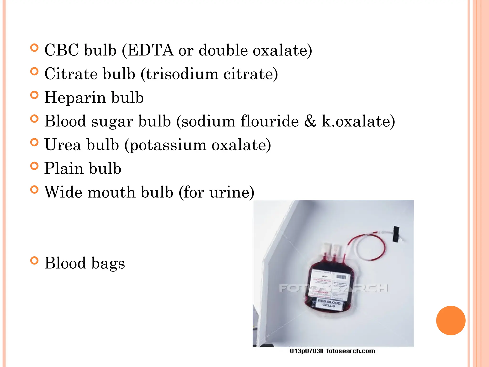
Specimen containers . Plain bulb & anticoagulant bulbs.
Request form
70 % alcohol or 0.5% chlorhexidine
Sterile gauze swabs
Adhesive dressings
Self sealing plastic bags
Rack to hold the specimen containers.
6.
What isthe Technique of blood collection..??
Identification.
Universal precautions.
Inform the patient of the procedure/consent.
Venous blood is best collected from the antecubital vein.
The skin should be cleaned with 70% alcohol or 0.5 %
chlorhexidine & allowed to dry spontaneously.
The tourniquetshould be tied just above the puncture site, which is
released once blood starts flowing into the syringe. Delay may lead to
haemoconcentration as a result of stagnation.
Insert the needle through the skin at a plane parallel to the vein & care
should be taken that the vein doesn't get counter punctured.
The piston of the syringe should not be withdrawn fast as it may cause
hemolysis.
Sufficient amount of blood should be withdrawn and dispensed in
appropriate bulbs.
The bulbs should be shaken for a while so that the blood gets mixed with
the anticoagulant properly.
Remove the needle and apply a sterile swab over the site & apply pressure
over it. After the bleeding stops , apply a small adhesive dressing.
BIO-MEDICAL WASTE MANAGEMENT..!!!!
Complications..!!!
What areANTICOAGULANTS..??
Agents which prevent the coagulation of blood.
DIFFERENT TYPES..
EDTA (Ethylenediaminetetra Acetic acid)
Trisodium Citrate
Double oxalate
Heparin
ACD - Acid-citrate-dextrose
CPD/CPDA - Citrate - phosphate - dextrose
/CPD+adenine
12.
EDTA (sequestrene/versene)-:
Commonly used anticoagulant in routine practice.
K/Na/Li salts of EDTA can be used.
Mechanism Of Action:
It acts by chelating the calcium molecules in the blood.
1.2 mg EDTA /ml of blood is required.
EDTA is used for Hb. estimation, TLC, DLC, PS, hematocrit,
sickling test, reticulocyte count, hemoglobin electrophoresis.
DISADVANTAGES:
• RBC morphology is hampered if the concentration is more than
the required.
• Not good for coagulation studies.
13.
Trisodium Citrate:
It is used for coagulation studies.
Mechanism of action :
It also works on the principle of calcium chelation.
9 volumes of blood are added to 1 volume of 32gm/l sodium citrate
solution.
For ESR estimation, 4 volumes of blood are added to 1 volume of
sodium citrate solution & well mixed.
14.
Double Oxalate:
It contains
ammonium oxalate ( 3 parts)
potassium oxalate ( 2 parts )
This combination minimizes the morphological changes that
occur in the erythrocytes.
Mechanism of action :
Calcium chelation
Uses - Routine hematological examinations & ESR
determination.
Disadvantage: Crenation of red cells - cannot be used for making
blood films.
15.
HEPARIN:
Itis used for chemistry , blood gas analysis & emergency tests.
It is the best anticoagulant for osmotic fragility tests &
immunophenotyping.
The lithium or sodium salt of heparin at a concentration of 10- 20
IU / ml blood is generally used.
It is not suitable for blood counts as it induces platelet &
leukocyte clumping & gives faint blue color on PS.