BONE MARROW ANDHEMATOPOIESIS
• Presented by – Maj (Dr) AP Singh (Retd)
• Guide – Lt Col Sudeep Kumar
2.
BONE MARROW
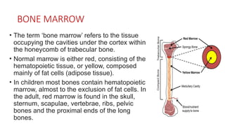
• Theterm ‘bone marrow’ refers to the tissue
occupying the cavities under the cortex within
the honeycomb of trabecular bone.
• Normal marrow is either red, consisting of the
hematopoietic tissue, or yellow, composed
mainly of fat cells (adipose tissue).
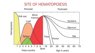
• In children most bones contain hematopoietic
marrow, almost to the exclusion of fat cells. In
the adult, red marrow is found in the skull,
sternum, scapulae, vertebrae, ribs, pelvic
bones and the proximal ends of the long
bones.
3.
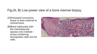
Fig.(A, B) Lowpower view of a bone marrow biopsy.
(A)Periosteal connective
tissue is seen external to
cortical bone.
(B)Bone trabeculae with
the intertrabecular
spaces (one marked –
arrow) containing
hemopoietic cells and fat
cells.
4.
STRUCTURE OF BONEMARROW
• Made up of Cellular elements and Stroma.
• CELLULAR ELEMENTS
• Erythroid, myeloid, lymphoid and platelet precursors, plasma cells,
macrophages, mast cells, dendritic cells, osteoblasts and
osteoclasts.
• STROMA – Consist of stromal cells and Extracellular matrix.
• Stromal cells - Fibroblasts, fat cells, macrophages, lymphocytes,
endothelial cells and reticulum cells.
• Extracellular matrix – Fibronectin, homonectin, laminin, collagen,
proteoglycans, acid mucopolysaccharides, chondroitin and heparan
sulphate.
5.
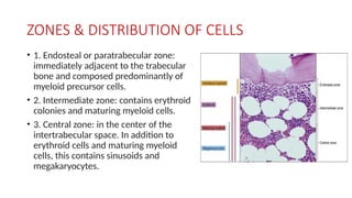
ZONES & DISTRIBUTIONOF CELLS
• 1. Endosteal or paratrabecular zone:
immediately adjacent to the trabecular
bone and composed predominantly of
myeloid precursor cells.
• 2. Intermediate zone: contains erythroid
colonies and maturing myeloid cells.
• 3. Central zone: in the center of the
intertrabecular space. In addition to
erythroid cells and maturing myeloid
cells, this contains sinusoids and
megakaryocytes.
6.
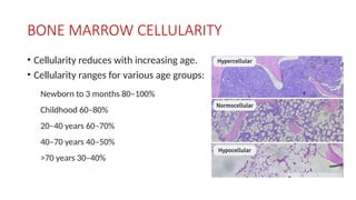
BONE MARROW CELLULARITY
•Cellularity reduces with increasing age.
• Cellularity ranges for various age groups:
Newborn to 3 months 80–100%
Childhood 60–80%
20–40 years 60–70%
40–70 years 40–50%
>70 years 30–40%
7.
HEMATOPOIESIS
• Process ofproduction of blood cells from hemopoietic stem cells
(HSC).
• These HSCs have extensive potential of proliferation to produce more
stem cells (Self renewal) and differentiate into progenitor cells
(Differentiation).
• Progenitor cells (Lineage committed cells) are committed to one
lineage, eg. myeloid and lymphoid.
• Proliferative potential of progenitor cells is limited as compared to
HSCs.
8.
HEMOPOIETIC STEM CELLS(HSCs)
• HSCs in the bone marrow reside in Stem cell niche and are
surrounded by proliferating and differentiating precursors alongwith
non-hemopoietic cells.
• Non-hemopoietic cells are stromal cells, macrophages, fibroblasts,
endothelial cells and fat cells, which form the microenvironment of
marrow which controls the self renewal and differentiation of HSCs.
• HSCs lies very close to endosteal osteoblasts which induce synthesis
of many cytokines.
9.
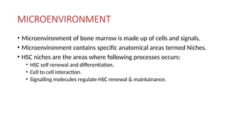
MICROENVIRONMENT
• Microenvironment ofbone marrow is made up of cells and signals,
• Microenvironment contains specific anatomical areas termed Niches.
• HSC niches are the areas where following processes occurs:
• HSC self renewal and differentiation.
• Cell to cell interaction.
• Signalling molecules regulate HSC renewal & maintainance.
10.
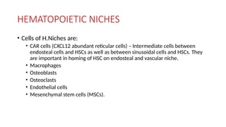
HEMATOPOIETIC NICHES
• Cellsof H.Niches are:
• CAR cells (CXCL12 abundant reticular cells) – Intermediate cells between
endosteal cells and HSCs as well as between sinusoidal cells and HSCs. They
are important in homing of HSC on endosteal and vascular niche.
• Macrophages
• Osteoblasts
• Osteoclasts
• Endothelial cells
• Mesenchymal stem cells (MSCs).
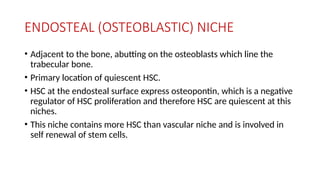
ENDOSTEAL (OSTEOBLASTIC) NICHE
•Adjacent to the bone, abutting on the osteoblasts which line the
trabecular bone.
• Primary location of quiescent HSC.
• HSC at the endosteal surface express osteopontin, which is a negative
regulator of HSC proliferation and therefore HSC are quiescent at this
niches.
• This niche contains more HSC than vascular niche and is involved in
self renewal of stem cells.
13.
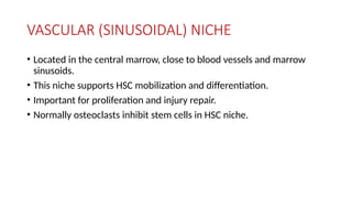
VASCULAR (SINUSOIDAL) NICHE
•Located in the central marrow, close to blood vessels and marrow
sinusoids.
• This niche supports HSC mobilization and differentiation.
• Important for proliferation and injury repair.
• Normally osteoclasts inhibit stem cells in HSC niche.
14.
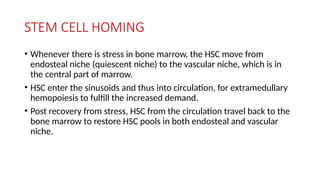
STEM CELL HOMING
•Whenever there is stress in bone marrow, the HSC move from
endosteal niche (quiescent niche) to the vascular niche, which is in
the central part of marrow.
• HSC enter the sinusoids and thus into circulation, for extramedullary
hemopoiesis to fulfill the increased demand.
• Post recovery from stress, HSC from the circulation travel back to the
bone marrow to restore HSC pools in both endosteal and vascular
niche.
15.
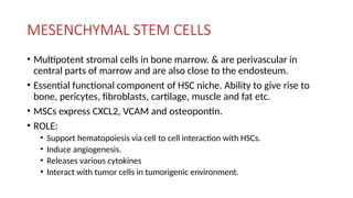
MESENCHYMAL STEM CELLS
•Multipotent stromal cells in bone marrow. & are perivascular in
central parts of marrow and are also close to the endosteum.
• Essential functional component of HSC niche. Ability to give rise to
bone, pericytes, fibroblasts, cartilage, muscle and fat etc.
• MSCs express CXCL2, VCAM and osteopontin.
• ROLE:
• Support hematopoiesis via cell to cell interaction with HSCs.
• Induce angiogenesis.
• Releases various cytokines
• Interact with tumor cells in tumorigenic environment.
16.
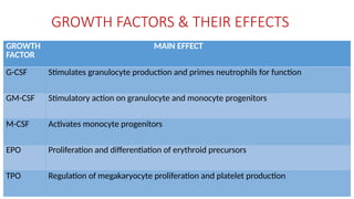
GROWTH FACTORS &THEIR EFFECTS
GROWTH
FACTOR
MAIN EFFECT
G-CSF Stimulates granulocyte production and primes neutrophils for function
GM-CSF Stimulatory action on granulocyte and monocyte progenitors
M-CSF Activates monocyte progenitors
EPO Proliferation and differentiation of erythroid precursors
TPO Regulation of megakaryocyte proliferation and platelet production
17.
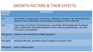
GROWTH FACTORS &THEIR EFFECTS
GROWTH
FACTOR
MAIN EFFECT
IL-1 Neutrophil, T-lymphocytes, monocyte, osteoclast activation, NK-cell proliferation,
Stromal cell & endothelial cell activation to produce G-CSF & GM-CSF
IL-6 Conversion of cytotoxic T-lymphocytes, conversion of B-lymphocytes to plasma
cells, proliferation and differentiation of stem cells into granulocytes, red cells,
monocytes and platelets
IFN-gamma Enhances NK-cell activity, inhibits apoptosis
IFN-alpha Activates NK-cells, promotes class-II antigens on B and T cells
TNF-alpha Role in inflammation
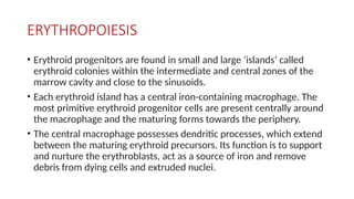
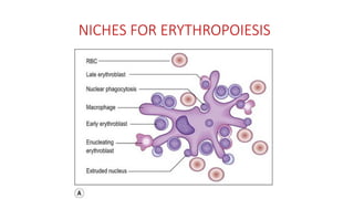
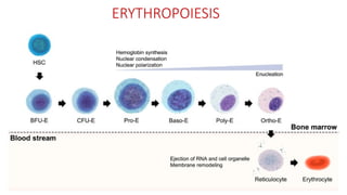
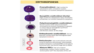
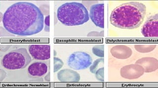
ERYTHROPOIESIS
• Erythroid progenitorsare found in small and large ‘islands’ called
erythroid colonies within the intermediate and central zones of the
marrow cavity and close to the sinusoids.
• Each erythroid island has a central iron-containing macrophage. The
most primitive erythroid progenitor cells are present centrally around
the macrophage and the maturing forms towards the periphery.
• The central macrophage possesses dendritic processes, which extend
between the maturing erythroid precursors. Its function is to support
and nurture the erythroblasts, act as a source of iron and remove
debris from dying cells and extruded nuclei.
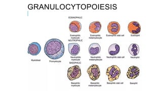
GRANULOCYTOPOIESIS
• Production ofgranular cells which exist in peripheral blood and
contain granules in their cytoplasm (Neutrophils, eosinophils and
basophils).
• Three basic requirements:
• Adequate number of myeloid stem cells.
• Suitable microenvironment provided by stromal cells.
• Adequate levels of growth factors (G-CSF, GM-CSF).
26.
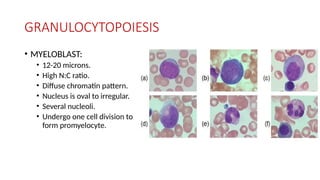
GRANULOCYTOPOIESIS
• MYELOBLAST:
• 12-20microns.
• High N:C ratio.
• Diffuse chromatin pattern.
• Nucleus is oval to irregular.
• Several nucleoli.
• Undergo one cell division to
form promyelocyte.
27.
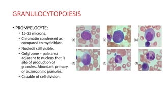
GRANULOCYTOPOIESIS
• PROMYELOCYTE:
• 15-25microns.
• Chromatin condensed as
compared to myeloblast.
• Nucleoli still visible.
• Golgi zone – pale area
adjacent to nucleus thet is
site of production of
granules. Abundant primary
or auzorophilic granules.
• Capable of cell division.
28.
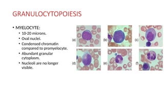
GRANULOCYTOPOIESIS
• MYELOCYTE:
• 10-20microns.
• Oval nuclei.
• Condensed chromatin
compared to promyelocyte.
• Abundant granular
cytoplasm.
• Nucleoli are no longer
visible.
29.
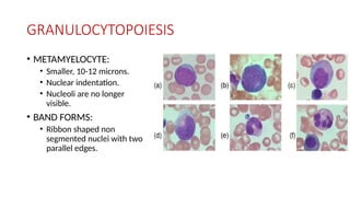
GRANULOCYTOPOIESIS
• METAMYELOCYTE:
• Smaller,10-12 microns.
• Nuclear indentation.
• Nucleoli are no longer
visible.
• BAND FORMS:
• Ribbon shaped non
segmented nuclei with two
parallel edges.
30.
GRANULOCYTOPOIESIS
• NEUTROPHILS:
• Segmentednuclei with
specific granules.
• EOSINOPHILS:
• Reddish orange granules.
• Bilobed nuclei.
• BASOPHILS:
• Large blue black granules
overlying the nucleus.
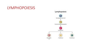
LYMPHOPOIESIS
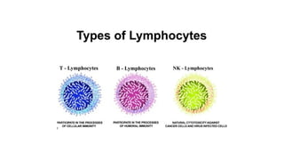
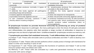
• Pluripotent stemcells give rise to Pre-B and Pre-T cells.
• Pre-T cells, as they pass through thymus become immunologically
competent T cells.
• Pre-B cells migrate to peripheral lymphoid organs and get
transformed to B-cells capable of transformation into plasma cells on
contact with antigens.
• Primary sites of lymphopoiesis are bone marrow and thymus and
secondary lymphatic organs are lymph nodes, spleen, and lymphoid
tissue in GIT.
33.
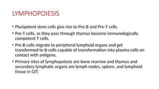
LYMPHOPOIESIS
• LYMPHOBLAST:
• 12-20microns.
• Centrally placed nuclei with 1-2 nucleoli.
• Chromatin is coarser than that of myeloblasts.
• Thin rim of pale blue cytoplasm.
34.
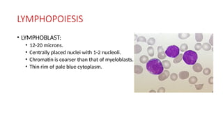
LYMPHOPOIESIS
• LARGE LYMPHOCYTE:
•10-15 microns.
• Central/eccentric nucleus.
• No nucleoli.
• Clear pale blue abundant cytoplasm.
• Nuclear chromatin is less condensed
than that of small lymphocytes.
35.
LYMPHOPOIESIS
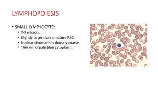
• SMALL LYMPHOCYTE:
•7-9 microns.
• Slightly larger than a mature RBC.
• Nuclear chromatin is densely coarse.
• Thin rim of pale blue cytoplasm.
36.
LYMPHOPOIESIS
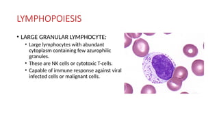
• LARGE GRANULARLYMPHOCYTE:
• Large lymphocytes with abundant
cytoplasm containing few azurophilic
granules.
• These are NK cells or cytotoxic T-cells.
• Capable of immune response against viral
infected cells or malignant cells.
39.
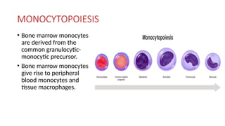
MONOCYTOPOIESIS
• Bone marrowmonocytes
are derived from the
common granulocytic-
monocytic precursor.
• Bone marrow monocytes
give rise to peripheral
blood monocytes and
tissue macrophages.
40.
MONOCYTOPOIESIS
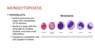
• MONOBLASTS:
• Earliestprecursors are
larger than myeloblasts,
13-15 microns.
• Nucleus is large with fine
chromatin & multiple
nucleoli, may have small
indentation.
• Cytoplasm is basophilic and
cell capable of mitosis.
41.
MONOCYTOPOIESIS
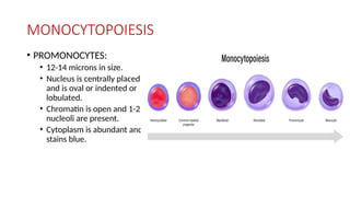
• PROMONOCYTES:
• 12-14microns in size.
• Nucleus is centrally placed
and is oval or indented or
lobulated.
• Chromatin is open and 1-2
nucleoli are present.
• Cytoplasm is abundant and
stains blue.
42.
MONOCYTOPOIESIS
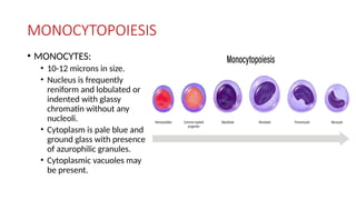
• MONOCYTES:
• 10-12microns in size.
• Nucleus is frequently
reniform and lobulated or
indented with glassy
chromatin without any
nucleoli.
• Cytoplasm is pale blue and
ground glass with presence
of azurophilic granules.
• Cytoplasmic vacuoles may
be present.
43.
MONOCYTOPOIESIS
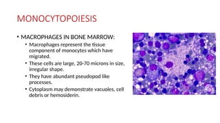
• MACROPHAGES INBONE MARROW:
• Macrophages represent the tissue
component of monocytes which have
migrated.
• These cells are large, 20-70 microns in size,
irregular shape.
• They have abundant pseudopod like
processes.
• Cytoplasm may demonstrate vacuoles, cell
debris or hemosiderin.
44.
THROMBOPOIESIS
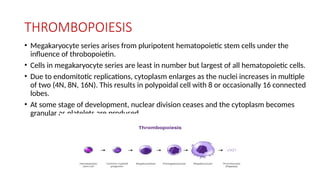
• Megakaryocyte seriesarises from pluripotent hematopoietic stem cells under the
influence of throbopoietin.
• Cells in megakaryocyte series are least in number but largest of all hematopoietic cells.
• Due to endomitotic replications, cytoplasm enlarges as the nuclei increases in multiple
of two (4N, 8N, 16N). This results in polypoidal cell with 8 or occasionally 16 connected
lobes.
• At some stage of development, nuclear division ceases and the cytoplasm becomes
granular as platelets are produced.
45.
THROMBOPOIESIS
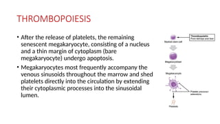
• After therelease of platelets, the remaining
senescent megakaryocyte, consisting of a nucleus
and a thin margin of cytoplasm (bare
megakaryocyte) undergo apoptosis.
• Megakaryocytes most frequently accompany the
venous sinusoids throughout the marrow and shed
platelets directly into the circulation by extending
their cytoplasmic processes into the sinusoidal
lumen.
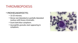
THROMBOPOIESIS
• PROMEGAKARYOCYTE:
• 15-25microns.
• Dense non lobulated or partially lobulated
nucleus with heavy chromatin.
• Scant dark blue cytoplasm.
• Azurophilic granules start appearing in
cytoplasm.
48.
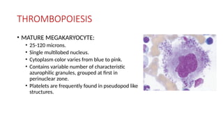
THROMBOPOIESIS
• MATURE MEGAKARYOCYTE:
•25-120 microns.
• Single multilobed nucleus.
• Cytoplasm color varies from blue to pink.
• Contains variable number of characteristic
azurophilic granules, grouped at first in
perinuclear zone.
• Platelets are frequently found in pseudopod like
structures.


![Hemangiblastoma CNS PPT.pptx [Autosaved].pptx](https://cdn.slidesharecdn.com/ss_thumbnails/hemangiblastomacnsppt-251005155615-644fd7bd-thumbnail.jpg?width=640&height=640&fit=bounds)


