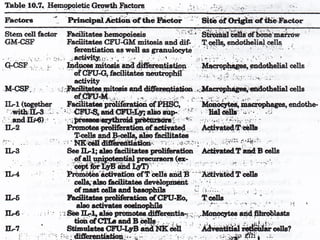
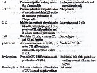
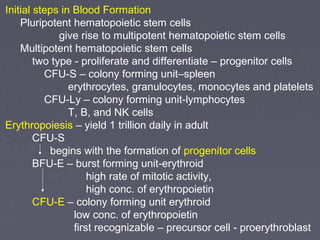
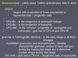
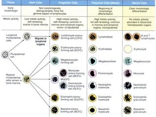
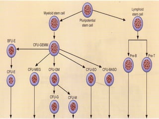
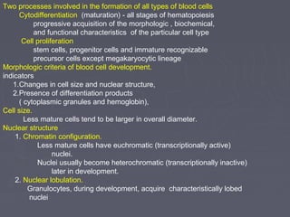
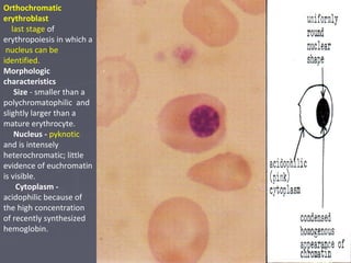
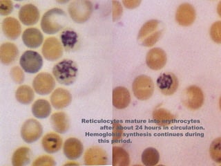
The document summarizes hematopoiesis, the process by which blood cells are formed from stem cells in the bone marrow. It discusses that hematopoiesis occurs through the simultaneous, continuous proliferation and differentiation of pluripotent stem cells in the bone marrow and lymphatic tissues. The structural organization of the hematopoietic marrow is also described, including the blood vessels, stroma, sinusoids, and cell types involved in hematopoiesis. The stages of development from stem cells to mature blood cells are outlined for erythropoiesis, granulopoiesis, monocytopoiesis, and thrombocytopoiesis.
The document discusses urine analysis, including chemical examination of urine. It describes tests for detecting ketone bodies, which indicate fatty acid breakdown. Causes of ketonuria include uncontrolled diabetes, starvation, and pregnancy. Tests are described for detecting bile pigments like bilirubin and bile salts, which can indicate liver or gallbladder issues. Tests for urobilinogen and blood are also summarized. The document provides details on chemical tests to detect significant bacteriuria through nitrite and leukocyte tests.
This document provides information about estimating serum urea levels, including:
- An overview of the urea cycle and how urea is produced from excess amino acids and used to excrete nitrogen from the body.
- Details two common enzymatic methods for quantifying urea levels: the urease method and diacetyl monoxime method.
- Discusses factors that influence serum urea levels and the clinical significance of elevated or decreased levels. elevated BUN:creatinine ratio indicates prerenal azotemia while a low ratio suggests renal failure.
Automated urine analysis systems can perform a complete urinalysis including physical, chemical, and microscopic components. They provide standardized, efficient results with improved turnaround times compared to manual methods. Key advantages include objective particle identification and classification through techniques like flow cytometry and digital image analysis, as well as automated test strip reading and sample handling for tests like glucose, protein, blood, and more. While providing productivity and consistency benefits, automated urine analysis aims to enhance urine sediment examination and reporting.
normal and abnormalities in red blood cellRfa Mohd
This document provides information on various types of red blood cell morphologies seen on blood smears, including normal and abnormal shapes and inclusions. It describes normocytic and normochromic red blood cells as well as variations such as microcytic, macrocytic, poikilocytes (variations in shape), anisocytosis (variation in size), and polychromasia (variation in color). Specific abnormal red blood cells and inclusions are defined such as sickle cells, target cells, Howell-Jolly bodies, basophilic stippling, malaria parasites, and reticulocytes. Causes and disorders are provided for each abnormal finding.
Fluid cytology in serous cavity effusionstashagarwal
The intrathoracic and intraperitoneal organs are covered by a single layer of mesothelial cells, which is continuous with the lining of the thoracic and peritoneal cavities. The potential space between the two layers of epithelium contains a small amount of lubricating fluid.
Serous fluid lies between the membranes lining the body cavities(parietal) and those covering the organs within the cavities(visceral).
Production and reabsorption are normally at a constant rate. They are influenced by
Changes in osmotic and hydrostatic pressure in the blood.
Concentration of chemical constituents in the plasma
Permeability of blood vessels and membranes.
An accumulation of fluid, called an effusion, results from an imbalance of fluid production and reabsorption. This fluid accumulation in the pleural, pericardial, and peritoneal cavities is known as serous effusion.
The document describes the procedure for performing an activated partial thromboplastin time (APTT) test using citrated plasma. The test involves incubating plasma with brain extract, kaolin, and calcium chloride before measuring the clotting time. Prolonged APTT results indicate deficiencies in the intrinsic coagulation pathway, such as issues with factors VIII, IX, XI, or XIII; liver disease; vitamin K deficiency; or disseminated intravascular coagulation.
Demonstration of different fixatives used in Histopathology
Demonstration of different Microtome used in Histopathology
To demonstrate the activity of enzyme
Demonstration of following enzymes activity in a Tissue
Demonstration of Laboratory method that uses antibodies
Demonstrate the FIC and FITC techniques
Demonstration of the technique used to separate DN
Demonstration of technique for rapidly producing
Demonstrate the Flow cytometer technique
My report . (wbc count)
Report to practical physiology .
......
University of AL_Ameed .
College of Dentistry .
________________________________
Telegram : @Goldenalzaidy
Instagram : goldenalzaidy
__________________________________
تقرير كامل ومفيد عن طريقة حساب عدد الكريات البيض تستطيع اعادة صياغته وتقديمه
---------------
This document provides information and instructions for making and examining a blood smear. There are three main types of blood smears: the cover glass smear, wedge smear, and spun smear. Additional types like the buffy coat smear are used for specific purposes. The document outlines the proper procedure for making a wedge smear from a blood sample and describes characteristics of a good smear. Common causes of a poor smear and biological factors that can affect the smear are also discussed. The document then covers slide fixation, staining using Leishman's stain, and examining the smear under the microscope to perform tasks like a manual differential count and assessing red blood cell morphology.
The document discusses the history and methods of bilirubin analysis. It describes how Ehrlich first described the diazo reaction for bilirubin detection in 1883 and how later methods by Malloy and Evelyn and Jendrassik and Grof improved on this. It outlines the fractions of bilirubin that are measured and calculated, including total, conjugated, and unconjugated bilirubin. Specimen collection factors and the Jendrassik-Grof method are summarized, along with reference ranges and urine bilirubin detection.
Synovial fluid is produced by synovial membranes in joint cavities and acts as a lubricant and shock absorber. It contains hyaluronic acid and filters interstitial fluid. During a joint aspiration procedure, fluid is extracted from a joint through a needle for analysis. Abnormal fluid characteristics can indicate conditions like gout, arthritis, or infection. Precautions are taken to prevent complications, and patients are monitored after the procedure. Laboratory tests of synovial fluid include examining appearance, cell count, crystals, and culturing for bacteria.
Bone marrow is found within bones and produces blood cells, containing two types - red marrow which produces blood cells throughout life and yellow marrow containing fat; it undergoes examination by aspiration and biopsy to evaluate blood cell production and detect abnormalities by analyzing cellular composition and bone marrow architecture.
Urinalysis- Methods, observations and clinical significanceNamrata Chhabra
The document provides information on urinalysis including urine specimen collection, preservation, composition, and various tests performed to examine the physical and chemical properties of normal and abnormal urine. It discusses urine color, volume, specific gravity, pH, and how to test for organic and inorganic constituents like urea, creatinine, chlorides, phosphates, and calcium. Abnormal results are interpreted to help diagnose underlying renal and systemic diseases.
This document summarizes hemostasis and the coagulation process. It discusses that hemostasis involves platelets, blood vessels, and plasma proteins interacting to maintain blood fluidity and prevent hemorrhage. The three main steps of hemostasis are primary hemostasis involving platelet plug formation, the coagulation cascade, and fibrinolysis. Disorders can occur from abnormalities in blood vessels, platelets, or coagulation factors, causing excessive bleeding. Several laboratory tests are used to monitor coagulation factors and platelet function, including prothrombin time, activated partial thromboplastin time, and thromboelastography.
The document provides information about cerebrospinal fluid (CSF) examination including CSF composition, functions, how it is formed and drained, significant features, indications for examination, collection process, and laboratory diagnosis. Key points include:
- CSF acts as a cushion and removes waste from the brain and spinal cord. It is produced by choroid plexus and contains glucose, electrolytes, and few cells.
- Laboratory diagnosis of CSF includes physical, microscopic, and chemical examination to detect cells, proteins, glucose and other analytes that can indicate conditions like meningitis.
- CSF is collected via lumbar puncture for diagnostic and therapeutic purposes and analyzed for appearance, pressure, cell count and differentials to identify issues like
Reticulocytes are immature red blood cells that are released from the bone marrow into circulation. They contain remnants of RNA and ribosomes. Reticulocyte counts are used to assess bone marrow response to anemia and erythropoietin therapy. Reticulocytes can be manually counted using supravital dyes like new methylene blue that stain the RNA, or automatically using cell counters that detect nucleic acid content. Normal ranges are 0.5-2.5% and increased counts indicate bone marrow response to anemia or therapy, while decreased counts suggest bone marrow suppression.
This document provides information on evaluating proteinuria in urine. It discusses the different types of proteinuria including glomerular, tubular, overflow and hemodynamic proteinuria. Glomerular proteinuria is caused by damage to the glomerular basement membrane and can be selective or non-selective. Tubular proteinuria occurs when low molecular weight proteins are excreted due to tubular damage. Tests for detecting and quantifying protein in urine include heat and acetic acid test, reagent strip, sulphosalicylic acid test, and 24-hour urine collection. The document provides normal ranges and indications for proteinuria testing.
This document summarizes key components of hemostasis including primary and secondary hemostasis. It describes platelet adhesion, activation, aggregation and secretion. Tests for evaluating hemostasis are outlined including bleeding time, platelet function analyzer, and assays for factors, fibrinogen, D-dimer and FDP. Causes and interpretation of abnormal results are provided for tests such as PT, APTT, TT and specific assays of platelet function and coagulation factors.
Leukocytes, or white blood cells, are divided into myelocytes and lymphocytes. Myelocytes include granulocytes like neutrophils, eosinophils, and basophils, as well as monocytes. Lymphocytes include B and T lymphocytes. Leukopoiesis is the process by which these cells develop from hematopoietic stem cells in the bone marrow. Morphological abnormalities that can occur include alterations in neutrophil nuclei like the Pelger-Huet anomaly and cytoplasmic changes such as May-Hegglin anomaly. Lymphocytes and monocytes can also demonstrate abnormal morphologies like Reed-Sternberg cells.
I have listed out the LE cells structure and Microscopical examinaton of LE CELLS, Difference between tart cells and le cells, clinical symptoms and diagnostic procedure.
special and routine stains in haematology 1Dr.SHAHID Raza
The document discusses various routine and special stains used in hematology. Routine stains like Leishman, Giemsa, and Wright stains are used to stain peripheral blood films and differentiate blood cells. Special stains require additional processing but can identify characteristics not seen with routine stains, such as periodic acid Schiff stain which detects carbohydrates like glycogen by oxidizing glycol groups and producing a red reaction. Proper staining techniques such as fixation, washing, and timing are important for preparing clear blood smears and accurately identifying blood components.
Romanowsky stains are commonly used stains for blood films that use a combination of basic dyes like methylene blue and acidic dyes like eosin. Some common Romanowsky stains discussed are Leishman, Giemsa, Wright, Field, and Jenner stains. Each stain has a specific composition and method of use to differentially stain structures in blood cells like nuclei, cytoplasm, and granules to aid in identification of cell types under a microscope. Proper staining technique and avoiding issues like overstaining or understaining are important for producing diagnostic quality blood films.
This document discusses several red blood cell indices used to characterize anemias, including mean cell volume (MCV), mean cell hemoglobin (MCH), mean cell hemoglobin concentration (MCHC), and red cell distribution width (RDW). It provides details on how each index is calculated and interpreted, and examples of abnormal red blood cell morphologies seen in different types of anemias that would affect the index values.
Urine analysis is an integral part of a clinical laboratory. automation techniques in urine biochemistry, their priniciplas and microscopy along with their advantages and disadvantages are outlined.
This document provides an overview of flow cytometry, including its history, components, principles, and applications. Flow cytometry involves passing cells in suspension through a laser beam to measure physical properties like size and granularity, as well as cell markers detected by fluorescent antibodies. This allows identification of cell types, lineages, and abnormalities. The document discusses sample preparation, common specimens analyzed, immunophenotyping using multiple fluorochromes, and applications like DNA content analysis, erythrocyte analysis, and reticulocyte counting.
El documento describe el tejido mieloide y la hematopoyesis. La médula ósea es el órgano principal de la formación de células sanguíneas, incluidos glóbulos rojos, plaquetas y diferentes tipos de glóbulos blancos como neutrófilos, eosinófilos, basófilos, linfocitos y monocitos. Todas estas células se originan a partir de células madre hematopoyéticas en un proceso llamado hematopoyesis.
Flow cytometry is a technique that uses lasers and fluorescence to count and examine microscopic particles like cells. It can measure multiple parameters of individual cells as they flow in a liquid stream past the laser beam at thousands of cells per second. Components include a flow cell to arrange cells in a stream, optical systems to generate light signals, detectors to convert light signals to electrical signals, and read-out devices to analyze the results. Flow cytometry is used widely in clinical laboratories for applications like immunophenotyping, DNA analysis for malignancy, detecting enzymatic deficiencies, genetic diseases, and hematology analysis.
Monophyletic theory of hematopoiesis. Stem cells.Iryna Nováková
There are three main theories of hematopoiesis or blood cell formation: monophyletic, dualistic, and polyphyletic. The monophyletic or unitary theory, first proposed over 100 years ago, suggests that all blood cells originate from a common stem cell. Stem cells are classified based on their differentiation potential as totipotent, pluripotent, multipotent, or unipotent. Hematopoiesis occurs primarily in the bone marrow and is regulated by growth factors. The process involves the proliferation, differentiation, and maturation of stem cells into the various mature blood cell types through multiple cellular stages. A complete blood count provides indicators of normal or abnormal hematopoiesis. Stem cells
TEJIDOS: hematopoyetico, mieloide, linfoide y sanguineo juan negrete
Este documento describe los principales tejidos relacionados con la sangre, incluyendo el tejido hematopoyético, el tejido mieloide, el tejido linfoide y el tejido sanguíneo. Explica que el tejido hematopoyético se encuentra principalmente en la médula ósea y es responsable de la producción de células sanguíneas a través de los procesos de diferenciación, proliferación y maduración. También describe las características y funciones del tejido mieloide y cómo se orig
This document discusses hematopoiesis, the formation of blood cells. It presents two main theories: the monoplastic theory which states all blood cells originate from one stem cell, and the polyplastic theory which suggests different blood cell types originate from different stem cells. The stages of blood cell development are described starting from the embryonic stage through the hepatic and medullary stages where development occurs in the bone marrow. The processes of erythropoiesis and granulopoiesis, leading to the production and maturation of red blood cells and white blood cells, are outlined in detail through their characteristic cell types.
This document provides an overview of the structure and cell types of the cerebral cortex. It begins with an introduction to the basic structure of neurons and the central nervous system. It then describes the six layers of the neocortex and the principal cell types found within each layer, including pyramidal cells, stellate cells, and others. The interconnections between neurons and various staining techniques used to visualize cortical components are also summarized. Finally, several common diseases that affect the cerebral cortex are briefly mentioned.
This document provides an introduction to flow cytometry. It defines flow cytometry as a method for sensing individual cells in a fluid stream as they pass through a laser beam, measuring light scattering and fluorescence. Key aspects of flow cytometry systems and methodology are described, including hydrodynamic focusing of cells, light scattering measurements, use of fluorescent markers, optical and electronic components, data acquisition and analysis techniques like gating and compensation. The history of technological developments in flow cytometry is also summarized.
This document summarizes hematopoiesis and the development of blood cells from stem cells in the bone marrow. It discusses the types of stem cells, the origins and development of blood cells in the embryo and fetus, the sites of hematopoiesis including the bone marrow, liver and spleen, and the cellular elements and maturation processes involved in erythropoiesis, granulopoiesis, lymphopoiesis, and megakaryopoiesis. It also covers hematopoietic growth factors, cytokines, and techniques for examining maturing blood cells.
This document summarizes hematopoiesis and the development of blood cells from stem cells in the bone marrow. It discusses the types of stem cells, the origins and development of blood cells in the embryo and fetus, the anatomical sites of blood cell development including the yolk sac, liver, spleen, and bone marrow. It also describes the cellular elements of the bone marrow including the different blood cell progenitors, erythropoiesis, granulopoiesis, lymphopoiesis, megakaryopoiesis, marrow stromal cells, mast cells, macrophages, bone cells, interleukins, and hematopoietic growth factors.
The document summarizes hematopoiesis, the formation of blood cells. It discusses prenatal hematopoiesis, which occurs in four phases: mesodermal, hepatic, splenic, and myeloid. Postnatal hematopoiesis takes place primarily in the bone marrow. The monophyletic theory proposes that all blood cells originate from a common hematopoietic stem cell. Key stages in erythropoiesis are also outlined, including the proerythroblast, basophilic erythroblast, and polychromatophilic erythroblast stages. Hematopoietic growth factors play an important role in regulating blood cell production.
The document discusses haematopoiesis, or blood cell formation. It defines the process and sites where it occurs, including the bone marrow, thymus, lymph nodes, spleen and liver. It describes the progression of haematopoiesis from embryonic stages through postnatal development when the bone marrow becomes the primary site. The functions of different haematopoietic organs are outlined. Stem cells differentiate into committed progenitor cells that give rise to mature blood cells through the influence of various growth factors and cytokines.
Development anatomy and physiology of haematopoiesis, hematological copySreemayee Kundu
Hematopoiesis is the process by which blood cells are formed from hematopoietic stem cells in the bone marrow. There are four main stages of development: stem cells differentiate into progenitor cells, then precursor cells, and finally mature blood cells. The major types of blood cells produced are red blood cells, white blood cells, and platelets. During development in the embryo and fetus, hematopoiesis occurs first in the yolk sac, then liver, and later primarily in the bone marrow. Different types of hemoglobin are expressed at various developmental stages.
Hematopoiesis is the formation of blood cells in the bone marrow. Hematopoietic stem cells originate in the yolk sac and migrate to the liver, spleen and bone marrow where they differentiate into various blood cell types. The major sites of hematopoiesis change throughout development - the liver is dominant during the fetal stage while the bone marrow becomes the permanent site of hematopoiesis after birth. In the bone marrow, stem cells differentiate through several stages into mature red blood cells, white blood cells or platelets through the action of growth factors and cytokines.
This document summarizes hematopoiesis, the process of blood cell production. It describes the hematopoietic growth factors that regulate blood cell differentiation and proliferation, including G-CSF, GM-CSF, M-CSF, EPO, thrombopoietin, and cytokines. The normal sites of hematopoiesis change from the yolk sac and liver during embryonic development to solely the bone marrow after birth. The document also outlines the development pathways and maturation stages of the major blood cell types: red blood cells, white blood cells, platelets, lymphocytes, monocytes, granulocytes, and megakaryocytes.
This document summarizes the process of leukopoiesis, or white blood cell formation. It describes the myeloid and lymphoid stem cell lineages that give rise to granulocytes, monocytes, and lymphocytes. The key stages of development are discussed, from pluripotent stem cells into committed progenitor cells, blast cells, promyelocytes/promonocytes, myelocytes, metamyelocytes, and band or segmented mature forms. Cytokines such as colony stimulating factors regulate white blood cell development and differentiation. Leukocytes mature over 10 days, spending half that time dividing and half maturing, before circulating and taking on tissue-specific functions.
Hemopoiesis is the process of blood cell formation in the bone marrow. It begins with pluripotent stem cells that give rise to progenitor cells committed to specific blood cell lineages. Progenitor cells further differentiate into precursor cells and eventually mature blood cells. Key events in hemopoiesis include the maturation of erythrocytes through the loss of organelles and nuclei, and the development of granulocytes marked by the production of granules in the cytoplasm. Growth factors regulate hemopoiesis by stimulating progenitor and precursor cell proliferation and differentiation.
Hemopoiesis is the process of blood cell formation in the bone marrow. It begins with pluripotent stem cells that give rise to progenitor cells committed to specific blood cell lineages. Progenitor cells further differentiate into precursor cells and eventually mature blood cells. Key events in hemopoiesis include the maturation of erythrocytes through the loss of organelles and nuclei, and the development of granulocytes marked by the production of granules in the cytoplasm. Disruptions in hemopoiesis can provide clinical insights, such as an increase in immature neutrophils signaling bacterial infection.
The document discusses hematopoiesis, the process of blood cell production. Key points include:
- Hematopoiesis occurs through hematopoietic stem cells which give rise to all other blood cell types.
- Early in development the yolk sac and liver are sites of hematopoiesis, but later it mainly occurs in the bone marrow.
- Hematopoietic stem cells self-renew and differentiate into progenitor cells through regulation by growth factors, transcription factors, adhesion molecules and other components of the bone marrow microenvironment.
- Hematopoiesis is controlled to balance cell production, differentiation and apoptosis/cell death.
This document discusses tumor angiogenesis and stromagenesis. It describes 4 main mechanisms of tumor angiogenesis: 1) sprouting angiogenesis, 2) angioblast recruitment, 3) cooption of existing blood vessels, and 4) formation of mosaic vessels. It also outlines 10 steps in the process of tumor stromagenesis, including vascular hyperpermeability, fibrin deposition, new blood vessel formation, and eventual formation of dense connective tissue. Finally, it compares wound healing and tumor stroma formation, noting that VEGF and vascular permeability are transient in wound healing but persistent indefinitely in tumor stromagenesis.
presented by HAFIZ M WASEEM
university of education LAHORE Pakistan
i am from mailsi vehari and studied in lahore
bsc in science college multan
msc from lahore
Hemopoiesis is the process of blood cell formation in the bone marrow and other hematopoietic tissues. Mature blood cells have short lifespans and must be continuously replaced. The document discusses the stages of development of red blood cells, granulocytes, and other blood cell types from hematopoietic stem cells through progenitor and precursor cells with the influence of growth factors in the bone marrow microenvironment. It also describes the structure and cellular components of red bone marrow.
Hematopoiesis is the process by which all blood cells are generated from hematopoietic stem cells located in the bone marrow. Erythropoiesis is the production of red blood cells. It involves the commitment of myeloid erythroid progenitors to the erythroid lineage and four mitotic divisions, resulting in enucleated reticulocytes. Erythropoiesis is regulated by erythropoietin and transcription factors like GATA-1 through signaling pathways and feedback loops involving oxygen levels. Erythroblasts develop within erythroblastic islands, where interactions with central macrophages support their development and enucleation.
HEMATOPOIESIS: ORIGIN OF BLOOD CELLS AND BLOOD PLASMAharshalshelke4
Hematopoiesis is the process in which all Blood lineages are produced from Hematopoietic Stem cells. Through series of Progenitor cells, Hematopoietic stem cells produce a large number of Adult Stem cells. Hematopoietic Stem cells have the ability of self-renewal and self-division This Divisional event results in the Differentiation of Hematopoietic Stem cells into –Lymphocytes, Granulocytes, Monocytes, etc. Progenitor cells also contain cells that act as our body's Defense mechanism. This Always active defense mechanism consists of T cells, B cells, and Natural Killer cells. These cells work together to fight against any viruses or any pathogen and destroy them or exclude them out of the body. By understanding the Hematopoietic process and Defense mechanism, development has been done in the formation of new therapies for curing specific diseases. The basic concept of Stem cell production and its classification are defined here. This paper includes the concept of the production of Progenitor cell lineages which are classified as Lymphoid Progenitor cells and Myeloid Progenitor cells. Here we discuss the Embryonic development and classification of each blood lineages.
Hematopoiesis is the process of blood cells being differentiated from hematopoietic stem cells. This process must be repeated on a regular basis in order to keep the body's circulating blood cell numbers stable. Blood cells are divided into three main linages:
Reticulocytes and erythrocytes make up the Erythroid Lineage (red blood cells).
Lymphocytes (B and T cells) and natural killer cells make up the lymphoid lineage.
Macrophages, dendritic cells, granulocytes, and megakaryocytes are all members of the myeloid lineage.
## Site Of Hematopoiesis
Yolk sac
Liver and spleen
Bone marrow
Gradual replacement of active (red) marrow by tissue inactive (fatty)
Expansion can occur during increased need for cell production
Hematopoiesis is the process by which all blood cells are produced from hematopoietic stem cells. It begins during the first weeks of embryonic development in the yolk sac and later transitions to the spleen, liver, lymph nodes, and finally the bone marrow, where it continues for life. Hematopoietic stem cells can differentiate into either common lymphoid or myeloid progenitor cells. The lymphoid lineage produces lymphocytes and myeloid produces red blood cells, platelets, and white blood cells. Cytokines precisely regulate hematopoiesis by activating transcription factors that control the differentiation of stem cells into specific blood cell types.
the presentation tells you about hematopoiesis which is the process of formation of blood cells i.e. RBC’S, WBC’S and platelets is called as hematopoiesis and the sites where it occurs are known as hematopoietic tissues or organs.
Osteoporosis - Definition , Evaluation and Management .pdfJim Jacob Roy
Osteoporosis is an increasing cause of morbidity among the elderly.
In this document , a brief outline of osteoporosis is given , including the risk factors of osteoporosis fractures , the indications for testing bone mineral density and the management of osteoporosis
Travel vaccination in Manchester offers comprehensive immunization services for individuals planning international trips. Expert healthcare providers administer vaccines tailored to your destination, ensuring you stay protected against various diseases. Conveniently located clinics and flexible appointment options make it easy to get the necessary shots before your journey. Stay healthy and travel with confidence by getting vaccinated in Manchester. Visit us: www.nxhealthcare.co.uk
Mercurius is named after the roman god mercurius, the god of trade and science. The planet mercurius is named after the same god. Mercurius is sometimes called hydrargyrum, means ‘watery silver’. Its shine and colour are very similar to silver, but mercury is a fluid at room temperatures. The name quick silver is a translation of hydrargyrum, where the word quick describes its tendency to scatter away in all directions.
The droplets have a tendency to conglomerate to one big mass, but on being shaken they fall apart into countless little droplets again. It is used to ignite explosives, like mercury fulminate, the explosive character is one of its general themes.
Kosmoderma Academy, a leading institution in the field of dermatology and aesthetics, offers comprehensive courses in cosmetology and trichology. Our specialized courses on PRP (Hair), DR+Growth Factor, GFC, and Qr678 are designed to equip practitioners with advanced skills and knowledge to excel in hair restoration and growth treatments.
Histololgy of Female Reproductive System.pptxAyeshaZaid1
Dive into an in-depth exploration of the histological structure of female reproductive system with this comprehensive lecture. Presented by Dr. Ayesha Irfan, Assistant Professor of Anatomy, this presentation covers the Gross anatomy and functional histology of the female reproductive organs. Ideal for students, educators, and anyone interested in medical science, this lecture provides clear explanations, detailed diagrams, and valuable insights into female reproductive system. Enhance your knowledge and understanding of this essential aspect of human biology.
Cell Therapy Expansion and Challenges in Autoimmune DiseaseHealth Advances
There is increasing confidence that cell therapies will soon play a role in the treatment of autoimmune disorders, but the extent of this impact remains to be seen. Early readouts on autologous CAR-Ts in lupus are encouraging, but manufacturing and cost limitations are likely to restrict access to highly refractory patients. Allogeneic CAR-Ts have the potential to broaden access to earlier lines of treatment due to their inherent cost benefits, however they will need to demonstrate comparable or improved efficacy to established modalities.
In addition to infrastructure and capacity constraints, CAR-Ts face a very different risk-benefit dynamic in autoimmune compared to oncology, highlighting the need for tolerable therapies with low adverse event risk. CAR-NK and Treg-based therapies are also being developed in certain autoimmune disorders and may demonstrate favorable safety profiles. Several novel non-cell therapies such as bispecific antibodies, nanobodies, and RNAi drugs, may also offer future alternative competitive solutions with variable value propositions.
Widespread adoption of cell therapies will not only require strong efficacy and safety data, but also adapted pricing and access strategies. At oncology-based price points, CAR-Ts are unlikely to achieve broad market access in autoimmune disorders, with eligible patient populations that are potentially orders of magnitude greater than the number of currently addressable cancer patients. Developers have made strides towards reducing cell therapy COGS while improving manufacturing efficiency, but payors will inevitably restrict access until more sustainable pricing is achieved.
Despite these headwinds, industry leaders and investors remain confident that cell therapies are poised to address significant unmet need in patients suffering from autoimmune disorders. However, the extent of this impact on the treatment landscape remains to be seen, as the industry rapidly approaches an inflection point.
8 Surprising Reasons To Meditate 40 Minutes A Day That Can Change Your Life.pptxHolistified Wellness
We’re talking about Vedic Meditation, a form of meditation that has been around for at least 5,000 years. Back then, the people who lived in the Indus Valley, now known as India and Pakistan, practised meditation as a fundamental part of daily life. This knowledge that has given us yoga and Ayurveda, was known as Veda, hence the name Vedic. And though there are some written records, the practice has been passed down verbally from generation to generation.
10 Benefits an EPCR Software should Bring to EMS Organizations Traumasoft LLC
The benefits of an ePCR solution should extend to the whole EMS organization, not just certain groups of people or certain departments. It should provide more than just a form for entering and a database for storing information. It should also include a workflow of how information is communicated, used and stored across the entire organization.
2. Hematopoiesis
process
specialized blood cells develop from pluripotent stem cells of
myeloid tissue in the bone marrow
result of simultaneous,
continuous proliferation and
differentiation – reduction in
the potential of the cells
Site - occurs in myeloid and lymphatic tissue.
Myeloid tissue
bone marrow
Lymphatic tissue
lymphatic organs
- not a rigidly compartmentalized process;
blood cells usually associated with myeloid tissue can
arise in lymphoid tissue, and vice versa
7. Structural Organization Of Hematopoietic Marrow
Cancellous bone – bony spicules or trabeculae lined by
endosteum and marrow filled with
hematopoietic and non-hematopoietic cells
Blood vessels of the marrow compartment
1. Nutrient arteries
periosteum pass through the compact bone to enter
the marrow space.
2. Longitudinal arteries
formed by the division of a nutrient artery - run
parallel to the long axis of a bone.
3. Radial arteries
spoke-like branches that arise from longitudinal
arteries to form thin-walled sinusoids in the
hematopoietic tissue.
8.
9.
10.
11. Bone marrow
very large and complex organ
cavities of the skeleton
total mass, adult – 1600 to 3000 grams
½ - hematopoietically inactive fatty (yellow) marrow
few microscopic foci of hematopoietic cells
½ - hematopoietically active (red) marrow
function - based on a high degree of structural organization
(organization - labile, altering rapidly in response to
many stimuli)
hematopoietic marrow
formation and release - blood cells
phagocytosis and degradation - microorganisms
and abnormal or senescent rbcs
antibody production
non-hematopoietic marrow
large store of reserve lipids
12. Sinusoids
endothelial cells
no basement membrane,
overlapping and may interdigitate extensively
adventitial layer
external discontinuous layer
Stroma of the Marrow
Cells
3-dimensional meshwork of reticular cells and a delicate
web of containing hematopoietic cells, macrophages, mast
cells, fat cells, lymphocytes and plasma cells.
reticular cells - form a loose net of reticular fibers
reinforce the sinusoidal capillaries and
internal support for the stroma
cytoplasmic processes – lie along the sinusoidal
surface and protrude outward in- between
hematopoietic cells
13.
14.
15. Matrix
contains collagen types I and III, fibronectin, laminin, and
proteoglycans. Laminin, fibronectin, and hemonectin - cell
binding substance
interact with cell receptors to bind cells
to the matrix
Other cells
osteoblast and osteoclast
fat cells
newborn - 0%
2 week-old infant – 15%
children between 18 months to 11 year-old – 20% to 65%
adult – 30% to 70%
70 year-old - >70%
Marrow cellularity - proportion of the area occupied by cells
other than fat cells
normocellular, hypocellular or
hypercellular
16. Macrophages
intracytoplasmic inclusion - refractile yellow-brown
hemosiderin - iron (+)granules
marrow fragments or smear semi-quantitatively to
assess total iron store
long cytoplasmic processes – protrude into the sinusoids and
phagocytozed senescent or damage rbcs, progranulocytes
and circulating microorganism
present in the erythroblastic islands, plasma cell island and
lymphatic nodules but may also occurs elsewhere
generate various neutrophilic growth factors
Mast cells
progenitor cells occur in the marrow, but proliferation and
maturation and acquisition of granules occurs in the tissues
associated with lymphoid nodule, wall of the arterioles,
adjacent to the endothelium of sinusoids and endosteal
cells of bone trabeculae
17. Characterization of Hematopoietic tissue
microscopically by differentiating blood cells.
1. Stained smear of bone marrow reveals a complex population
comprising several types of blood cells and their precursors
2. These cell types can be sorted into several developmental
sequences, each sequence culminating in one of the several
types of mature blood cells – M:E ratio
3. Bone marrow biopsy
cellularity
architecture – structural relationship of the components
tumor
18. Hematopoietic Cords - Cell Associations in Red Bone Marrow
Histologic sections of bone marrow show the following
relationships.
a. Nests of erythroblasts and myelocytes.
developing blood cells are often seen clumped into nests or
islets - cells clump when mitotic events increase their
numbers and the daughter cells remain restricted to
the immediate vicinity.
b. Normoblasts
orthochromatic erythroblasts and macrophages.
Macrophages are found in close association
with nests of normoblasts, where they
phagocytize nuclei expelled by the
normoblasts during erythropoiesis.
c. Megakaryocytes and the sinusoidal wall.
Megakaryocytes are found in close proximity to the walls of
marrow blood capillaries(sinusoids) - facilitates the release of
platelets into the blood stream
19. During preparation of a bone marrow smear,
these normal cellular relationships are demolished.
20.
21.
22. Abnormal Increase in Hematopoiesis
Young children – rapid increase in hematpoietic tissue
accomodated mainly by a
reduction in the proportion of marrow space
occupied by sinusoids
skeletal abnormalities –
frontal and parietal bossing, dental
deformation, and malocclusion of the teeth,
thinning of the cortex – fracture
if the increase is substantial - extramedullary
hematopoies
Adult
initially associated with the replacement of fat cells in the red
marrow by hematopoietic cells and also with the spread of red
marrow into marrow cavities normally containing yellow marrow
if the increase is gross – extramedullary hematopoiesis
23. Postnatal Hematopoiesis
involved three classes of cells
1. Pluripotent stem cells - primitive hematopoietic
two properties – undergo enormous proliferation
a. differentiate into multiple cell lineage - ability to mature
into several types of blood cells
give rise to progenitor cells
lymphoid progenitor cells
multipotent myeloid progenitor cells
b. self- renewal - extensive capacity to generate new stem
cells
present – blood circulation and BM
no identifiable morphologic feature – resemble large
lymphocytes
24. 2. Progenitor cells
proliferating and differentiating stem cells to form daughter cell
with reduced potentiality.
committed to a single cell lineage
unipotential or bipotential progenitor cell generate
precursor cells (blasts)
produce both progenitor and precursor cells,
morphologically indistinguishable from stem cells
3. Precursor cells
display distinct morphologic characteristics
produce only mature blood cells.
25. Hematopoiesis depends on favorable
1. Microenvironmental conditions
2. Presence of growth factors.
Microenvironmental conditions
furnished by cells of the stroma of hematopoietic organs,
which produce an adequate extracellular matrix.
conditions are present
development of blood cells depend on factors that affect cell
proliferation and differentiation.
Growth factors,
act mainly
by stimulating proliferation (mitogenic activity) of
immature
(mostly progenitor and precursor) cell
supporting the differentiation of
maturing cells and enhancing the functions of mature cells.
26.
27.
28. Rate of cell division is accelerated
in both progenitor cells and precursor cells,
and
large numbers of differentiated, mature cells are produced
3 X 10 erythrocytes
0.85 X 10 granulocytes/kg/day in human bone marrow).
29.
30. Initial steps in Blood Formation
Pluripotent hematopoietic stem cells
give rise to multipotent hematopoietic stem cells
Multipotent hematopoietic stem cells
two type - proliferate and differentiate – progenitor cells
CFU-S – colony forming unit–spleen
erythrocytes, granulocytes, monocytes and platelets
CFU-Ly – colony forming unit-lymphocytes
T, B, and NK cells
Erythropoiesis – yield 1 trillion daily in adult
CFU-S
begins with the formation of progenitor cells
BFU-E – burst forming unit-erythroid
high rate of mitotic activity,
high conc. of erythropoietin
CFU-E – colony forming unit erythroid
low conc. of erythropoietin
first recognizable – precursor cell - proerythroblast
31. Granulopoiesis – yields about 1million granulocytes daily in adult
CFU-S
begins with production of three unipotential or
bipotential cells – progenitor cells
CFU-Eo – is the progenitor of eosinophil lineage
CFU-Ba – is the progenitor of basophils
CFU-NM – is the common progenitor of neutrophils and
monocytes - give rise to CFU-N and CFU-M
give rise to histologically identical - in the early stage of all three
lineages
myeloblast (precursor) and promyelocytes - develop
characteristic granules unique to each cell type
during the myelocytes stage and a distinctive
nuclear shape during stab stage
CFU-NM – give rise to CFU-N – progenitor cells of neutrophils
give rise to precursor cells
32. Monocytopoiesis – yields about 10 trillion daily in adult
CFU-NM – progenitor for both neutrophils and monocytes
begins with the formation of
CFU-M – progenitor of monoocyte
give rise to monoblast – precursor cell
Thrombocytopoiesis
CFU-S
begins with the progenitor cells
CFU-Meg
give rise to precursor cells
megakaryoblast
33.
34.
35. Lymphopoiesis
CFU-Ly
begins with differentiation into immunoincompetent
progenitor cells
CFU-LyB and CFU-LyT
give rise to progenitor cells
pre-B pre-T
lymphocytes lymphocytes
stay migrate
Bone marrow Thymus
lymphoblast –precursor cells
proliferation and differentiation
immunocompetent – mature lymphocytes
leave and circulate to the Peripheral lymphatic tissues and
organs
36.
37. Release of mature bone cells from the marrow
controlled by releasing factors produced in response to
the needs of the organism.
Substances with releasing activity
C3 component of complement, some bacterial toxins.
hormones (glucocorticoids and androgens),
38. Two processes involved in the formation of all types of blood cells
Cytodifferentiation (maturation) - all stages of hematopoiesis
progressive acquisition of the morphologic , biochemical,
and functional characteristics of the particular cell type
Cell proliferation
stem cells, progenitor cells and immature recognizable
precursor cells except megakaryocytic lineage
Morphologic criteria of blood cell development.
indicators
1.Changes in cell size and nuclear structure,
2.Presence of differentiation products
( cytoplasmic granules and hemoglobin),
Cell size.
Less mature cells tend to be larger in overall diameter.
Nuclear structure
1. Chromatin configuration.
Less mature cells have euchromatic (transcriptionally active)
nuclei.
Nuclei usually become heterochromatic (transcriptionally inactive)
later in development.
2. Nuclear lobulation.
Granulocytes, during development, acquire characteristically lobed
nuclei
39. 3. Nuclear loss.
Erythrocytes, during development, extrude the nucleus that is
present in an immature cell.
4. Nucleolar loss.
intranuclear organelle may be visible in immature blood cells but
disappears from cells nearing completion of development.
Differentiation Products
Cytoplasmic granules.
In granulocytes,
presence and staining characteristics of azurophilic granules
and specific granules are important developmental criteria.
In erythrocytes,
gradual changes in cytoplasmic staining caused by accumulating
hemoglobin are important developmental criteria
40.
41. ERYTHROPOIESIS.
red blood cells develop through
several well-defined stages from
a progenitor cell to a mature
erythrocyte.
Proerythroblast.
first developmental stage
not an easily recognized cell
Morphologic characteristics
Size - large cell (18-25 um)
Nucleus - euchromatic and
usually has one or
two nucleoli.
Cytoplasm - exhibits
basophilia.
42. Basophilic erythroblast
Morphologic characteristics
Size - (15-18 um) smaller than
a proerythroblast.
Nucleus - spheroidal and
becomes increasingly
heterochromatic with successive
mitoses.
Cytoplasm - distinctly
basophilic
Significance of cytoplasmic
basophilia - large number of
polyribosomes, assembled in
preparation for the synthesis of
hemoglobin.
43. Polychromatophilic erythroblast.
(normoblast)
shows evidence of Hgb accumulation.
Morphologic characteristics
Size - (12-15 um) slightly smaller
than a basophilic erythroblast.
Nucleus - Coarse heterochromatin
and alternating euchromatic regions
"checkerboard" arrangement in a
spherical nucleus - useful identifying
feature.
Cytoplasm - acquires a
polychromatophilic staining
characteristic- acidophilic and
basophilic.
Significance of cytoplasmic
polychromatophilia
Polyribosomes- basophilic component
Hemoglobin - acidophilic component
- accumulates in stainable
amounts - Acidophilia of accumulating
hemoglobin gradually dilutes the
basophilia of the polyribosomes
44. Orthochromatic
erythroblast
last stage of
erythropoiesis in which a
nucleus can be
identified.
Morphologic
characteristics
Size - smaller than a
polychromatophilic and
slightly larger than a
mature erythrocyte.
Nucleus - pyknotic
and is intensely
heterochromatic; little
evidence of euchromatin
is visible.
Cytoplasm -
acidophilic because of
the high concentration
of recently synthesized
hemoglobin.
45. Expulsion of the nucleus from the cell
a. normoblast stage ends when the condensed, kernel-like nucleus is cast out
of the cell.
b. nucleus is not viable - phagocytized in the macrophage-rich hematopoietic
compartment.
Reticulocyte
nearly mature red cells
found in circulating blood.
Morphologic characteristics
Size - is approximately the same size as the mature erythrocyte.
Nucleus - does not have a nucleus.
Cytoplasm
routine blood stains - strongly acidophilic and has essentially the same
staining characteristics as the mature erythrocyte.
supravital staining by brilliant cresyl blue - stains the remaining
polyribosomes of the cell, producing the basophilic
reticulum
Circulating reticulocytes
released into the peripheral blood; therefore, developing red blood cells circulate
before erythropoiesis is completed.
peripheral blood
comprise about 1% to 2% of the circulating red blood cells
46. Reticulocytes mature into erythrocytes
after about 24 hours in circulation.
Hemoglobin synthesis continues during
this period.
47. Kinetics of red blood cell development
Mitotic and postmitotic phases of erythropoiesis
mitosis occurs
erythroblasts up to and including the polychromatophilic erythroblast.
postmitotic cells
orthochromatophilic , reticulocytes, and mature erythrocytes
Distribution of the erythrocyte population
Virtually all erythrocytes are released into the circulation as soon as they are
formed.
Normally released into circulation are reticulocytes, not fully mature cells
Erythropoiesis - completed as the cells circulate through the body.
Bone marrow - not a site of red blood cell storage
Apparently mature erythrocytes observed in routinely stained bone marrow smears
are either
1. reticulocytes about to be released into the circulation
or
2. intravascular cells that were passing through the marrow at the time of biopsy
48. Duration of erythropoiesis and Life-span of the mature erythrocyte
Basophilic erythroblast
appear as mature erythrocytes in about 1 week.
Mature erythrocyte
functions in the peripheral blood for about 120 days before it is removed
by macrophages in the spleen
51. GRANULOPOIESIS.
Dev. of granulocytes
(neutrophils, eosinophils,
and basophils) passes
through several well-defined
stages.
Myeloblast
first developmental
stage
not an easily recognized
cell
Morphologic
characteristic
Size- large cell (about
14-18 um in
diameter),
approximately twice
the
diameter of an
erythrocyte.
Nucleus - euchromatic
and nucleoli are usually
52. Promyelocyte
Morphologic
characteristics
Size - (18-20 um)
slightly larger than the
myeloblast and is much
larger than an RBC
Nucleus - large and
euchromatic, and nucleoli
may
be identified.
No indentation of the
nuclear
surface
Cytoplasmic granules
(1) Azurophilic, or
primary, granules are
present important
indicator of this stage of
granulopoiesis.
These granules are stained
by the azure dye that is
one of the components of
53. Characteristics of promyelocyte granules
Azurophilic granules (and in later granulocyte stages)
Type of lysosome,
contain both lysosomal enzymes and peroxidase – myeloperoxidase to
emphasize its presence in myeloid cells.
synthesized only by promyelocytes, and not by cells in later stages of
granulopoiesis.
Hence,
number of azurophilic granules per developing granulocyte
diminishes with each cell division of the promyelocyte and its
progeny.
Multipotential nature of promyelocytes
a. Promyelocytes cannot be divided into neutrophilic, eosinophilic, or
basophilic subtypes.
become recognizable at the myelocyte stage - specific granules
54. Myelocyte.
commonly encountered
cell type in bone marrow.
Neutrophilic myelocytes,
eosinophilic myelocytes,
basophilic myelocytes
recognized on the basis of
the staining of their specific
granules.
These secondary granules
are first seen at this stage of
granulopoiesis.
Morphologic characteristics
Size - iapproximately the
size of the mature
granulocyte (12-15 um).
Nucleus
acquires an indentation
on its surface facing the
interior of the cell.
becomes more
heterochromatic, and
nucleoli are usually not
visible.
55. Cytoplasmic granules
Two populations of granules - recognized in myelocytes.
Specific granules
characteristic staining reactions (neutrophilia, acidophilia, or basophilia),
first appear in myelocytes.
Azurophilic granules
form a decreasing fraction of the total number of granules in the
developing granulocyte.
eosinophils and basophils tend to be obscured by the larger, more
numerous, more intensely stained, and more electron-opaque
specific granules.
"dawn of neutrophilia"
characteristic of the developing neutrophilic myelocyte.
56. Specificity of secondary granules - Specific granules
impart to the developing granulocyte.
functional specificity AND morphologic specificity
a. Neutrophilic specific granules
contain bacteriostatic and bactericidal substances such as lysozyme,
lactoferrin, and alkaline phosphatase
act in concert with the lysosomal azurophilic granules during the
phagocytic function of neutrophils.
b. Eosinophilic specific granules
containing a paracrystalline, arginine-rich protein.
gives the granule its characteristic acidophilia,
its refractility, and its unique fine structure
c. Basophilic specific granules
contain histamine and heparan sulfate
57.
58.
59. Metamyelocyte
next developmental stage beyond the myelocyte.
Nucleus
a. indentation of the nucleus deepens
and the nucleus becomes kidney-shaped.
b. chromatin - slightly more condensed
(heterochromatic) than in the myelocyte stage.
Cytoplasmic granules
a. few hundred granules with specific granules outnumbering
azurophilic granules by 3 or 4 to 1.
b. No new azurophilic or specific granules are formed
60.
61. Band cell
developmentally closest to the mature neutrophil.
stab cell
no comparable stage for developing eosinophils and basophils.
Morphologic characteristics
Nuclear shape - band-like, horseshoe-shaped structure.
Nuclear lobulation
(1) first indications of nuclear lobe formation
(2) Only when nuclear lobulation is complete and when, typically, 3-5
distinct segmented lobes are apparent is the cell considered a
mature polymorphonuclear leukocyte (PMN - neutrophil)
62.
63.
64.
65. KINETIC OF NEUTROPHIL PRODUCTION
total time taken for a myeloblast to emerge as a mature neutrophil in
the circulation is about 11days.
Under normal circumstances,
5 mitotic divisions occur in the myeloblast, promyelocyte, and
neutrophilic myelocyte stages of development.
Neutrophils Pass Through Several Functionally and Anatomically Defined
Compartments
Medullary Formation Compartment
subdivided into a
1.Mitotic compartment (-3 days) and a
2.Maturation compartment (-4 days).
Remain in this compartment for about 4 days.
Medullary Storage Compartment
acts as a Buffer System, capable of releasing large numbers of
mature neutrophils on demand.
66. Circulating Compartment
consists of neutrophils suspended in plasma and circulating in blood
vessels.
Marginating Compartment
composed of neutrophils that are present in blood but do not circulate,
are in capillaries and are temporarily excluded from the circulation
by vasoconstriction, or especially in the lungs – they may be at the
periphery of vessels, adhering to the endothelium, and not in the
main bloodstream.
Marginating and Circulating compartments
are of about equal size, and there is a constant interchange of cells
between them.
half-life of a neutrophil in these two compartments is 6-7 hours.
67. Medullary Formation and Storage Compartments
together are about 10 times as large as the Circulating and Marginating
compartments.
Neutrophils and Other Granulocytes
enter the connective tissues by passing through intercellular junctions
found between endothelial cells of capillaries and postcapilIary venules
(diapedesis).
Connective Tissues Compartment
size is not known.
Neutrophils reside here for 1-4 days and then die by apoptosis, whether
or not they have performed their major function of phagocytosis.
69. Circulating Band Cells.
small number of band cells may be found in normal blood smears.
number in peripheral blood is elevated under conditions that place demands
on the neutrophil population.
Kinetics of neutrophil development
1. Mitotic and postmitotic phases of granulopoiesis
a. Cell divisions cease by the late myelocyte stage.
b. Postmitotic cells.
Metamyelocytes, band cells, and mature neutrophils
do not divide.
2. Distribution of the neutrophil population
a. Approximately 15 times more mature neutrophils and nearly mature
neutrophils (band cells) are found in the Marrow than in the
Peripheral blood.
b. Large numbers are stored in the marrow and enter the circulation
in response to injury and infection.
c. leave the circulation to enter the perivascular connective tissue.
70. Duration of granulopoiesis and life-span of a mature peripheral neutrophil
a. mitotic phase,
myeloblast to late myelocyte - lasts about 1 week.
b. postmitotic phase
late myelocyte to mature neutrophil - lasts about 1 week.
c. Neutrophils
circulate for 6 to 12 hours in peripheral blood before they enter the
perivascular connective tissue.
After 1 to 2 days in the perivascular compartment
neutrophils are phagocytized and destroyed by macrophages.
71. Numbers of neutrophils and erythropoietic cells in bone marrow
1. Neutrophils are the predominant cell type in bone marrow.
a. Immature neutrophils (metamyelocytes and band cells) and mature
neutrophils account for approximately 50% of the cells in a bone
marrow smear.
b. Erythropoietic cells, from early basophilic erythroblasts to
normoblasts, account for only about 18% of the cells in a marrow
smear.
2. Therefore, while erythrocytes vastly outnumber granulocytes in
circulating blood, their immature forms are a distinct minority in
the marrow compartment.
72. MEGAKARYOCYTOPOIESIS.
Megakaryocytes
bone marrow cells that give rise to platelets.
Megakaryoblast
immature cell derived from the pluripotential CFC.
Morphologic characteristics
large cell (about 30 um in diameter) with a nonlobulated nucleus.
No evidence is seen of platelet formation by the megakaryoblast.
Megakaryoblast-megakaryocyte transition
1. Successive endomitoses occur in the megakaryoblast.
a. DNA replicates and the number of chromosomes increases.
b. Neither karyokinesis nor cytokinesis takes place, however, so that
the chromosomes remain within one enlarging nucleus and
ploidy increases from 2N to 32N or 64N.
2. Once the cell becomes large and polyploid - considered a
megakaryocyte.
73.
74. Megakaryocyte.
mature, platelet-forming cell.
Chromosome replication does not occur - postendomitotic cell.
Morphologic characteristics
Size
(1) vary in size from 50 to 100 um in diameter.
largest cell in normal marrow.
(2) Both the cell and its nucleus have increased in size over the
megakaryoblast, in proportion to the ploidy of the cell.
Cytoplasm.
electron microscope,
superficial cytoplasm - divided into small compartments by
multiple invaginations of the plasma
membrane.
platelet demarcation channels and
they define future platelets.
75. Cell surface.
smears of bone marrow examined by light microscopy,
clusters of platelets, about to be released, are often seen at the surface of
megakaryocytes.
Platelet formation and release
a. Each cytoplasmic compartment
defined by platelet demarcation channels in the megakaryocyte,
corresponds to a developing platelet.
b. platelet is released from the megakaryocyte when its surrounding
demarcation channels become continuous with one another.
c. Platelets are shed from the surface of the megakaryocyte as small,
membrane-bounded cytoplasmic packets.
76.
77.
78. MONOCYTOPOIESIS.
development of the monocyte-macrophage cell line takes place in
three sites.
Bone Marrow,
monocyte develops from the CFC through intermediate stages.
monoblasts and promonocytes,
Peripheral Blood,
monocytes can be recognized in this location, however, they are
not fully differentiated cells.
Perivascular Connective Tissue,
final differentiation occurs.
Monocytes leave the blood by crossing the vessel wall to
enter the connective tissue around the blood vessel.
differentiate several types of Mononuclear - Phagocytic Cells
macrophage - final stage of development of a monocyte.