Bone graft
•Download as PPT, PDF•
8 likes•909 views

The document provides information on bone grafting and reconstruction. It discusses the historical background of bone grafting gaining experience from wars. It then describes the three biological mechanisms involved in bone grafting: osteogenesis, osteoconduction, and osteoinduction. The document proceeds to provide detailed information on the surgical anatomy of potential bone graft harvesting sites, including the rib, iliac crest, and tibia. It describes the structural features and harvesting procedures for each bone.

Recommended
More Related Content
What's hot
What's hot (20)
Viewers also liked
Viewers also liked (20)
Similar to Bone graft
Similar to Bone graft (20)
More from Selva Arockiam
More from Selva Arockiam (13)
Recently uploaded
Recently uploaded (20)
Bone graft
- 1. Bone Grafting and Reconstruction A.SELVA AROCKIAM CRI CSICDSR
- 2. Introduction Historical background: Surgeons have gained their experience in reconstruction from the numerous wars Civilian injuries produces the largest number and the most extensive tissue loss almost indistinguishable from ware injuries
- 3. Introduction It started in WW I and concentrated around reconstruction of the mandible but without antibiotic support In WW II distant bone blocks were transplanted from the ilium, rib and tibia with routine use of antibiotic No cancellous cellular marrow
- 4. Introduction Mowlem in 1944, introduced the concept of “Iliac cancellous bone chips” beginning the evolution of predictable bony reconstruction of the jaw bone Boyne brought about the “use of particulate bone and cancellous marrow” with metallic trays splinted to large acellular cortical bone
- 5. Biology of bone grafting Three biological mechanism are involved: Osteogenesis: Is the production of new bone by proliferation, osteoid production and mineralization Osteoconduction: Is the production of new bone and migration of local osteocompetent cells along a conduit e.g. fibrin, blood vessel or even certain alloplast material like hydroxyapatite Originate from the endostium or residual periostium of the host bone Osteoinduction: Is the formation of bone by stem cells transforming into osteocompetent cells by BMP It induct the recipient tissue cells to form periostium and endostium
- 6. The Rib
- 7. Surgical anatomy The Rib The first, eleventh and twelfth ribs are atypical A typical rib has a head, a neck and a shaft. The shaft slopes down and laterally to an angle and then curve forward The upper border is blunt and lateral to the angle the lower border form a sharp ridge sheltering a costal groove This feature identify right from left ribs
- 8. Surgical anatomy The Rib Typical rib: The head: Bevelled by two articular facets that slope away from a dividing ridge. The lower one is vertical and articulate with the upper border of its own vertebra The upper facets faces up and articulate with the lower border of the vertebra above Each form a synovial joint separated by a ligament attached to the ridge
- 9. Surgical anatomy The Rib The neck: Is flattened with its upper border curving into a thin, prominent ridge, the crest The tubercle: Shows two small facets lying medial and lateral The medial one is covered with hyaline cartilage and form synovial joint with the transverse process of its vertebra The lateral facet is smooth surfaced and receive the costotransverse ligament
- 10. Surgical anatomy The Rib Costal cartilages: They form a primary cartilaginous joints at the extremities of all twelve ribs The first is short and articulate with the manubrium and the clavicle They increase in length below and the seventh has the longest. They are bend from a downward slope with the rib to upward slope toward the sternum
- 11. Surgical anatomy The Rib Rib harvesting: Indicated for costochondral graft to restore pseudoarticulation of the TMJ, or to replace a missing part of the anterior mandible to reconstruct a functional articulation The rib is usually 5th or 6th typical one Incision is placed in the infra-mammary crease, to hide the scar
- 12. Surgical anatomy The Rib Right rib is always preferred because: It could be contoured to fit either side of the mandible or facial bones Postoperative pain is less likely to be confused with cardiogenic pain The 6th rib is where the distal origin of the pectoralis major muscle, dissection transect the muscle minimally Sharp dissection is carried through full thickness of skin, subcutaneous tissues and the muscle, to expose the rib periostium, the chest wall cortex
- 13. Surgical anatomy The Rib The periostium is incised from 1 cm onto the rib cartilage to the full desired length, the anterior border of the latissimus dorsi muscle, about 12 cm. Reflected carefully from the chest wall cortex around the inferior and superior rib edges to the pleural cortex periostium, using a maxillofacial surgery periosteal elevator rather than Doyen rib stripper
- 14. Surgical anatomy The Rib This is to avoid creating pleural tear, because of the irregularities and bony projection to which periostium and lung pleura are firmly attached, leading to pneumothorax A releasing incision made at right angle to the rib incision carried to the rib edges help in reflecting the perichondrium and gaining access to the cartilage
- 15. Surgical anatomy The Rib The cartilage is separated first by scalpel blade and the proximal part is cut with a saw or rib cutter after lifting the rib and carefully separating any adherent periosteal membrane from the pleural cortex The closure is layered, periostium, subcutaneous tissue, dermis and lastly skin Drain is not necessary
- 16. Surgical anatomy The Rib The length of the cartilage is related to the growth of the graft not to the prevention of bony ankylosis Disadvantages: Longer length create a longer lever arm, promoting separation (2-3 mm) Associated with overgrowth Incorporation of the perichondrium or periostium sleeve, in the graft does not enhance survival or stability of the graft In children the cartilage is easily separated from bone, sleeve reduce the chance of separation In adult the cartilage is firmly incorporated to bone Increases the probability of pneumothorax
- 17. Surgical anatomy The Rib It is recommended, a 2 – 3 mm of cartilage length without adherent periostium of perichondrium for both costochondral growth grafts in children and articulation graft in adult
- 18. The Iliac crest
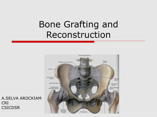
- 19. Surgical Anatomy: Iliac crest Hip Bone: Made of three bones fused in a Y-shaped epiphysis involving the acetabulum, (hip joint socket), a concave hemisphere, Pubis and ischium form incomplete bony wall for pelvic cavity, their outer surface gives attachment to the thigh muscles The ilium forms a brim between the hip joint and the joint with the sacrum
- 20. Surgical Anatomy: Iliac crest The anterior 2/3 is thin bone forming the iliac fossa, posterior abdominal wall The posterior 1/3 is thick bone and carries the articular surface for the sacrum The ilium is nearly at right angle to the other two bones
- 21. Surgical Anatomy: Iliac crest The outer surface rises wedge-shaped along an anterior border to the anterior superior iliac spine Behind the acetabulum, it passes up as a thick bar of weight-bearing bone and curve back to the posterior superior iliac spine It is the attachment of the muscles of the buttock, Gluteus minimus, medius and maximus
- 22. Surgical Anatomy: Iliac crest The upper border between the anterior and posterior superior iliac spines , the iliac crest, has a bold upward convexity and curve from front backward in a sinuous bend The anterior part is curved outwards and it’s external rim has a more prominent convexity behind the anterior superior iliac crest spine, the iliac tubercle
- 23. Surgical Anatomy: Iliac crest The gluteal surface: Convex in front, concave behind, conforming to the curvature of the iliac crest The anterior border: Shows a gentle S- shaped bend Sartorius muscle is attached a finger breadth below the anterior spine The posterior part of the crest is thicker than the rest
- 24. Surgical Anatomy: Iliac crest The inner surface: The iliac fossa, shows a gentle concavity and is paper thin in it’s deepest part The lower 2/3 is bare bone The iliacus muscle and fascia are attached to the inner lip of the crest over the whole area
- 25. Surgical Anatomy: Iliac crest Bone harvesting: The lateral approach to the anterior ilium affect the gait the most The medial anterior approach involve the large iliacus muscle which is not necessary for normal gait but large medial haematoma might produce gait disturbances
- 26. Surgical Anatomy: Iliac crest Surgical access: Incision should be placed 1 cm posterior to the anterior superior spine and extend to the iliac tubercle It should be placed lateral to the bony prominence to prevent irritation by tight cloths or belt Proceed down to bone medial to the muscles, tensor fascia lata and gluteus medius and lateral to the iliacus and the external abdominal muscles
- 27. Surgical Anatomy: Iliac crest Cancellous bone is available in the anterior ilium within the upper 2 – 3 cm and between the tubercle and the anterior superior spine, IliacIliac crest graftcrest graft. “Trap door” is one of the most common osteotomy used for anterior ilium harvest During closure, strict attention should be followed in order to reorient and reposition the muscles in their original positions A drain is required to because of the dead space and should be placed within the bony cavity
- 28. The tibia
- 29. Surgical Anatomy: The tibia Is the largest and medial bone of the lower leg, has a large upper end and a smaller lower one The shaft is vertical and triangular in cross-section Its anterior and posterior borders with the medial surface between them are subcutaneous
- 30. Surgical Anatomy: The tibia The anterior border is sharp above and blunt below where it continue with medial malleolus The posterior border is blunt and run down into the posterior border of the medial malleolus On the fibular side it has a sharp interosseous border
- 31. Surgical Anatomy: The tibia The upper end: Expand widely with prominent tuberosity projecting anteriorly from its lower part The surface bone is a very thin compact-type which is fragile around the margins
- 32. Surgical Anatomy: The tibia The superior articular surface or plateau shows a pair of condylar concavity to articulate with meniscus and the condyle of the femur Between the condylar surfaces, the plateau is elevated into intercondylar eminence and grooved by the medial and lateral tubercles
- 33. Surgical Anatomy: The tibia The lower end: Is rectangular in section Medially, it is subcutaneous, anteriorly, it is bare bone Laterally, the surface is triangular and articulate with the fibula The extensive subcutaneous surface of the tibia makes it an accessible donor site for bone grafts
- 34. Surgical Anatomy: The tibia Bone harvesting: The tibial plateau is an excellent reservoir for cancellous bone It can provide up to 40 cc of bone without affecting the structural support of the tibia Indication: Small bony defects Non-union, Osteotomy defects Dentoalveolar defects Sinus lift procedure
- 35. Surgical Anatomy: The tibia Surgical access: Could be done under local anaesthesia and conscious sedation Incision over the lateral tubercle best accomplished by flexing the leg at the knee joint It is 6 – 10 mm from the skin and dissection is made through the thin subcutaneous tissue Sharp dissection to reflect the tensor fascia lata band and make 1 cm opening into the cortex and the cancellous bone could be harvested lateral and inferior to the midline to avoid damage to the knee
- 36. THANK YOU