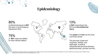
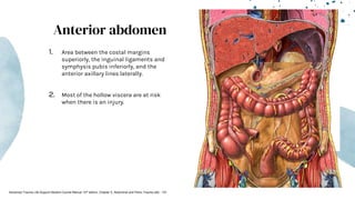
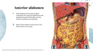
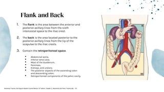
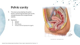
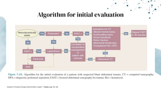
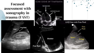
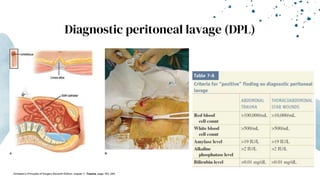
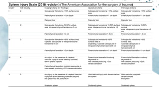
Blunt Abdominal
Trauma
Done by: Dr. Faisal Rawagah
General Surgeon and Intensivist
Jordan University Hospital
Outline
01
Introduction and
Epidemiology
02
Anatomy of the
abdomen
03
Pathophysiologic
mechanism
04
Management
• Initial evaluation
• Liver
• Spleen
05
Bleeding and
Coagulopathy
Introduction
Blunt abdominal trauma (BAT) can be hidden;
presence of distracting injuries, altered mental
status, head injury or intoxication.
01
Epidemiology
75%
of BAT cases are related
to motor vehicle collision.
The spleen and liver are the most
commonly injured.
The pancreas, bowel and
mesentery, bladder, and
diaphragm, as well as
retroperitoneal structures (kidneys,
abdominal aorta), are less common.
13%
of BAT presenting to the
ED have intra-abdominal
injury.
80%
of abdominal injuries is BAT
that seen in the Emergency
Department (ED).
-Blunt abdominal traumaInitial evaluation and management of blunt abdominal trauma in adults Authors:Deborah B Diercks, MD, MScSamuel O Clarke, MD, MASSection Editors:Maria E Moreira, MDBharti Khurana,
MDDeputy Editor:Jonathan Grayzel, MD, FAAEM (Uptodate Aug 11, 2021)
Anatomy of the Abdomen
02
Advanced Trauma Life Support Student Course Manual 10th edition. Chapter 5, Abdominal and Pelvic Trauma p82 - 101
Diaphragm
Left kidney
Left suprarenal gland
Spleen
Splenic artery
Diaphragm
Thoracoabdomen
● Area inferior to the nipple
line anteriorly and the
infrascapular line
posteriorly, and superior to
the costal margins.
● Diaphragm, Liver, Spleen,
and Stomach.
Advanced Trauma Life Support Student Course Manual 10th edition. Chapter 5, Abdominal and Pelvic Trauma p82 - 101
Body of pancreas
Thoracoabdomen
● Area inferior to the nipple
line anteriorly and the
infrascapular line
posteriorly, and superior to
the costal margins.
● Diaphragm, Liver, Spleen,
and Stomach.
Advanced Trauma Life Support Student Course Manual 10th edition. Chapter 5, Abdominal and Pelvic Trauma p82 - 101
Diaphragm
Transverse mesocolon
Transverse colon
Diaphragm
Thoracoabdomen
● Area inferior to the nipple
line anteriorly and the
infrascapular line
posteriorly, and superior to
the costal margins.
● Diaphragm, Liver, Spleen,
and Stomach.
Advanced Trauma Life Support Student Course Manual 10th edition. Chapter 5, Abdominal and Pelvic Trauma p82 - 101
Spleen
Transverse colon
Liver
Stomach
Thoracoabdomen
● Area inferior to the nipple
line anteriorly and the
infrascapular line
posteriorly, and superior to
the costal margins.
● Diaphragm, Liver, Spleen,
and Stomach.
Advanced Trauma Life Support Student Course Manual 10th edition. Chapter 5, Abdominal and Pelvic Trauma p82 - 101
Spleen
Liver
Diaphragm
Stmach
Thoracoabdomen
● Area inferior to the nipple
line anteriorly and the
infrascapular line
posteriorly, and superior to
the costal margins.
● Diaphragm, Liver, Spleen,
and Stomach.
Advanced Trauma Life Support Student Course Manual 10th edition. Chapter 5, Abdominal and Pelvic Trauma p82 - 101
Liver
Anterior abdominal wall
Stomach
Thoracoabdomen
● Area inferior to the nipple
line anteriorly and the
infrascapular line
posteriorly, and superior to
the costal margins.
● Diaphragm, Liver, Spleen,
and Stomach.
Advanced Trauma Life Support Student Course Manual 10th edition. Chapter 5, Abdominal and Pelvic Trauma p82 - 101
Anterior abdomen
1. Area between the costal margins
superiorly, the inguinal ligaments and
symphysis pubis inferiorly, and the
anterior axillary lines laterally.
2. Most of the hollow viscera are at risk
when there is an injury.
Advanced Trauma Life Support Student Course Manual 10th edition. Chapter 5, Abdominal and Pelvic Trauma p82 - 101
Flank and Back
1. The flank is the area between the anterior and
posterior axillary lines from the sixth
intercostal space to the iliac crest.
2. The back is the area located posterior to the
posterior axillary lines from the tip of the
scapulae to the iliac crests.
3. Contain the retroperitoneal space.
• Abdominal aorta;
• Inferior vena cava;
• Most of the duodenum,
• Pancreas,
• Kidneys, and ureters;
• The posterior aspects of the ascending colon
and descending colon;
• Retroperitoneal components of the pelvic cavity.
Advanced Trauma Life Support Student Course Manual 10th edition. Chapter 5, Abdominal and Pelvic Trauma p82 - 101
Pelvic cavity
1. The area surrounded by the pelvic
bones, containing the lower part of the
retroperitoneal and intraperitoneal
spaces.
2. Contains:
• Rectum
• Bladder,
• Iliac vessels,
• Female internal reproductive organs.
Advanced Trauma Life Support Student Course Manual 10th edition. Chapter 5, Abdominal and Pelvic Trauma p82 - 101
Pathophysiologic
mechanism
03
Direct blow
1. Compression and crushing injuries
to abdominopelvic viscera and
pelvic bones.
2. Deform solid and hollow organs
Advanced Trauma Life Support Student Course Manual 10th edition. Chapter 5, Abdominal and Pelvic Trauma p82 - 101
Deceleration injuries
1. Differential movement of fixed and
mobile parts of the body.
2. lacerations of the liver and spleen, both
movable organs that are fixed at the sites
of their supporting ligaments.
3. Bucket handle injuries.
-Schwartz’s Principles of Surgery Eleventh Edition: chapter 7- Trauma, page 183- 249
-Advanced Trauma Life Support Student Course Manual 10th edition. Chapter 5, Abdominal and Pelvic Trauma p82 - 101
High energy transfer injuries vs Low
energy transfer injuries.
1. High energy transfer injuries: Motor vehicle collisions in which the car’s change of velocity (ΔV)
exceeds 20 mph (32 km/h) or in which the patient has been ejected, motorcycle collisions, and falls
from heights >20 ft (6 m).
2. Low energy transfer injuries: Club or falling from a bicycle.
-Schwartz’s Principles of Surgery Eleventh Edition: chapter 7- Trauma, page 183- 249
Management
04
Algorithm for initial evaluation
-Schwartz’s Principles of Surgery Eleventh Edition: chapter 7- Trauma, page 183- 249
Focused
assessment with
sonography in
trauma (FAST)
Diagnostic peritoneal lavage (DPL)
-Schwartz’s Principles of Surgery Eleventh Edition: chapter 7- Trauma, page 183- 249
Contrast-enhanced ultrasound (CEUS)
-Spahn DR, Bouillon B, Cerny V, Duranteau J, Filipescu D, Hunt BJ, Komadina R,
Maegele M, Nardi G, Riddez L, Samama CM, Vincent JL, Rossaint R. The
European guideline on management of major bleeding and coagulopathy following
trauma: fifth edition. Crit Care. 2019 Mar 27;23(1):98. doi: 10.1186/s13054-019-
2347-3. PMID: 30917843; PMCID: PMC6436241.
-https://www.semanticscholar.org/paper/Contrast-enhanced-ultrasound-(CEUS)-
in-blunt-Miele-Piccolo/7eb01553b5cae61d4451facdad5bea1900385b71/figure/3
Liver
1. Nonoperative management of solid organ injuries is pursued in hemodynamically stable patients
who do not have overt peritonitis or other indications for laparotomy.
2. Patients with >grade II injuries should be admitted to the SICU with:
a. Hemodynamic monitoring,
b. Determination of hemoglobin,
c. Serial Abdominal examination
3. The only absolute contraindication to nonoperative management is hemodynamic instability from
intraperitoneal hemorrhage.
4. Factors may predict complications or failure of nonoperative management:
a. High injury grade,
b. Large hemoperitoneum,
c. Contrast extravasation,
d. Pseudoaneurysms.
5. Angioembolization and endoscopic retrograde cholangiopancreatography (ERCP) are useful adjuncts
that can improve the success rate of nonoperative management
6. The indication for angiography (Angioembolization): Transfusion of 4 units of RBCs in 6 hours or 6
units of RBCs in 24 hours.
-Schwartz’s Principles of Surgery Eleventh Edition: chapter 7- Trauma, page 183- 249
Liver Injury Scale (2018 revision) (The American Association for the surgery of trauma)
AAST
Grade
AIS
Severity
Imaging Criteria (CT Findings) Operative Criteria Pathologic Criteria
I 2 Subcapsular hematoma <10% surface area Subcapsular hematoma <10% surface area Subcapsular hematoma <10% surface area
Parenchymal laceration <1 cm depth Parenchymal laceration <1 cm depth Parenchymal laceration <1 cm depth
Capsular tear Capsular tear
II 2 Subcapsular hematoma 10-50% surface area; intraparenchymal
hematoma <10 cm in diameter
Subcapsular hematoma 10-50% surface area;
intraparenchymal hematoma <10 cm in
diameter
Subcapsular hematoma 10-50% surface
area; intraparenchymal hematoma
<10 cm in diameter
Laceration 1-3 cm in depth and
≤10 cm length
Laceration 1-3 cm in depth and
≤10 cm length
Laceration 1-3 cm in depth and
≤10 cm length
III 3 Subcapsular hematoma >50% surface area; ruptured
subcapsular or parenchymal hematoma
Subcapsular hematoma >50% surface area or
expanding; ruptured subcapsular or
parenchymal hematoma
Subcapsular hematoma >50% surface area;
ruptured subcapsular or intraparenchymal
hematoma
Intrparenchymal laceration >10 cm Intraparenchymal hematoma >10 cm Intraparenchymal hematoma >10 cm
Laceration >3 cm depth Laceration >3 cm depth Laceration >3 cm depth
Any injury in the presence of a liver vascular injury or active
bleeding contained within liver parenchyma
IV 4 Parenchymal disruption involving 25-75% of a hepatic lobe Parenchymal disruption involving
25-75% of a hepatic lobe
Parenchymal disruption involving
25-75% of a hepatic lobe
Active bleeding extending beyond the liver parenchyma into the
peritoneum
V 5 Parenchymal disruption >75% of hepatic lobe Parenchymal disruption >75% of hepatic lobe Parenchymal disruption >75% of hepatic lobe
Juxtahepatic venous injury to include
retrohepatic vena cava and central
major hepatic veins
Juxtahepatic venous injury to include
retrohepatic vena cava and central
major hepatic veins
Juxtahepatic venous injury to include
retrohepatic vena cava and central
major hepatic veins
Spleen
1. Nonoperative management has become the preferred means of splenic salvage for all patients.
2. The success of nonoperative management is reported to be 70%–90% for children and 40%–50% for
adults.
3. Criteria for nonoperative management:
a. Hemodynamic stability,
b. Documented CT classification of injury,
c. Absence of additional injuries necessitating surgery,
d. Transfusion of two or fewer units of red blood cells
4. Risk factor for failure of nonoperative management: Contrast extravasation.
5. Angioembolization warranted in grade III and higher injuries, particularly those with contrast blush.
6. Indications for early intervention in adults include initiation of blood transfusion within the first 12
hours and hemodynamic instability.
7. Delayed hemorrhage or rupture of the spleen can occur up to weeks after injury.
-Schwartz’s Principles of Surgery Eleventh Edition: chapter 7- Trauma, page 183- 249
-Fracker M. The spleen. In: Mulholland MW, Greenfield LJ, eds. Greenfield’s Surgery: Scientific Principles & Practice. 5th ed. Philadelphia, PA: Wolters Kluwer/Lippincott Williams & Wilkins; 2011.
-Demetriades D, Lam L. Splenectomy and splenorrhaphy. In: Fisher JE, Bland KE, eds. Mastery of Surgery. 6th ed. Philadelphia: Lippincott Williams & Wilkins; 2012.
Operative management
1. Operative management: splenectomy, partial splenectomy, or splenic repair (splenorrhaphy).
2. Splenectomy: in unstable patients or those with severe head injuries.
3. Immediate postsplenectomy increase in platelets and WBCs is normal.
4. Beyond postoperative day 5: WBC count above 15,000/mm3 and a platelet/WBC ratio of <20 are
associated with sepsis.
5. Postsplenectomy complication:
a. Subphrenic abscess
b. pancreatic tail injury (pancreatic ascites or fistula)
6. Overwhelming post-splenectomy infection (OPSI)
a. Encapsulated bacteria: Streptococcus pneumoniae, Haemophilus influenzae, and Neisseria meningitidis
b. Vaccines administered optimally at >14 days postinjury.
-Schwartz’s Principles of Surgery Eleventh Edition: chapter 7- Trauma, page 183- 249
-Fracker M. The spleen. In: Mulholland MW, Greenfield LJ, eds. Greenfield’s Surgery: Scientific Principles & Practice. 5th ed. Philadelphia, PA: Wolters Kluwer/Lippincott Williams & Wilkins; 2011.
-Demetriades D, Lam L. Splenectomy and splenorrhaphy. In: Fisher JE, Bland KE, eds. Mastery of Surgery. 6th ed. Philadelphia: Lippincott Williams & Wilkins; 2012.
How to minimize the risk of OPSI?
1. Only one-third of the spleen is required for retention of its immunologic benefit.
2. Autologous splenic transplantation
3. Partial splenectomy, or splenic repair (splenorrhaphy).
4. No grade restriction for performing splenorrhaphy after injury, and it may be done as long as one-
third of the spleen remains viable.
-Schwartz’s Principles of Surgery Eleventh Edition: chapter 7-
Trauma, page 183- 249
-Fracker M. The spleen. In: Mulholland MW, Greenfield LJ, eds.
Greenfield’s Surgery: Scientific Principles & Practice. 5th ed.
Philadelphia, PA: Wolters Kluwer/Lippincott Williams & Wilkins;
2011.
-Demetriades D, Lam L. Splenectomy and splenorrhaphy. In: Fisher
JE, Bland KE, eds. Mastery of Surgery. 6th ed. Philadelphia:
Lippincott Williams & Wilkins; 2012.
Spleen Injury Scale (2018 revision) (The American Association for the surgery of trauma)
Grade* AIS Severity Imaging Criteria (CT Findings) Operative Criteria Pathologic Criteria
I 2 Subcapsular hematoma <10% surface area Subcapsular hematoma <10% surface
area
Subcapsular hematoma <10% surface
area
Parenchymal laceration <1 cm depth Parenchymal laceration <1 cm depth Parenchymal laceration <1 cm depth
Capsular tear Capsular tear Capsular tear
II 2 Subcapsular hematoma 10-50% surface
area; intraparenchymal hematoma <5 cm
Subcapsular hematoma 10-50% surface
area; intraparenchymal hematoma <5
cm
Subcapsular hematoma 10-50%
surface
area; intraparenchymal hematoma <5
cm
Parenchymal laceration 1-3 cm Parenchymal laceration 1-3 cm Parenchymal laceration 1-3 cm
III 3 Subcapsular hematoma >50% surface area;
ruptured subcapsular or intraparenchymal
hematoma ≥5 cm
Subcapsular hematoma >50%
surface area or
expanding; ruptured subcapsular or
intraparenchymal hematoma ≥5 cm
Subcapsular hematoma >50%
surface area;
ruptured subcapsular or
intraparenchymal
hematoma ≥5 cm
Parenchymal laceration >3 cm depth Parenchymal laceration >3 cm depth Parenchymal laceration >3 cm depth
IV 4 Any injury in the presence of a splenic
vascular injury or active bleeding confined
within splenic capsule
Parenchymal laceration involving
segmental or
hilar vessels producing >25%
devascularization
Parenchymal laceration involving
segmental or
hilar vessels producing >25%
devascularization
Parenchymal laceration involving segmental or
hilar vessels producing >25% devascularization
V 5 Any injury in the presence of a splenic vascular
injury with active bleeding extended beyond
the spleen into the peritoneum
Hilar vascular injury with devascularizes
the spleen
Hilar vascular injury with
devascularizes
the spleen
Shattered spleen Shattered spleen Shattered spleen
Bleeding and
Coagulopathy
05
Goal-directed therapy coagulation
management
1. Fresh frozen plasma- based management
Guided by standard laboratory coagulation screening parameters (PT and/or APTT > 1.5 times normal and/or
viscoelastic evidence of a coagulation factor deficiency)
2. Coagulation factor concentrate- based management (PCC)
INR 2–4.0 ; 25 U/kg
INR 4–6.0 ; 35 U/kg
INR is > 6.0 ; 50 U/kg
3. Fibrinogen supplementation (fibrinogen level ≤ 1.5 g/L)
15–20 single-donor units of cryoprecipitate
3–4 g fibrinogen concentrate
4. Platelets Keep platelet count above - 50 × 10⁹/L.
patients with ongoing bleeding and/or TBI - 100 × 10⁹/L
5. Calcium (Ionized calcium levels be monitored)
Keep in the normal range (4.1-4.9 mg/dL)
6. Recombinant activated coagulation factor VII
not recommend as first-line treatment
Spahn DR, Bouillon B, Cerny V, Duranteau J, Filipescu D, Hunt BJ, Komadina R, Maegele M, Nardi G, Riddez L, Samama CM, Vincent JL, Rossaint R. The European guideline on management of major bleeding and coagulopathy following trauma: fifth edition.
Crit Care. 2019 Mar 27;23(1):98. doi: 10.1186/s13054-019-2347-3. PMID: 30917843; PMCID: PMC6436241.
-Schwartz’s Principles of Surgery Eleventh Edition: chapter 7- Trauma, page 183- 249
Thromboelastogram
1. R-time (5-10 min)
Coltting Factars / Anticoagulants
2. K(Kinetics) (1-3min)
Fibrinogen
3. α-Angle (50°-70°)
Rate of Clot formation
4. MA (Max. Amplitude)(55-73 mm)
Platelets / Fibrin / Factar II,III
5. Ly30 (0-8%)
Fibrinolysis
Algorithm for the management of trauma-induced hemorrhage
without viscoelastic testing
-Casu S Simplified treatment algorithm for the management of trauma-induced hemorrhage without viscoelastic testing Trauma Surgery & Acute Care Open 2021;6:e000779. doi: 10.1136/tsaco-2021-000779
References
• Blunt abdominal traumaInitial evaluation and management of blunt abdominal trauma in adults Authors:Deborah B
Diercks, MD, MScSamuel O Clarke, MD, MASSection Editors:Maria E Moreira, MDBharti Khurana, MDDeputy Editor:Jonathan
Grayzel, MD, FAAEM (Uptodate Aug 11, 2021)
• Advanced Trauma Life Support Student Course Manual 10th edition. Chapter 5, Abdominal and Pelvic Trauma p82 - 101
• Schwartz’s Principles of Surgery Eleventh Edition: chapter 7- Trauma, page 183- 249
• Spahn DR, Bouillon B, Cerny V, Duranteau J, Filipescu D, Hunt BJ, Komadina R, Maegele M, Nardi G, Riddez L, Samama CM,
Vincent JL, Rossaint R. The European guideline on management of major bleeding and coagulopathy following trauma: fifth
edition. Crit Care. 2019 Mar 27;23(1):98. doi: 10.1186/s13054-019-2347-3. PMID: 30917843; PMCID: PMC6436241.
• https://www.semanticscholar.org/paper/Contrast-enhanced-ultrasound-(CEUS)-in-blunt-Miele-
Piccolo/7eb01553b5cae61d4451facdad5bea1900385b71/figure/3
• The American Association for the surgery of trauma
• Fracker M. The spleen. In: Mulholland MW, Greenfield LJ, eds. Greenfield’s Surgery: Scientific Principles & Practice. 5th ed.
Philadelphia, PA: Wolters Kluwer/Lippincott Williams & Wilkins; 2011.
• Demetriades D, Lam L. Splenectomy and splenorrhaphy. In: Fisher JE, Bland KE, eds. Mastery of Surgery. 6th ed. Philadelphia:
Lippincott Williams & Wilkins; 2012.
• Casu S Simplified treatment algorithm for the management of trauma-induced hemorrhage without viscoelastic testing
Trauma Surgery & Acute Care Open 2021;6:e000779. doi: 10.1136/tsaco-2021-000779
Blunt Abdominal Trauma

Blunt Abdominal Trauma

  • 1.
    Blunt Abdominal Trauma Done by:Dr. Faisal Rawagah General Surgeon and Intensivist Jordan University Hospital
  • 2.
    Outline 01 Introduction and Epidemiology 02 Anatomy ofthe abdomen 03 Pathophysiologic mechanism 04 Management • Initial evaluation • Liver • Spleen 05 Bleeding and Coagulopathy
  • 3.
    Introduction Blunt abdominal trauma(BAT) can be hidden; presence of distracting injuries, altered mental status, head injury or intoxication. 01
  • 4.
    Epidemiology 75% of BAT casesare related to motor vehicle collision. The spleen and liver are the most commonly injured. The pancreas, bowel and mesentery, bladder, and diaphragm, as well as retroperitoneal structures (kidneys, abdominal aorta), are less common. 13% of BAT presenting to the ED have intra-abdominal injury. 80% of abdominal injuries is BAT that seen in the Emergency Department (ED). -Blunt abdominal traumaInitial evaluation and management of blunt abdominal trauma in adults Authors:Deborah B Diercks, MD, MScSamuel O Clarke, MD, MASSection Editors:Maria E Moreira, MDBharti Khurana, MDDeputy Editor:Jonathan Grayzel, MD, FAAEM (Uptodate Aug 11, 2021)
  • 5.
    Anatomy of theAbdomen 02 Advanced Trauma Life Support Student Course Manual 10th edition. Chapter 5, Abdominal and Pelvic Trauma p82 - 101
  • 6.
    Diaphragm Left kidney Left suprarenalgland Spleen Splenic artery Diaphragm Thoracoabdomen ● Area inferior to the nipple line anteriorly and the infrascapular line posteriorly, and superior to the costal margins. ● Diaphragm, Liver, Spleen, and Stomach. Advanced Trauma Life Support Student Course Manual 10th edition. Chapter 5, Abdominal and Pelvic Trauma p82 - 101
  • 7.
    Body of pancreas Thoracoabdomen ●Area inferior to the nipple line anteriorly and the infrascapular line posteriorly, and superior to the costal margins. ● Diaphragm, Liver, Spleen, and Stomach. Advanced Trauma Life Support Student Course Manual 10th edition. Chapter 5, Abdominal and Pelvic Trauma p82 - 101
  • 8.
    Diaphragm Transverse mesocolon Transverse colon Diaphragm Thoracoabdomen ●Area inferior to the nipple line anteriorly and the infrascapular line posteriorly, and superior to the costal margins. ● Diaphragm, Liver, Spleen, and Stomach. Advanced Trauma Life Support Student Course Manual 10th edition. Chapter 5, Abdominal and Pelvic Trauma p82 - 101 Spleen
  • 9.
    Transverse colon Liver Stomach Thoracoabdomen ● Areainferior to the nipple line anteriorly and the infrascapular line posteriorly, and superior to the costal margins. ● Diaphragm, Liver, Spleen, and Stomach. Advanced Trauma Life Support Student Course Manual 10th edition. Chapter 5, Abdominal and Pelvic Trauma p82 - 101 Spleen
  • 10.
    Liver Diaphragm Stmach Thoracoabdomen ● Area inferiorto the nipple line anteriorly and the infrascapular line posteriorly, and superior to the costal margins. ● Diaphragm, Liver, Spleen, and Stomach. Advanced Trauma Life Support Student Course Manual 10th edition. Chapter 5, Abdominal and Pelvic Trauma p82 - 101
  • 11.
    Liver Anterior abdominal wall Stomach Thoracoabdomen ●Area inferior to the nipple line anteriorly and the infrascapular line posteriorly, and superior to the costal margins. ● Diaphragm, Liver, Spleen, and Stomach. Advanced Trauma Life Support Student Course Manual 10th edition. Chapter 5, Abdominal and Pelvic Trauma p82 - 101
  • 12.
    Anterior abdomen 1. Areabetween the costal margins superiorly, the inguinal ligaments and symphysis pubis inferiorly, and the anterior axillary lines laterally. 2. Most of the hollow viscera are at risk when there is an injury. Advanced Trauma Life Support Student Course Manual 10th edition. Chapter 5, Abdominal and Pelvic Trauma p82 - 101
  • 13.
    Anterior abdomen 1. Areabetween the costal margins superiorly, the inguinal ligaments and symphysis pubis inferiorly, and the anterior axillary lines laterally. 2. Most of the hollow viscera are at risk when there is an injury. Advanced Trauma Life Support Student Course Manual 10th edition. Chapter 5, Abdominal and Pelvic Trauma p82 - 101
  • 14.
    Anterior abdomen 1. Areabetween the costal margins superiorly, the inguinal ligaments and symphysis pubis inferiorly, and the anterior axillary lines laterally. 2. Most of the hollow viscera are at risk when there is an injury. Advanced Trauma Life Support Student Course Manual 10th edition. Chapter 5, Abdominal and Pelvic Trauma p82 - 101
  • 15.
    Anterior abdomen 1. Areabetween the costal margins superiorly, the inguinal ligaments and symphysis pubis inferiorly, and the anterior axillary lines laterally. 2. Most of the hollow viscera are at risk when there is an injury. Advanced Trauma Life Support Student Course Manual 10th edition. Chapter 5, Abdominal and Pelvic Trauma p82 - 101
  • 16.
    Flank and Back 1.The flank is the area between the anterior and posterior axillary lines from the sixth intercostal space to the iliac crest. 2. The back is the area located posterior to the posterior axillary lines from the tip of the scapulae to the iliac crests. 3. Contain the retroperitoneal space. • Abdominal aorta; • Inferior vena cava; • Most of the duodenum, • Pancreas, • Kidneys, and ureters; • The posterior aspects of the ascending colon and descending colon; • Retroperitoneal components of the pelvic cavity. Advanced Trauma Life Support Student Course Manual 10th edition. Chapter 5, Abdominal and Pelvic Trauma p82 - 101
  • 17.
    Pelvic cavity 1. Thearea surrounded by the pelvic bones, containing the lower part of the retroperitoneal and intraperitoneal spaces. 2. Contains: • Rectum • Bladder, • Iliac vessels, • Female internal reproductive organs. Advanced Trauma Life Support Student Course Manual 10th edition. Chapter 5, Abdominal and Pelvic Trauma p82 - 101
  • 18.
  • 19.
    Direct blow 1. Compressionand crushing injuries to abdominopelvic viscera and pelvic bones. 2. Deform solid and hollow organs Advanced Trauma Life Support Student Course Manual 10th edition. Chapter 5, Abdominal and Pelvic Trauma p82 - 101
  • 20.
    Deceleration injuries 1. Differentialmovement of fixed and mobile parts of the body. 2. lacerations of the liver and spleen, both movable organs that are fixed at the sites of their supporting ligaments. 3. Bucket handle injuries. -Schwartz’s Principles of Surgery Eleventh Edition: chapter 7- Trauma, page 183- 249 -Advanced Trauma Life Support Student Course Manual 10th edition. Chapter 5, Abdominal and Pelvic Trauma p82 - 101
  • 21.
    High energy transferinjuries vs Low energy transfer injuries. 1. High energy transfer injuries: Motor vehicle collisions in which the car’s change of velocity (ΔV) exceeds 20 mph (32 km/h) or in which the patient has been ejected, motorcycle collisions, and falls from heights >20 ft (6 m). 2. Low energy transfer injuries: Club or falling from a bicycle. -Schwartz’s Principles of Surgery Eleventh Edition: chapter 7- Trauma, page 183- 249
  • 22.
  • 23.
    Algorithm for initialevaluation -Schwartz’s Principles of Surgery Eleventh Edition: chapter 7- Trauma, page 183- 249
  • 24.
  • 25.
    Diagnostic peritoneal lavage(DPL) -Schwartz’s Principles of Surgery Eleventh Edition: chapter 7- Trauma, page 183- 249
  • 26.
    Contrast-enhanced ultrasound (CEUS) -SpahnDR, Bouillon B, Cerny V, Duranteau J, Filipescu D, Hunt BJ, Komadina R, Maegele M, Nardi G, Riddez L, Samama CM, Vincent JL, Rossaint R. The European guideline on management of major bleeding and coagulopathy following trauma: fifth edition. Crit Care. 2019 Mar 27;23(1):98. doi: 10.1186/s13054-019- 2347-3. PMID: 30917843; PMCID: PMC6436241. -https://www.semanticscholar.org/paper/Contrast-enhanced-ultrasound-(CEUS)- in-blunt-Miele-Piccolo/7eb01553b5cae61d4451facdad5bea1900385b71/figure/3
  • 27.
    Liver 1. Nonoperative managementof solid organ injuries is pursued in hemodynamically stable patients who do not have overt peritonitis or other indications for laparotomy. 2. Patients with >grade II injuries should be admitted to the SICU with: a. Hemodynamic monitoring, b. Determination of hemoglobin, c. Serial Abdominal examination 3. The only absolute contraindication to nonoperative management is hemodynamic instability from intraperitoneal hemorrhage. 4. Factors may predict complications or failure of nonoperative management: a. High injury grade, b. Large hemoperitoneum, c. Contrast extravasation, d. Pseudoaneurysms. 5. Angioembolization and endoscopic retrograde cholangiopancreatography (ERCP) are useful adjuncts that can improve the success rate of nonoperative management 6. The indication for angiography (Angioembolization): Transfusion of 4 units of RBCs in 6 hours or 6 units of RBCs in 24 hours. -Schwartz’s Principles of Surgery Eleventh Edition: chapter 7- Trauma, page 183- 249
  • 28.
    Liver Injury Scale(2018 revision) (The American Association for the surgery of trauma) AAST Grade AIS Severity Imaging Criteria (CT Findings) Operative Criteria Pathologic Criteria I 2 Subcapsular hematoma <10% surface area Subcapsular hematoma <10% surface area Subcapsular hematoma <10% surface area Parenchymal laceration <1 cm depth Parenchymal laceration <1 cm depth Parenchymal laceration <1 cm depth Capsular tear Capsular tear II 2 Subcapsular hematoma 10-50% surface area; intraparenchymal hematoma <10 cm in diameter Subcapsular hematoma 10-50% surface area; intraparenchymal hematoma <10 cm in diameter Subcapsular hematoma 10-50% surface area; intraparenchymal hematoma <10 cm in diameter Laceration 1-3 cm in depth and ≤10 cm length Laceration 1-3 cm in depth and ≤10 cm length Laceration 1-3 cm in depth and ≤10 cm length III 3 Subcapsular hematoma >50% surface area; ruptured subcapsular or parenchymal hematoma Subcapsular hematoma >50% surface area or expanding; ruptured subcapsular or parenchymal hematoma Subcapsular hematoma >50% surface area; ruptured subcapsular or intraparenchymal hematoma Intrparenchymal laceration >10 cm Intraparenchymal hematoma >10 cm Intraparenchymal hematoma >10 cm Laceration >3 cm depth Laceration >3 cm depth Laceration >3 cm depth Any injury in the presence of a liver vascular injury or active bleeding contained within liver parenchyma IV 4 Parenchymal disruption involving 25-75% of a hepatic lobe Parenchymal disruption involving 25-75% of a hepatic lobe Parenchymal disruption involving 25-75% of a hepatic lobe Active bleeding extending beyond the liver parenchyma into the peritoneum V 5 Parenchymal disruption >75% of hepatic lobe Parenchymal disruption >75% of hepatic lobe Parenchymal disruption >75% of hepatic lobe Juxtahepatic venous injury to include retrohepatic vena cava and central major hepatic veins Juxtahepatic venous injury to include retrohepatic vena cava and central major hepatic veins Juxtahepatic venous injury to include retrohepatic vena cava and central major hepatic veins
  • 29.
    Spleen 1. Nonoperative managementhas become the preferred means of splenic salvage for all patients. 2. The success of nonoperative management is reported to be 70%–90% for children and 40%–50% for adults. 3. Criteria for nonoperative management: a. Hemodynamic stability, b. Documented CT classification of injury, c. Absence of additional injuries necessitating surgery, d. Transfusion of two or fewer units of red blood cells 4. Risk factor for failure of nonoperative management: Contrast extravasation. 5. Angioembolization warranted in grade III and higher injuries, particularly those with contrast blush. 6. Indications for early intervention in adults include initiation of blood transfusion within the first 12 hours and hemodynamic instability. 7. Delayed hemorrhage or rupture of the spleen can occur up to weeks after injury. -Schwartz’s Principles of Surgery Eleventh Edition: chapter 7- Trauma, page 183- 249 -Fracker M. The spleen. In: Mulholland MW, Greenfield LJ, eds. Greenfield’s Surgery: Scientific Principles & Practice. 5th ed. Philadelphia, PA: Wolters Kluwer/Lippincott Williams & Wilkins; 2011. -Demetriades D, Lam L. Splenectomy and splenorrhaphy. In: Fisher JE, Bland KE, eds. Mastery of Surgery. 6th ed. Philadelphia: Lippincott Williams & Wilkins; 2012.
  • 30.
    Operative management 1. Operativemanagement: splenectomy, partial splenectomy, or splenic repair (splenorrhaphy). 2. Splenectomy: in unstable patients or those with severe head injuries. 3. Immediate postsplenectomy increase in platelets and WBCs is normal. 4. Beyond postoperative day 5: WBC count above 15,000/mm3 and a platelet/WBC ratio of <20 are associated with sepsis. 5. Postsplenectomy complication: a. Subphrenic abscess b. pancreatic tail injury (pancreatic ascites or fistula) 6. Overwhelming post-splenectomy infection (OPSI) a. Encapsulated bacteria: Streptococcus pneumoniae, Haemophilus influenzae, and Neisseria meningitidis b. Vaccines administered optimally at >14 days postinjury. -Schwartz’s Principles of Surgery Eleventh Edition: chapter 7- Trauma, page 183- 249 -Fracker M. The spleen. In: Mulholland MW, Greenfield LJ, eds. Greenfield’s Surgery: Scientific Principles & Practice. 5th ed. Philadelphia, PA: Wolters Kluwer/Lippincott Williams & Wilkins; 2011. -Demetriades D, Lam L. Splenectomy and splenorrhaphy. In: Fisher JE, Bland KE, eds. Mastery of Surgery. 6th ed. Philadelphia: Lippincott Williams & Wilkins; 2012.
  • 31.
    How to minimizethe risk of OPSI? 1. Only one-third of the spleen is required for retention of its immunologic benefit. 2. Autologous splenic transplantation 3. Partial splenectomy, or splenic repair (splenorrhaphy). 4. No grade restriction for performing splenorrhaphy after injury, and it may be done as long as one- third of the spleen remains viable. -Schwartz’s Principles of Surgery Eleventh Edition: chapter 7- Trauma, page 183- 249 -Fracker M. The spleen. In: Mulholland MW, Greenfield LJ, eds. Greenfield’s Surgery: Scientific Principles & Practice. 5th ed. Philadelphia, PA: Wolters Kluwer/Lippincott Williams & Wilkins; 2011. -Demetriades D, Lam L. Splenectomy and splenorrhaphy. In: Fisher JE, Bland KE, eds. Mastery of Surgery. 6th ed. Philadelphia: Lippincott Williams & Wilkins; 2012.
  • 32.
    Spleen Injury Scale(2018 revision) (The American Association for the surgery of trauma) Grade* AIS Severity Imaging Criteria (CT Findings) Operative Criteria Pathologic Criteria I 2 Subcapsular hematoma <10% surface area Subcapsular hematoma <10% surface area Subcapsular hematoma <10% surface area Parenchymal laceration <1 cm depth Parenchymal laceration <1 cm depth Parenchymal laceration <1 cm depth Capsular tear Capsular tear Capsular tear II 2 Subcapsular hematoma 10-50% surface area; intraparenchymal hematoma <5 cm Subcapsular hematoma 10-50% surface area; intraparenchymal hematoma <5 cm Subcapsular hematoma 10-50% surface area; intraparenchymal hematoma <5 cm Parenchymal laceration 1-3 cm Parenchymal laceration 1-3 cm Parenchymal laceration 1-3 cm III 3 Subcapsular hematoma >50% surface area; ruptured subcapsular or intraparenchymal hematoma ≥5 cm Subcapsular hematoma >50% surface area or expanding; ruptured subcapsular or intraparenchymal hematoma ≥5 cm Subcapsular hematoma >50% surface area; ruptured subcapsular or intraparenchymal hematoma ≥5 cm Parenchymal laceration >3 cm depth Parenchymal laceration >3 cm depth Parenchymal laceration >3 cm depth IV 4 Any injury in the presence of a splenic vascular injury or active bleeding confined within splenic capsule Parenchymal laceration involving segmental or hilar vessels producing >25% devascularization Parenchymal laceration involving segmental or hilar vessels producing >25% devascularization Parenchymal laceration involving segmental or hilar vessels producing >25% devascularization V 5 Any injury in the presence of a splenic vascular injury with active bleeding extended beyond the spleen into the peritoneum Hilar vascular injury with devascularizes the spleen Hilar vascular injury with devascularizes the spleen Shattered spleen Shattered spleen Shattered spleen
  • 33.
  • 34.
    Goal-directed therapy coagulation management 1.Fresh frozen plasma- based management Guided by standard laboratory coagulation screening parameters (PT and/or APTT > 1.5 times normal and/or viscoelastic evidence of a coagulation factor deficiency) 2. Coagulation factor concentrate- based management (PCC) INR 2–4.0 ; 25 U/kg INR 4–6.0 ; 35 U/kg INR is > 6.0 ; 50 U/kg 3. Fibrinogen supplementation (fibrinogen level ≤ 1.5 g/L) 15–20 single-donor units of cryoprecipitate 3–4 g fibrinogen concentrate 4. Platelets Keep platelet count above - 50 × 10⁹/L. patients with ongoing bleeding and/or TBI - 100 × 10⁹/L 5. Calcium (Ionized calcium levels be monitored) Keep in the normal range (4.1-4.9 mg/dL) 6. Recombinant activated coagulation factor VII not recommend as first-line treatment Spahn DR, Bouillon B, Cerny V, Duranteau J, Filipescu D, Hunt BJ, Komadina R, Maegele M, Nardi G, Riddez L, Samama CM, Vincent JL, Rossaint R. The European guideline on management of major bleeding and coagulopathy following trauma: fifth edition. Crit Care. 2019 Mar 27;23(1):98. doi: 10.1186/s13054-019-2347-3. PMID: 30917843; PMCID: PMC6436241.
  • 35.
    -Schwartz’s Principles ofSurgery Eleventh Edition: chapter 7- Trauma, page 183- 249
  • 36.
    Thromboelastogram 1. R-time (5-10min) Coltting Factars / Anticoagulants 2. K(Kinetics) (1-3min) Fibrinogen 3. α-Angle (50°-70°) Rate of Clot formation 4. MA (Max. Amplitude)(55-73 mm) Platelets / Fibrin / Factar II,III 5. Ly30 (0-8%) Fibrinolysis
  • 37.
    Algorithm for themanagement of trauma-induced hemorrhage without viscoelastic testing -Casu S Simplified treatment algorithm for the management of trauma-induced hemorrhage without viscoelastic testing Trauma Surgery & Acute Care Open 2021;6:e000779. doi: 10.1136/tsaco-2021-000779
  • 38.
    References • Blunt abdominaltraumaInitial evaluation and management of blunt abdominal trauma in adults Authors:Deborah B Diercks, MD, MScSamuel O Clarke, MD, MASSection Editors:Maria E Moreira, MDBharti Khurana, MDDeputy Editor:Jonathan Grayzel, MD, FAAEM (Uptodate Aug 11, 2021) • Advanced Trauma Life Support Student Course Manual 10th edition. Chapter 5, Abdominal and Pelvic Trauma p82 - 101 • Schwartz’s Principles of Surgery Eleventh Edition: chapter 7- Trauma, page 183- 249 • Spahn DR, Bouillon B, Cerny V, Duranteau J, Filipescu D, Hunt BJ, Komadina R, Maegele M, Nardi G, Riddez L, Samama CM, Vincent JL, Rossaint R. The European guideline on management of major bleeding and coagulopathy following trauma: fifth edition. Crit Care. 2019 Mar 27;23(1):98. doi: 10.1186/s13054-019-2347-3. PMID: 30917843; PMCID: PMC6436241. • https://www.semanticscholar.org/paper/Contrast-enhanced-ultrasound-(CEUS)-in-blunt-Miele- Piccolo/7eb01553b5cae61d4451facdad5bea1900385b71/figure/3 • The American Association for the surgery of trauma • Fracker M. The spleen. In: Mulholland MW, Greenfield LJ, eds. Greenfield’s Surgery: Scientific Principles & Practice. 5th ed. Philadelphia, PA: Wolters Kluwer/Lippincott Williams & Wilkins; 2011. • Demetriades D, Lam L. Splenectomy and splenorrhaphy. In: Fisher JE, Bland KE, eds. Mastery of Surgery. 6th ed. Philadelphia: Lippincott Williams & Wilkins; 2012. • Casu S Simplified treatment algorithm for the management of trauma-induced hemorrhage without viscoelastic testing Trauma Surgery & Acute Care Open 2021;6:e000779. doi: 10.1136/tsaco-2021-000779