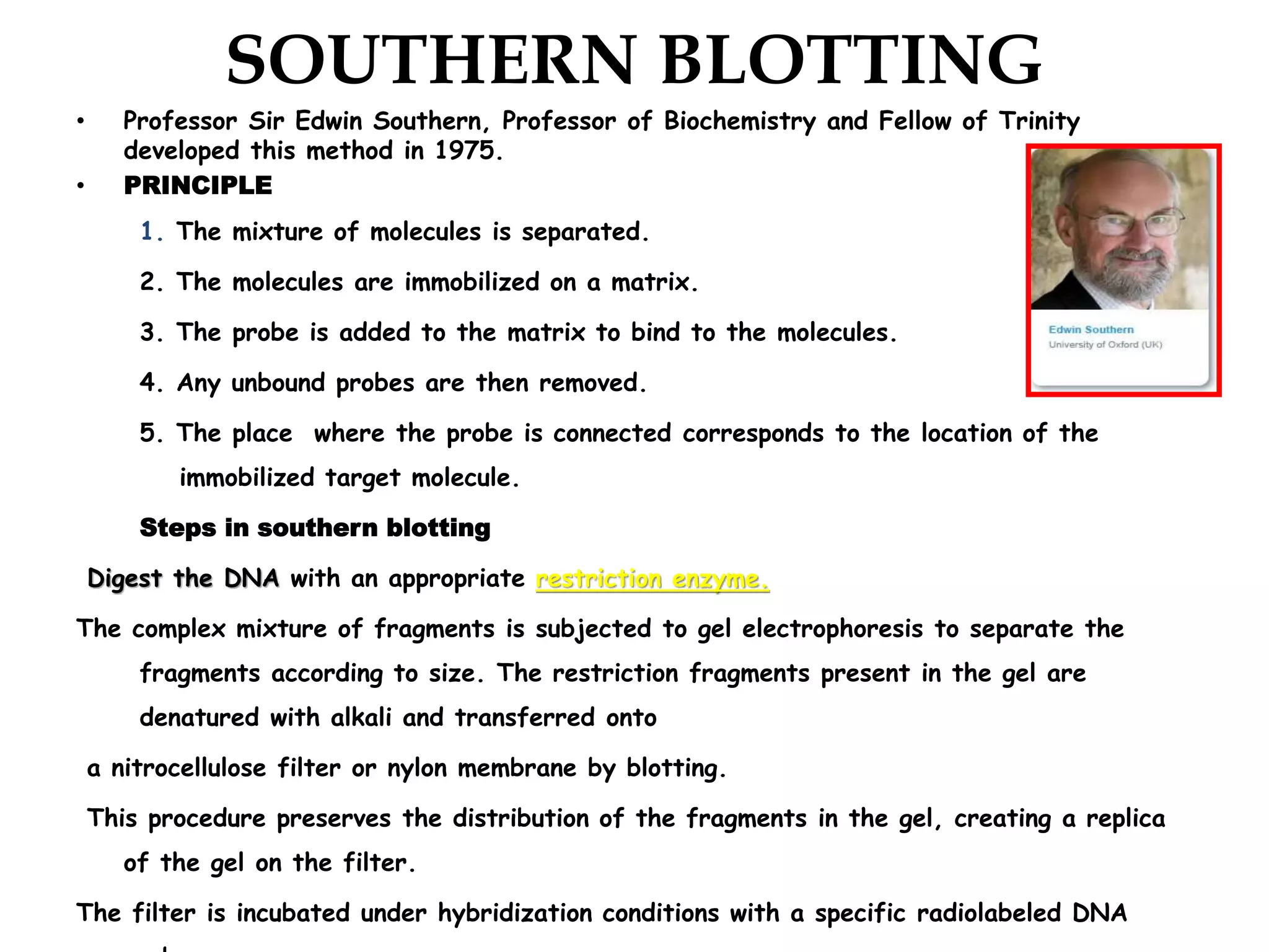
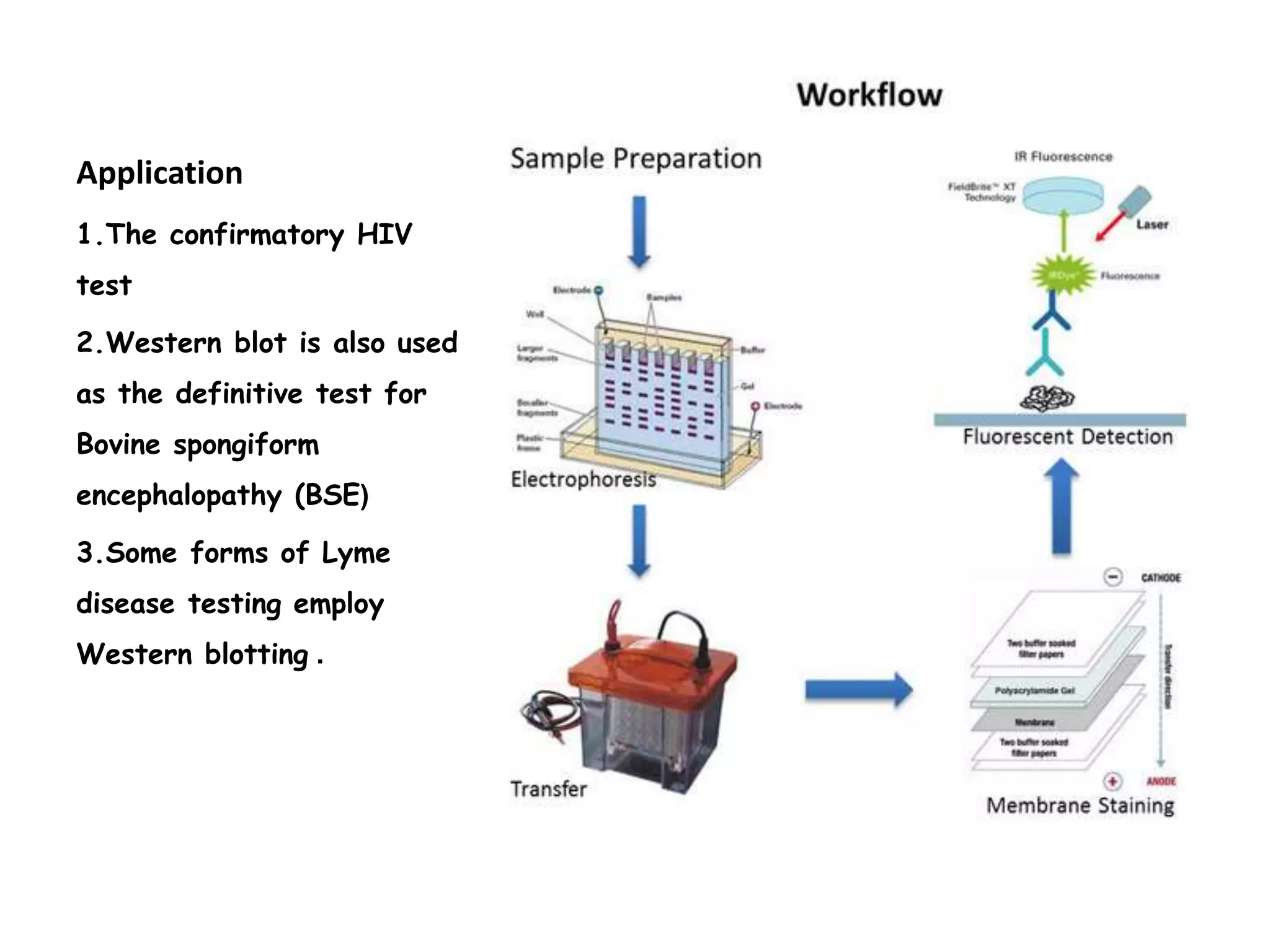
Southern blotting is a technique developed by Professor Edwin Southern in 1975 to detect specific DNA sequences. It involves separating DNA fragments by size, transferring them to a membrane, then using a probe to detect the bound fragment. It is used for gene mapping, evolution studies, and DNA fingerprinting. Northern blotting detects specific RNA sequences and was developed in 1979. RNA is separated by size and transferred to a membrane, then a probe detects bound RNA fragments. Western blotting detects specific proteins and was developed in 1981. Proteins are separated by gel electrophoresis, transferred to a membrane, and a primary antibody binds the target protein, which is then detected by a secondary antibody.
Nucleic Acid Quantification Methods - DNA / RNA Quantificationajithnandanam
Nucleic acids are quantified to check the concentration and purity of DNA/RNA present in the solution mixture.it is important to know the concentration and purity of the nucleic acid for the use in further applications like PCR, restriction digestion etc. Spectrophotometric analysis is the most commonly used method of quantifying DNA, agarose gel electrophoresis can also be used to analyse the DNA sample for purity.
Restriction Fragment Length Polymorphism (RFLP)
These are bacterial enzymes used by scientists to cut DNA molecules at known locations. RFLPs (pronounced "rif lips") are used as markers on genetic maps. Typically, gel electrophoresis is used to visualize RFLPs.
Techniques of DNA Extraction, Purification and QuantificationBHUMI GAMETI
Introduction
The overall process…
Uses of isolated genomic DNA
Extraction of DNA from plant material
Components of DNA extraction solutions
Cell Lysis or Cell disruption :
Purification of DNA
CTAB Method
Phenol–chloroform extraction
PROTEINASE K
Salting out
Silica adsorption method
Magnetic beads
FTA Paper
Nucleic acid quantification
Agarose Gel Electrophoresis
UV spectroscopy
DNA quantification using NanoDrop
Sanger sequencing is one of the DNA sequencing methods used to identify and determine the sequence (Nucleotide) of DNA .This is an enzymatic method of sequencing developed by Fred Sanger.
Concept: reannealing nucleic acids to identify sequence of interest.
Separates DNA/RNA in an agarose gel, then detects specific bands using probe and hybridization.
Hybridization takes advantage of the ability of a single stranded DNA or RNA molecule to find its complement, even in the presence of large amounts of unrelated DNA.
Allows detection of specific bands (DNA fragments or RNA molecules) that have complementary sequence to the probe.
Size bands and quantify abundance of molecule.
Define DNA fingerprint and DNA fingerprinting.
Explain some terms related to DNA fingerprinting.
Describe the method of collection and preservation of biological samples.
Describe the uses of DNA fingerprinting.
Describe the types of DNA fingerprinting.
Describe the steps of DNA fingerprinting.
Cancer cell metabolism: special Reference to Lactate PathwayAADYARAJPANDEY1
Normal Cell Metabolism:
Cellular respiration describes the series of steps that cells use to break down sugar and other chemicals to get the energy we need to function.
Energy is stored in the bonds of glucose and when glucose is broken down, much of that energy is released.
Cell utilize energy in the form of ATP.
The first step of respiration is called glycolysis. In a series of steps, glycolysis breaks glucose into two smaller molecules - a chemical called pyruvate. A small amount of ATP is formed during this process.
Most healthy cells continue the breakdown in a second process, called the Kreb's cycle. The Kreb's cycle allows cells to “burn” the pyruvates made in glycolysis to get more ATP.
The last step in the breakdown of glucose is called oxidative phosphorylation (Ox-Phos).
It takes place in specialized cell structures called mitochondria. This process produces a large amount of ATP. Importantly, cells need oxygen to complete oxidative phosphorylation.
If a cell completes only glycolysis, only 2 molecules of ATP are made per glucose. However, if the cell completes the entire respiration process (glycolysis - Kreb's - oxidative phosphorylation), about 36 molecules of ATP are created, giving it much more energy to use.
IN CANCER CELL:
Unlike healthy cells that "burn" the entire molecule of sugar to capture a large amount of energy as ATP, cancer cells are wasteful.
Cancer cells only partially break down sugar molecules. They overuse the first step of respiration, glycolysis. They frequently do not complete the second step, oxidative phosphorylation.
This results in only 2 molecules of ATP per each glucose molecule instead of the 36 or so ATPs healthy cells gain. As a result, cancer cells need to use a lot more sugar molecules to get enough energy to survive.
introduction to WARBERG PHENOMENA:
WARBURG EFFECT Usually, cancer cells are highly glycolytic (glucose addiction) and take up more glucose than do normal cells from outside.
Otto Heinrich Warburg (; 8 October 1883 – 1 August 1970) In 1931 was awarded the Nobel Prize in Physiology for his "discovery of the nature and mode of action of the respiratory enzyme.
WARNBURG EFFECT : cancer cells under aerobic (well-oxygenated) conditions to metabolize glucose to lactate (aerobic glycolysis) is known as the Warburg effect. Warburg made the observation that tumor slices consume glucose and secrete lactate at a higher rate than normal tissues.
Professional air quality monitoring systems provide immediate, on-site data for analysis, compliance, and decision-making.
Monitor common gases, weather parameters, particulates.
Introduction:
RNA interference (RNAi) or Post-Transcriptional Gene Silencing (PTGS) is an important biological process for modulating eukaryotic gene expression.
It is highly conserved process of posttranscriptional gene silencing by which double stranded RNA (dsRNA) causes sequence-specific degradation of mRNA sequences.
dsRNA-induced gene silencing (RNAi) is reported in a wide range of eukaryotes ranging from worms, insects, mammals and plants.
This process mediates resistance to both endogenous parasitic and exogenous pathogenic nucleic acids, and regulates the expression of protein-coding genes.
What are small ncRNAs?
micro RNA (miRNA)
short interfering RNA (siRNA)
Properties of small non-coding RNA:
Involved in silencing mRNA transcripts.
Called “small” because they are usually only about 21-24 nucleotides long.
Synthesized by first cutting up longer precursor sequences (like the 61nt one that Lee discovered).
Silence an mRNA by base pairing with some sequence on the mRNA.
Discovery of siRNA?
The first small RNA:
In 1993 Rosalind Lee (Victor Ambros lab) was studying a non- coding gene in C. elegans, lin-4, that was involved in silencing of another gene, lin-14, at the appropriate time in the
development of the worm C. elegans.
Two small transcripts of lin-4 (22nt and 61nt) were found to be complementary to a sequence in the 3' UTR of lin-14.
Because lin-4 encoded no protein, she deduced that it must be these transcripts that are causing the silencing by RNA-RNA interactions.
Types of RNAi ( non coding RNA)
MiRNA
Length (23-25 nt)
Trans acting
Binds with target MRNA in mismatch
Translation inhibition
Si RNA
Length 21 nt.
Cis acting
Bind with target Mrna in perfect complementary sequence
Piwi-RNA
Length ; 25 to 36 nt.
Expressed in Germ Cells
Regulates trnasposomes activity
MECHANISM OF RNAI:
First the double-stranded RNA teams up with a protein complex named Dicer, which cuts the long RNA into short pieces.
Then another protein complex called RISC (RNA-induced silencing complex) discards one of the two RNA strands.
The RISC-docked, single-stranded RNA then pairs with the homologous mRNA and destroys it.
THE RISC COMPLEX:
RISC is large(>500kD) RNA multi- protein Binding complex which triggers MRNA degradation in response to MRNA
Unwinding of double stranded Si RNA by ATP independent Helicase
Active component of RISC is Ago proteins( ENDONUCLEASE) which cleave target MRNA.
DICER: endonuclease (RNase Family III)
Argonaute: Central Component of the RNA-Induced Silencing Complex (RISC)
One strand of the dsRNA produced by Dicer is retained in the RISC complex in association with Argonaute
ARGONAUTE PROTEIN :
1.PAZ(PIWI/Argonaute/ Zwille)- Recognition of target MRNA
2.PIWI (p-element induced wimpy Testis)- breaks Phosphodiester bond of mRNA.)RNAse H activity.
MiRNA:
The Double-stranded RNAs are naturally produced in eukaryotic cells during development, and they have a key role in regulating gene expression .
Richard's aventures in two entangled wonderlandsRichard Gill
Since the loophole-free Bell experiments of 2020 and the Nobel prizes in physics of 2022, critics of Bell's work have retreated to the fortress of super-determinism. Now, super-determinism is a derogatory word - it just means "determinism". Palmer, Hance and Hossenfelder argue that quantum mechanics and determinism are not incompatible, using a sophisticated mathematical construction based on a subtle thinning of allowed states and measurements in quantum mechanics, such that what is left appears to make Bell's argument fail, without altering the empirical predictions of quantum mechanics. I think however that it is a smoke screen, and the slogan "lost in math" comes to my mind. I will discuss some other recent disproofs of Bell's theorem using the language of causality based on causal graphs. Causal thinking is also central to law and justice. I will mention surprising connections to my work on serial killer nurse cases, in particular the Dutch case of Lucia de Berk and the current UK case of Lucy Letby.
Multi-source connectivity as the driver of solar wind variability in the heli...Sérgio Sacani
The ambient solar wind that flls the heliosphere originates from multiple
sources in the solar corona and is highly structured. It is often described
as high-speed, relatively homogeneous, plasma streams from coronal
holes and slow-speed, highly variable, streams whose source regions are
under debate. A key goal of ESA/NASA’s Solar Orbiter mission is to identify
solar wind sources and understand what drives the complexity seen in the
heliosphere. By combining magnetic feld modelling and spectroscopic
techniques with high-resolution observations and measurements, we show
that the solar wind variability detected in situ by Solar Orbiter in March
2022 is driven by spatio-temporal changes in the magnetic connectivity to
multiple sources in the solar atmosphere. The magnetic feld footpoints
connected to the spacecraft moved from the boundaries of a coronal hole
to one active region (12961) and then across to another region (12957). This
is refected in the in situ measurements, which show the transition from fast
to highly Alfvénic then to slow solar wind that is disrupted by the arrival of
a coronal mass ejection. Our results describe solar wind variability at 0.5 au
but are applicable to near-Earth observatories.
(May 29th, 2024) Advancements in Intravital Microscopy- Insights for Preclini...Scintica Instrumentation
Intravital microscopy (IVM) is a powerful tool utilized to study cellular behavior over time and space in vivo. Much of our understanding of cell biology has been accomplished using various in vitro and ex vivo methods; however, these studies do not necessarily reflect the natural dynamics of biological processes. Unlike traditional cell culture or fixed tissue imaging, IVM allows for the ultra-fast high-resolution imaging of cellular processes over time and space and were studied in its natural environment. Real-time visualization of biological processes in the context of an intact organism helps maintain physiological relevance and provide insights into the progression of disease, response to treatments or developmental processes.
In this webinar we give an overview of advanced applications of the IVM system in preclinical research. IVIM technology is a provider of all-in-one intravital microscopy systems and solutions optimized for in vivo imaging of live animal models at sub-micron resolution. The system’s unique features and user-friendly software enables researchers to probe fast dynamic biological processes such as immune cell tracking, cell-cell interaction as well as vascularization and tumor metastasis with exceptional detail. This webinar will also give an overview of IVM being utilized in drug development, offering a view into the intricate interaction between drugs/nanoparticles and tissues in vivo and allows for the evaluation of therapeutic intervention in a variety of tissues and organs. This interdisciplinary collaboration continues to drive the advancements of novel therapeutic strategies.
The increased availability of biomedical data, particularly in the public domain, offers the opportunity to better understand human health and to develop effective therapeutics for a wide range of unmet medical needs. However, data scientists remain stymied by the fact that data remain hard to find and to productively reuse because data and their metadata i) are wholly inaccessible, ii) are in non-standard or incompatible representations, iii) do not conform to community standards, and iv) have unclear or highly restricted terms and conditions that preclude legitimate reuse. These limitations require a rethink on data can be made machine and AI-ready - the key motivation behind the FAIR Guiding Principles. Concurrently, while recent efforts have explored the use of deep learning to fuse disparate data into predictive models for a wide range of biomedical applications, these models often fail even when the correct answer is already known, and fail to explain individual predictions in terms that data scientists can appreciate. These limitations suggest that new methods to produce practical artificial intelligence are still needed.
In this talk, I will discuss our work in (1) building an integrative knowledge infrastructure to prepare FAIR and "AI-ready" data and services along with (2) neurosymbolic AI methods to improve the quality of predictions and to generate plausible explanations. Attention is given to standards, platforms, and methods to wrangle knowledge into simple, but effective semantic and latent representations, and to make these available into standards-compliant and discoverable interfaces that can be used in model building, validation, and explanation. Our work, and those of others in the field, creates a baseline for building trustworthy and easy to deploy AI models in biomedicine.
Bio
Dr. Michel Dumontier is the Distinguished Professor of Data Science at Maastricht University, founder and executive director of the Institute of Data Science, and co-founder of the FAIR (Findable, Accessible, Interoperable and Reusable) data principles. His research explores socio-technological approaches for responsible discovery science, which includes collaborative multi-modal knowledge graphs, privacy-preserving distributed data mining, and AI methods for drug discovery and personalized medicine. His work is supported through the Dutch National Research Agenda, the Netherlands Organisation for Scientific Research, Horizon Europe, the European Open Science Cloud, the US National Institutes of Health, and a Marie-Curie Innovative Training Network. He is the editor-in-chief for the journal Data Science and is internationally recognized for his contributions in bioinformatics, biomedical informatics, and semantic technologies including ontologies and linked data.
Seminar of U.V. Spectroscopy by SAMIR PANDASAMIR PANDA
Spectroscopy is a branch of science dealing the study of interaction of electromagnetic radiation with matter.
Ultraviolet-visible spectroscopy refers to absorption spectroscopy or reflect spectroscopy in the UV-VIS spectral region.
Ultraviolet-visible spectroscopy is an analytical method that can measure the amount of light received by the analyte.
Nutraceutical market, scope and growth: Herbal drug technologyLokesh Patil
As consumer awareness of health and wellness rises, the nutraceutical market—which includes goods like functional meals, drinks, and dietary supplements that provide health advantages beyond basic nutrition—is growing significantly. As healthcare expenses rise, the population ages, and people want natural and preventative health solutions more and more, this industry is increasing quickly. Further driving market expansion are product formulation innovations and the use of cutting-edge technology for customized nutrition. With its worldwide reach, the nutraceutical industry is expected to keep growing and provide significant chances for research and investment in a number of categories, including vitamins, minerals, probiotics, and herbal supplements.
Richard's entangled aventures in wonderlandRichard Gill
Since the loophole-free Bell experiments of 2020 and the Nobel prizes in physics of 2022, critics of Bell's work have retreated to the fortress of super-determinism. Now, super-determinism is a derogatory word - it just means "determinism". Palmer, Hance and Hossenfelder argue that quantum mechanics and determinism are not incompatible, using a sophisticated mathematical construction based on a subtle thinning of allowed states and measurements in quantum mechanics, such that what is left appears to make Bell's argument fail, without altering the empirical predictions of quantum mechanics. I think however that it is a smoke screen, and the slogan "lost in math" comes to my mind. I will discuss some other recent disproofs of Bell's theorem using the language of causality based on causal graphs. Causal thinking is also central to law and justice. I will mention surprising connections to my work on serial killer nurse cases, in particular the Dutch case of Lucia de Berk and the current UK case of Lucy Letby.
2. SOUTHERN BLOTTING
• Professor Sir Edwin Southern, Professor of Biochemistry and Fellow of Trinity
developed this method in 1975.
• PRINCIPLE
1. The mixture of molecules is separated.
2. The molecules are immobilized on a matrix.
3. The probe is added to the matrix to bind to the molecules.
4. Any unbound probes are then removed.
5. The place where the probe is connected corresponds to the location of the
immobilized target molecule.
Steps in southern blotting
Digest the DNA with an appropriate restriction enzyme.
The complex mixture of fragments is subjected to gel electrophoresis to separate the
fragments according to size. The restriction fragments present in the gel are
denatured with alkali and transferred onto
a nitrocellulose filter or nylon membrane by blotting.
This procedure preserves the distribution of the fragments in the gel, creating a replica
of the gel on the filter.
The filter is incubated under hybridization conditions with a specific radiolabeled DNA
3. The probe hybridizes to the complementary
DNA restriction fragment.
Excess probe is washed away and the
probe bound the filter is detected by
autoradiography, which reveals the to
which the probe hybridizes.
• APPLICATIONS
• Southern blots are used in gene
discovery , mapping, evolution and
development studies, diagnostics and
forensics (It is used for DNA
fingerprinting, preparation of RFLP
maps)
• Southern blots allow investigators to
determine the molecular weight of a
restriction fragment and to measure
relative amounts in different samples.
• Analyze the genetic patterns which
4. Northern Blotting
• Northern blotting is a technique for detection of specific RNA
sequences. Northern blotting was developed by James Alwine and
George Stark at Stanford University (1979) and was named such
by analogy to Southern blotting
• Steps
1. Isolation of intact mRNA
2. Separation of RNA according to size (through a denaturing agarose
gel e.g. with Glyoxal/formamide)
3. Transfer of the RNA to a solid support
4. Fixation of the RNA to the solid matrix
5. Hybridization of the immobilized RNA to probes complementary to
the sequences of interest
6. Removal of probe molecules that are nonspecifically bound to the
solid matrix
7. Detection, capture, & analysis of an image of the specifically bound
probe molecules.
5. APPLICATIONS
1,A standard for the study
of gene expression at the
level of mRNA .(messenger
RNA transcripts)
2.Detection of mRNA
transcript size .
3.Study RNA degradation .
4.Study RNA splicing
Study RNA half-life .
6. Western blotting
• Western blotting (1981) is an Immunoblotting technique which rely on the
specificity of binding between a protein of interest and a probe (antibody raised
against that particular protein) to allow detection of the protein of interest in a
mixture of many other similar molecules
• The SDS PAGE technique is a prerequisite for Western blotting .
• steps:-
1. A protein sample is subjected to electrophoresis on an SDS-polyacrylamide gel.
2. Electroblotting transfers the separated proteins from the gel to the surface of a
nitrocellulose membrane.
3. The blot is incubated with a generic protein (such as milk proteins or BSA) which binds
to any remaining sticky places on the nitrocellulose.
4. An antibody that is specific for the protein of interest (the primary antibody - Ab1) is
added to the nitrocellulose sheet and reacts with the antigen. Only the band
containing the protein of interest binds the antibody, forming a layer of antibody
molecules.
5. After washing for removal of non-specifically bound Ab1, second antibody (Ab2)is
added, which specifically recognizes the Fc domain of the primary antibody and binds