It consists of slides about blood, various blood groups , pre-transfusion testing , blood products , conditions where blood transfusion is indicated and the various complications of blood transfusion in the field of oral and maxillofacial surgery.
Blood, Blood transfusion and Blood products bijay19
This presentation give idea about blood, blood transfusion importance and things to note during transfusion...It shows various blood products, its indications and contraindications. the complication of blood transfusion
Blood, Blood transfusion and Blood products bijay19
This presentation give idea about blood, blood transfusion importance and things to note during transfusion...It shows various blood products, its indications and contraindications. the complication of blood transfusion
Autologous Blood Transfusion (ABT) means reinfusion of blood or blood products taken from the same patient
ABT is not a new concept, fear of transfusion- transmitted diseases stimulated the growth of autologous programme
Blood transfusion - components , procedure , pre transfusion testing and comp...prasanna lakshmi sangineni
blod transfusion- introduction , procedure , pre transfusion tests , complications , characteristics of components and components usually used like packed red cells, FFP, platelet rich plasma, cryoprecipitate, albumin and other plasma derivatives
Blood groups,blood transfusion,hazards,blood bankRanadhi Das
Austrian Karl Landsteiner(1901) discovered –
Human blood possess different antigenic and immune properties
Blood clumping was an immunological reaction.
Nobel Prize in Physiology and Medicine in 1930.
Adriano Sturli and Alfred von Decastello who were working under
Landsteiner discovered type AB a year later in 1902.
Janský is credited with the first classification of blood into the four types
(A, B, AB, O)in 1907, which remains in use today
Blood groups. There are 4 main blood groups (types of blood) – A, B, AB and O. Your blood group is determined by the genes you inherit from your parents. Each group can be either RhD positive or RhD negative, which means in total there are 8 blood groups
Anti ulcer drugs and their Advance pharmacology ||
Anti-ulcer drugs are medications used to prevent and treat ulcers in the stomach and upper part of the small intestine (duodenal ulcers). These ulcers are often caused by an imbalance between stomach acid and the mucosal lining, which protects the stomach lining.
||Scope: Overview of various classes of anti-ulcer drugs, their mechanisms of action, indications, side effects, and clinical considerations.
These lecture slides, by Dr Sidra Arshad, offer a quick overview of physiological basis of a normal electrocardiogram.
Learning objectives:
1. Define an electrocardiogram (ECG) and electrocardiography
2. Describe how dipoles generated by the heart produce the waveforms of the ECG
3. Describe the components of a normal electrocardiogram of a typical bipolar leads (limb II)
4. Differentiate between intervals and segments
5. Enlist some common indications for obtaining an ECG
Study Resources:
1. Chapter 11, Guyton and Hall Textbook of Medical Physiology, 14th edition
2. Chapter 9, Human Physiology - From Cells to Systems, Lauralee Sherwood, 9th edition
3. Chapter 29, Ganong’s Review of Medical Physiology, 26th edition
4. Electrocardiogram, StatPearls - https://www.ncbi.nlm.nih.gov/books/NBK549803/
5. ECG in Medical Practice by ABM Abdullah, 4th edition
6. ECG Basics, http://www.nataliescasebook.com/tag/e-c-g-basics
Explore natural remedies for syphilis treatment in Singapore. Discover alternative therapies, herbal remedies, and lifestyle changes that may complement conventional treatments. Learn about holistic approaches to managing syphilis symptoms and supporting overall health.
Couples presenting to the infertility clinic- Do they really have infertility...Sujoy Dasgupta
Dr Sujoy Dasgupta presented the study on "Couples presenting to the infertility clinic- Do they really have infertility? – The unexplored stories of non-consummation" in the 13th Congress of the Asia Pacific Initiative on Reproduction (ASPIRE 2024) at Manila on 24 May, 2024.
Lung Cancer: Artificial Intelligence, Synergetics, Complex System Analysis, S...Oleg Kshivets
RESULTS: Overall life span (LS) was 2252.1±1742.5 days and cumulative 5-year survival (5YS) reached 73.2%, 10 years – 64.8%, 20 years – 42.5%. 513 LCP lived more than 5 years (LS=3124.6±1525.6 days), 148 LCP – more than 10 years (LS=5054.4±1504.1 days).199 LCP died because of LC (LS=562.7±374.5 days). 5YS of LCP after bi/lobectomies was significantly superior in comparison with LCP after pneumonectomies (78.1% vs.63.7%, P=0.00001 by log-rank test). AT significantly improved 5YS (66.3% vs. 34.8%) (P=0.00000 by log-rank test) only for LCP with N1-2. Cox modeling displayed that 5YS of LCP significantly depended on: phase transition (PT) early-invasive LC in terms of synergetics, PT N0—N12, cell ratio factors (ratio between cancer cells- CC and blood cells subpopulations), G1-3, histology, glucose, AT, blood cell circuit, prothrombin index, heparin tolerance, recalcification time (P=0.000-0.038). Neural networks, genetic algorithm selection and bootstrap simulation revealed relationships between 5YS and PT early-invasive LC (rank=1), PT N0—N12 (rank=2), thrombocytes/CC (3), erythrocytes/CC (4), eosinophils/CC (5), healthy cells/CC (6), lymphocytes/CC (7), segmented neutrophils/CC (8), stick neutrophils/CC (9), monocytes/CC (10); leucocytes/CC (11). Correct prediction of 5YS was 100% by neural networks computing (area under ROC curve=1.0; error=0.0).
CONCLUSIONS: 5YS of LCP after radical procedures significantly depended on: 1) PT early-invasive cancer; 2) PT N0--N12; 3) cell ratio factors; 4) blood cell circuit; 5) biochemical factors; 6) hemostasis system; 7) AT; 8) LC characteristics; 9) LC cell dynamics; 10) surgery type: lobectomy/pneumonectomy; 11) anthropometric data. Optimal diagnosis and treatment strategies for LC are: 1) screening and early detection of LC; 2) availability of experienced thoracic surgeons because of complexity of radical procedures; 3) aggressive en block surgery and adequate lymph node dissection for completeness; 4) precise prediction; 5) adjuvant chemoimmunoradiotherapy for LCP with unfavorable prognosis.
Flu Vaccine Alert in Bangalore Karnatakaaddon Scans
As flu season approaches, health officials in Bangalore, Karnataka, are urging residents to get their flu vaccinations. The seasonal flu, while common, can lead to severe health complications, particularly for vulnerable populations such as young children, the elderly, and those with underlying health conditions.
Dr. Vidisha Kumari, a leading epidemiologist in Bangalore, emphasizes the importance of getting vaccinated. "The flu vaccine is our best defense against the influenza virus. It not only protects individuals but also helps prevent the spread of the virus in our communities," he says.
This year, the flu season is expected to coincide with a potential increase in other respiratory illnesses. The Karnataka Health Department has launched an awareness campaign highlighting the significance of flu vaccinations. They have set up multiple vaccination centers across Bangalore, making it convenient for residents to receive their shots.
To encourage widespread vaccination, the government is also collaborating with local schools, workplaces, and community centers to facilitate vaccination drives. Special attention is being given to ensuring that the vaccine is accessible to all, including marginalized communities who may have limited access to healthcare.
Residents are reminded that the flu vaccine is safe and effective. Common side effects are mild and may include soreness at the injection site, mild fever, or muscle aches. These side effects are generally short-lived and far less severe than the flu itself.
Healthcare providers are also stressing the importance of continuing COVID-19 precautions. Wearing masks, practicing good hand hygiene, and maintaining social distancing are still crucial, especially in crowded places.
Protect yourself and your loved ones by getting vaccinated. Together, we can help keep Bangalore healthy and safe this flu season. For more information on vaccination centers and schedules, residents can visit the Karnataka Health Department’s official website or follow their social media pages.
Stay informed, stay safe, and get your flu shot today!
TEST BANK for Operations Management, 14th Edition by William J. Stevenson, Ve...kevinkariuki227
TEST BANK for Operations Management, 14th Edition by William J. Stevenson, Verified Chapters 1 - 19, Complete Newest Version.pdf
New Directions in Targeted Therapeutic Approaches for Older Adults With Mantl...i3 Health
i3 Health is pleased to make the speaker slides from this activity available for use as a non-accredited self-study or teaching resource.
This slide deck presented by Dr. Kami Maddocks, Professor-Clinical in the Division of Hematology and
Associate Division Director for Ambulatory Operations
The Ohio State University Comprehensive Cancer Center, will provide insight into new directions in targeted therapeutic approaches for older adults with mantle cell lymphoma.
STATEMENT OF NEED
Mantle cell lymphoma (MCL) is a rare, aggressive B-cell non-Hodgkin lymphoma (NHL) accounting for 5% to 7% of all lymphomas. Its prognosis ranges from indolent disease that does not require treatment for years to very aggressive disease, which is associated with poor survival (Silkenstedt et al, 2021). Typically, MCL is diagnosed at advanced stage and in older patients who cannot tolerate intensive therapy (NCCN, 2022). Although recent advances have slightly increased remission rates, recurrence and relapse remain very common, leading to a median overall survival between 3 and 6 years (LLS, 2021). Though there are several effective options, progress is still needed towards establishing an accepted frontline approach for MCL (Castellino et al, 2022). Treatment selection and management of MCL are complicated by the heterogeneity of prognosis, advanced age and comorbidities of patients, and lack of an established standard approach for treatment, making it vital that clinicians be familiar with the latest research and advances in this area. In this activity chaired by Michael Wang, MD, Professor in the Department of Lymphoma & Myeloma at MD Anderson Cancer Center, expert faculty will discuss prognostic factors informing treatment, the promising results of recent trials in new therapeutic approaches, and the implications of treatment resistance in therapeutic selection for MCL.
Target Audience
Hematology/oncology fellows, attending faculty, and other health care professionals involved in the treatment of patients with mantle cell lymphoma (MCL).
Learning Objectives
1.) Identify clinical and biological prognostic factors that can guide treatment decision making for older adults with MCL
2.) Evaluate emerging data on targeted therapeutic approaches for treatment-naive and relapsed/refractory MCL and their applicability to older adults
3.) Assess mechanisms of resistance to targeted therapies for MCL and their implications for treatment selection
Title: Sense of Smell
Presenter: Dr. Faiza, Assistant Professor of Physiology
Qualifications:
MBBS (Best Graduate, AIMC Lahore)
FCPS Physiology
ICMT, CHPE, DHPE (STMU)
MPH (GC University, Faisalabad)
MBA (Virtual University of Pakistan)
Learning Objectives:
Describe the primary categories of smells and the concept of odor blindness.
Explain the structure and location of the olfactory membrane and mucosa, including the types and roles of cells involved in olfaction.
Describe the pathway and mechanisms of olfactory signal transmission from the olfactory receptors to the brain.
Illustrate the biochemical cascade triggered by odorant binding to olfactory receptors, including the role of G-proteins and second messengers in generating an action potential.
Identify different types of olfactory disorders such as anosmia, hyposmia, hyperosmia, and dysosmia, including their potential causes.
Key Topics:
Olfactory Genes:
3% of the human genome accounts for olfactory genes.
400 genes for odorant receptors.
Olfactory Membrane:
Located in the superior part of the nasal cavity.
Medially: Folds downward along the superior septum.
Laterally: Folds over the superior turbinate and upper surface of the middle turbinate.
Total surface area: 5-10 square centimeters.
Olfactory Mucosa:
Olfactory Cells: Bipolar nerve cells derived from the CNS (100 million), with 4-25 olfactory cilia per cell.
Sustentacular Cells: Produce mucus and maintain ionic and molecular environment.
Basal Cells: Replace worn-out olfactory cells with an average lifespan of 1-2 months.
Bowman’s Gland: Secretes mucus.
Stimulation of Olfactory Cells:
Odorant dissolves in mucus and attaches to receptors on olfactory cilia.
Involves a cascade effect through G-proteins and second messengers, leading to depolarization and action potential generation in the olfactory nerve.
Quality of a Good Odorant:
Small (3-20 Carbon atoms), volatile, water-soluble, and lipid-soluble.
Facilitated by odorant-binding proteins in mucus.
Membrane Potential and Action Potential:
Resting membrane potential: -55mV.
Action potential frequency in the olfactory nerve increases with odorant strength.
Adaptation Towards the Sense of Smell:
Rapid adaptation within the first second, with further slow adaptation.
Psychological adaptation greater than receptor adaptation, involving feedback inhibition from the central nervous system.
Primary Sensations of Smell:
Camphoraceous, Musky, Floral, Pepperminty, Ethereal, Pungent, Putrid.
Odor Detection Threshold:
Examples: Hydrogen sulfide (0.0005 ppm), Methyl-mercaptan (0.002 ppm).
Some toxic substances are odorless at lethal concentrations.
Characteristics of Smell:
Odor blindness for single substances due to lack of appropriate receptor protein.
Behavioral and emotional influences of smell.
Transmission of Olfactory Signals:
From olfactory cells to glomeruli in the olfactory bulb, involving lateral inhibition.
Primitive, less old, and new olfactory systems with different path
micro teaching on communication m.sc nursing.pdfAnurag Sharma
Microteaching is a unique model of practice teaching. It is a viable instrument for the. desired change in the teaching behavior or the behavior potential which, in specified types of real. classroom situations, tends to facilitate the achievement of specified types of objectives.
Pulmonary Thromboembolism - etilogy, types, medical- Surgical and nursing man...VarunMahajani
Disruption of blood supply to lung alveoli due to blockage of one or more pulmonary blood vessels is called as Pulmonary thromboembolism. In this presentation we will discuss its causes, types and its management in depth.
2. CONTENTS
Introduction
Components of blood
Functions of blood
History of blood transfusion
ABO blood groups
Rh blood group
Other common significant blood groups
General indications for blood transfusion
Pre-transfusion testing
Principles of blood transfusion
Precautions to be taken while blood transfusion
Blood products
Complications of blood transfusion
Newer modalities
3. INTRODUCTION
Blood is a connective tissue in liquid form . It is
considered to be the fluid of life as it supplies oxygen
to various parts of the body.
Blood transfusion can be defined as the transfusion of
the whole blood or its components from one person to
the other.
(Or)
Transfusion is simply the transplantation of a tissue
consisting of a suspension of cells in a serum
It involves the collection of blood from the donor and
administration of the blood to the patient
6. HISTORY OF BLOOD TRANSFUSION
As early as the 17th century, blood has been
used as a therapy for a variety of ailments..
Here is a look at some of the bigger milestones
related to blood transfusion over the years.
1665 – First recorded blood transfusion in
England , R Lower revived a dog by transfusing
blood from another dog via a tied artery
1818 James Blundell performs the first
successful blood transfusion of human blood to
treat postpartum hemorrhage.
1840 The first whole blood transfusion to treat
hemophilia is successfully completed.
1900 Karl Landsteiner discovers the first three
human blood groups, A, B and O.
1902 Landsteiner’s colleagues, Alfred
Decastello and Adriano Sturli, add a fourth
blood type, AB.
7. 1907 Blood typing and cross matching
between donors and patients is attempted
to improve the safety of transfusions. The
universality of the O blood group is
identified.
1914 Adolf Hustin discovers that sodium
citrate can anticoagulate blood for
transfusion, allowing it to be stored and
later transfused safely to patients on the
battlefield
1940 The Rh blood group is discovered
when RBCs of monkeys were injected
into rabbits .
1961 Platelet concentrates are
recognized to reduce mortality from
hemorrhaging in cancer patients.
1972 The process of apheresis is
discovered, allowing the extraction of one
component of blood, returning the rest to
the donor.
1985 The first HIV blood-screening test is
licensed and implemented by blood
banks.
8. ABO BLOOD GROUPS
The ABO antigens (agglutinogen) are carbohydrate
structures carried on large oligosaccharide molecules,
which are attached to glycoproteins and glycolipids in
the RBC membrane
The RBC membranes have over 2 million ABO antigens
Surface of RBC
when viewed under
Electron microscope
9. LANDSTEINER'S LAW
Based on his observations Karl
Landsteiner in 1900 framed a law
called Landsteiner's Law
It has 2 major components , they are :
If an agglutinogen is present in the
RBCs of an individual , the
corresponding agglutinin must be
absent from the plasma
If the agglutinogen is absent in the
individual RBCs , the corresponding
agglutinin must be present in the
plasma
The agglutinins are gamma globulins
as are other antibodies . Most of
them are IgM molecules
10.
11. RHESUS BLOOD GROUP
The Rh system, which includes the D, C, c, E, and e
antigens, differs from the ABO system in several ways
It is second only to the ABO system in importance in
transfusion medicine.
The Rh antigens are highly immunogenic, especially the D
antigen since these antigens are membrane-spanning
proteins, in contrast to polysaccharide moieties.
In the Rh system the antibodies are of IgG type and antigen
–antibody reaction occurs best at body temperature .[warm
antibodies]
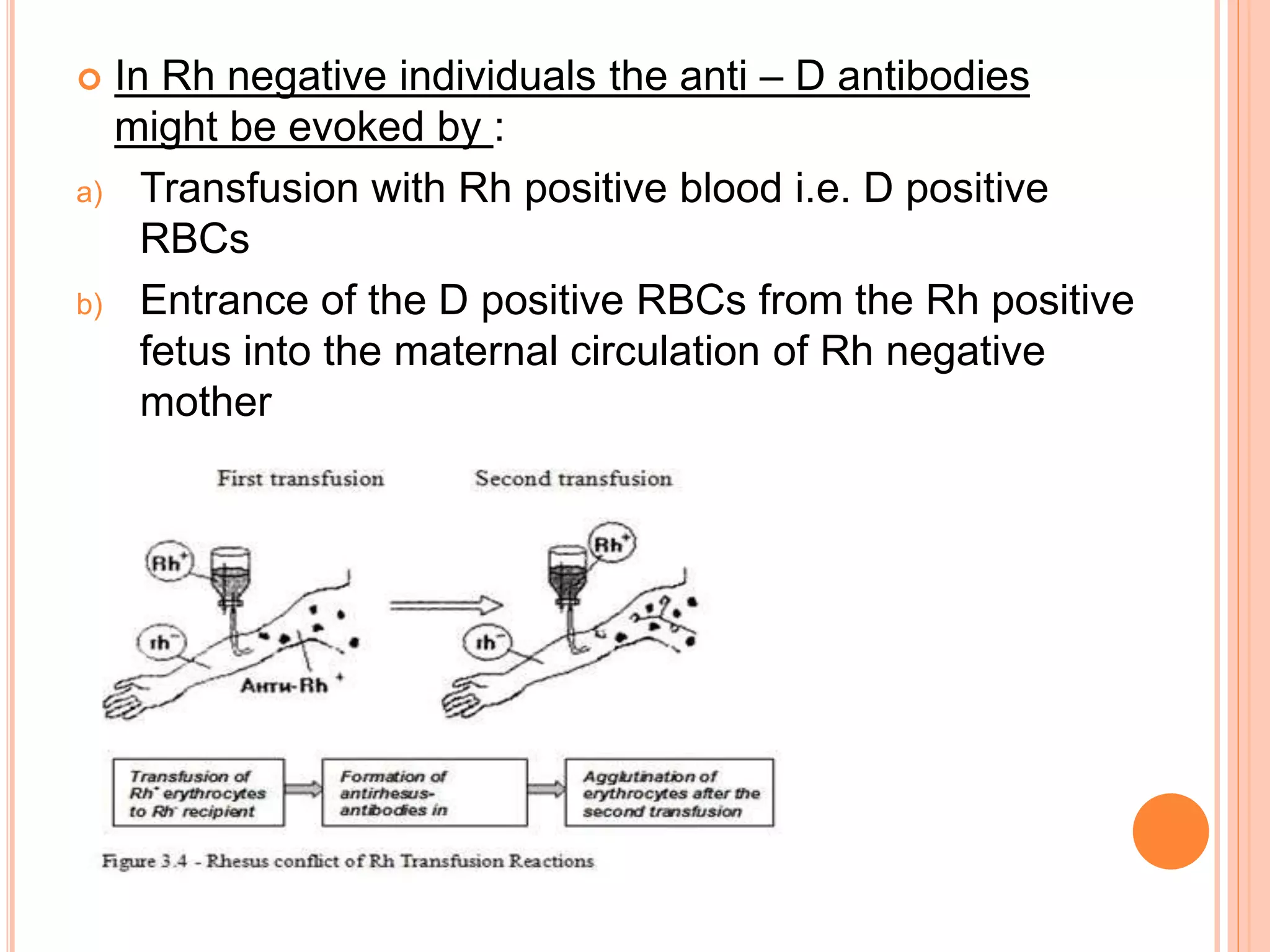
In Rh negative individuals , anti – D antibodies are not
naturally present in the plasma
12. In Rh negative individuals the anti – D antibodies
might be evoked by :
a) Transfusion with Rh positive blood i.e. D positive
RBCs
b) Entrance of the D positive RBCs from the Rh positive
fetus into the maternal circulation of Rh negative
mother
13. HEMOLYTIC DISEASE OR ERYTHROBLASTOSIS
FETALIS
Erythroblastosis fetalis is a disease of the fetus and new born infant
characterised by the progressive agglutination and subsequent
phagocytosis of the RBCs
If the mother is Rh negative and fetus is Rh positive serious
complications may occur.
RBCs containing D antigen may cross the placenta from the fetus to
the mother, either during pregnancy or a small amount of fetal blood
leaks into the maternal circulation at the time of the delivery.
The mother reacts by forming anti D which returns to the fetal
circulation and tends to destroy the fetal RBCs
16. Treatment of erythroblastosis fetalis
Transfusion of Rh –ve blood is done i.e. 400ml for 1.5 hrs
or more
It is repeated several times for the first few weeks of life
This is done so as to keep the blirubin levels low
Prevention of the Rh hemolytic disease
a. Destruction of Rh positive fetal cells in the maternal
blood can be brought about by administering a single
dose of anti Rh antibodies in the form of Rh
immunoglobulins soon after child birth
b. This prevents the formation of active antibodies by the
mother
17. OTHER COMMON SIGNIFICANT BLOOD
GROUPS
There are 34 other known blood groups systems
with more than 300 known variants . These are all
classified by the antigens found on the surface of
our red blood cells.
The “MNS blood group antigens” were discovered
in the 1920s by Karl Landsteiner.It’s common to find
antibodies to the M blood group in the plasma of
patients, as these are sometimes formed after
infection, and testing is required to ensure the
patient’s anti-M antibodies do not destroy donated
red blood cells.
18. Another blood group, the “S/s variants”, are named
after Sydney, where the blood group was
discovered. This blood group is signified by a
particular type of molecule on the red blood cells that
is a target of the malaria parasite.
A blood group known as Duffy is also associated
with infection by malaria .When this protein is absent
from the red blood cells, the cells are resistant to
infection by the malaria parasite. This protein is
absent from the blood cells of 90% of sub-Saharan
Africans, conferring malaria resistance on this
population. Antibodies to the Duffy antigens are
commonly found in a patient’s plasma and are a
cause of transfusion reactions if carefully matched
antigen negative blood is not given.
19. The K antigen was first detected in the 1940s as a
result of a woman without the K antigen on her red
blood cells being pregnant with a baby with the K
antigen on the red blood cells. While almost all
women post-partum have antibodies to some
antigens found on the baby’s white blood cells, red
cell antibodies are less common.
Another blood group, Kidd ( Jk) was named after the
patient in whom it was discovered. The Kidd proteins
are related to proteins in the kidney that help get rid
of waste from the body. For the Kidd blood group it’s
very important to avoid damaging reactions, and
therefore carefully matched antigen negative blood is
given.
20. BOMBAY BLOOD GROUP[OH GROUP]
o Rare individuals also lack the H antigen and
are designated as the “Bombay” phenotype (group
Oh). They make potent anti-H in addition to anti-A
and anti-B and must be transfused blood only from
other individuals with the Bombay phenotype.
It is observed to occur in 1 out
of every 250,000 people
It was discovered by Y.M
Bhende
22. ANTICOAGULANTS
1916 - First anticoagulant preservative was
discovered by Rous and turner – Citrate glucose
1943 – Acid citrate dextrose was introduced by
Loutit and Mollison
1957 - Gibson et al developed citrate phosphate
dextrose (CPD)
1978 – citrate phosphate with adenine (CPDA-1)
15ml o ACD or 14 ml of CPD/CPDA-1 is used in
preserving 100 ml o blood
PURPOSE: a. To prevent coagulation.
b. To preserve the life and survival of RBCs so as to
have the maximum post transfusion survival.
23. DONOR SELECTION
Donor history and risk factor assessment
Infectious disease testing
ABO and Rh typing
Cross matching
Noting post donation information
24. DONOR HISTORY
GENERAL PHYSICAL EXAMINATION :
General Appearance : should appear to be in good
health.
Age : between 18 and 65 years.
Weight : 45-55 Kg - 350 ml blood
55 Kg & above - 450 ml.
Temperature : should not exceed 37.5 C / 99.5 F
Pulse : 60 to 100 beats/min & regular pulse
Blood Pressure : SBP : b/w 100 and 160 mm of Hg
DBP : b/w 60-90 mm of Hg
Skin : free of any skin lesion or infections
25. MEDICAL HISTORY :
History of malaria : accepted after 3 months.
History of jaundice : deferred up to 1 year.
History of being HIV, HBsAg / HCV antibody positive
: permanently deferred.
Intimate contact with HIV, HBsAg / HCV antibody
positive individual : deferred for 1 year.
History of measles/mumps/chickenpox : deferred for
8 weeks
History of influenza : deferred till 1 week after
treatment
Having history of diarrhoea in preceding week
particularly if associated with fever should be
deferred
26. HISTORY OF VACCINATION
vaccination against TAB/TT/ Cholera/Hepatitis-A :
accepted if free of symptoms.
Hepatitis B vaccination : accepted after 7 days of
vaccination.
Yellow fever/measles/polio : deferred for 2 weeks
Rabies vaccination : deferred for 1 year.
Those bitten by any animal : deferred for one year.
Hepatitis B Immunoglobulin : should be deferred for 1
year
PREGNANCY
not be accepted during period of pregnancy and till 12
months after full term delivery and also during
lactation.
27. ASPIRIN INGESTION
Ingestion of Aspirin or any related medicine within 3
days prior to donation should preclude use of donor as
a source of platelet preparation.
SURGICAL PROCEDURES
Major : one year after the recovery
Minor : 6months
LABORATORY EXAMINATION :
Haemoglobin : not less than 12.0 gm/dl
Hematocrit : not less than 36%
28. DONOR INFECTIOUS DISEASE TESTING
Hepatitis B, HbsAg and anti-core antibody
•Hepatitis C antibody
•HIV 1 and 2 antibodies
•HTLV [Human T-cell lymphotropic virus] 1 and 2
antibodies
•Serologic Test for Syphilis
•Nucleic Acid Testing (NAT) for HIV, HCV
•Detection of Bacteria in platelet products
•CMV [Cytomegalo virus] antibody for select
recipients
29.
30. CROSS MATCHING
Blood matching between a patient and a donor is a direct
compatibility test
RBCs and plasma are crossmatched through major and minor
crossmatching process
“Major” crossmatch is comparing donor erythrocytes to
recipient serum where as the “minor” crossmatch is designed
to test opposite compatibility which is the donor's
serum/plasma with the recipient's red cells.
Minor cross match has almost been eliminated in most blood
banks, because the donor samples are screened before hand
for antibodies
31. GENERAL INDICATIONS OF BLOOD
TRANSFUSION
1. External bleeding
2. Internal bleeding (i) non-traumatic
(ii) traumatic
3. RBC lysis : e.g. malaria, HIV
4. Anaemia
5. Bleeding disorders
6. Burns
6. Anticipated need for blood
32. PHLEBOTOMY
The maximum volume of blood that may be collected is 10.5 mL/kg
of body weight
About 350- 450 ml is taken each time
The withdrawal of blood takes 10-15 mins
33. APHERESIS
•Apheresis refers to the process of separating the
cellular and soluble components of blood using a
machine.
• Apheresis is often done on donors where whole
blood is centrifuged to obtain individual
components ( RBCs, platelets, plasma based on
specific gravity) to use for transfusion in different
patients.
•Here the required component is collected and the
rest is returned to the donor
•Selective collection of RBCs/WBCs/platelets is
called cytapheresis
•Selective collection of plasma is called
plasmapheresis
•Here the anticoagulants such as citrate and
heparin is used
35. BANKED WHOLE BLOOD
No components have been removed
Contains RBCs ,WBCs ,platelets and Plasma
Can be stored for 5 weeks
Transfusions of whole blood are rarely required
They might be necessary in cases of acute blood
loss in major surgeries > 15% blood loss
It is a poor source of platelets and clotting factor 5
and 8
36. PACKED RED CELLS
Red cells from a donor unit diluted with plasma , to
a hematocrit of 75%
Volume is about 200ml
Storing red cells just above freezing allows survival
for about 42 days
It is the product of choice for most clinical situations
37. INDICATIONS FOR PACKED RED CELLS
In the field of orofacial surgery, a red blood cell
transfusion (RBCT) is occasionally required during
double jaw and oral cancer surgery
In the field of orofacial surgery, transfusion is performed
for the purpose of oxygen transfer to hypoxic tissues
and plasma volume expansion when there is bleeding.
RBCT can be a life-saving procedure for most patients
with acute anemia caused by perioperative bleeding
RBCT is the fastest way to increase the oxygen carrying
capacity of blood
A unit of RBCT will increase the Hct by 3% and Hb by 1-
1.5 gm/dl
38. TRANSFUSION STRATEGY & TRIGGER
The indications and triggers for RBCT are on-going
issues. There have been many studies and there are still
on-going studies in search of an answer. Based on
studies to date, there are two strategies :
a) In 1988, the “10/30 Rule”( liberal strategy) was
presented at the National Institutes of Health
Consensus Development Conference, which presented
the level of RBCT during perioperative period to be less
than Hb 10 g/dL and Hct 30% and transfusions were
performed based on those values
b) Recently, the restrictive strategy (Hb level below 7 g/dL)
has become more accepted due to the accumulation of
evidence regarding the negative impact on prognoses
following RBCT per the liberal strategy as well as the
complications and costs associated with RBCT
39. FROZEN RED CELLS
Concentrations of red blood
cells preserved frozen at -80ºC.
It reduces the risk of transfusing antigens or foreign
bodies that the body might regard as potentially
dangerous in previously sensitized patients
Not available for use in emergency situations
RBC viability is improved
ADP and 2,3 DPG(2,3-diphosphoglycerate) is
maintained
40. PLATELET CONCENTRATES
Composed of platelets and 50 ml plasma
Contains cellular components that help in the clotting
process
Platelets can be stored up to 5 days in room temperature
Indicated in :
a) Platelet disorders
b) When massive blood loss has occurred
One unit will usually raise the count to 5-10k / micro liter
41. FRESH FROZEN PLASMA
Obtained from freshly donated blood
Source of vit k dependent clotting factors
Only source of factor 5
Indicated for coagulopathy and different clotting factors
1 unit FPP = 3% increase in CF
42. CRYOPRECIPITATED ANTIHAEMOPHYLIC FACTOR
Its an antihaemophyllic concentrate, Cryoprecipitate
which is produced by allowing FFP to thaw slowly at 1–
6°C
It is prepared from plasma and rich in clotting factors
It is used in people with haemophyllia and Von
willebrand disease or other major abnormalities to
control bleeding
Its contents are major portion of factor 8 and fibronectin
which is present in freshly drawn and separated plasma
Indications for transfusion of cryoprecipitate include
repletion of fibrinogen levels activation of platelets;
emergent replacement of factor VIII, vWF, or factor XIII
when recombinant factors are unavailable; and as part
of a massive transfusion protocol
43.
44. PRINCIPLES OF BLOOD TRANSFUSION
1. Transfusion is only one part of the patient’s
management.
2. Blood loss should be minimized to reduce
the patient’s need for transfusion.
3. Acute blood loss should be given effective
resuscitation while the need for transfusion is
being assessed.
4. The patient’s haemoglobin value, although
important, should not be the sole deciding factor
for transfusion.
5. The clinician should be aware of the risks of
transfusion- transmissible infections
45. PRECAUTIONS TO BE TAKEN DURING BLOOD
TRANSFUSION
1. Use of Sterile Apparatus.
2. Blood bag should be checked
3. Temperature of blood to be
transfused must be same as body
temperature.
4. Transfusion rate must be slow in
order to prevent increase load on
heart.
5. Care full watch on the recipients
condition for 10 mins
46. DONATION INTERVAL
The interval b/w 2 donations : at least 12 weeks.
At least 48 hours must elapse after plasmapheresis
or cytapheresis before whole blood is collected from
a donor.
Apheresis should be done only after 90 days of
whole blood collection or in an event when red cells
are not returned at the end of apheresis.
47. Even though blood can supply a range
of products useful in a variety of
situations
Perioperative blood loss and
anaemia is best dealt with by
reducing the amount of blood lost at
surgery through minimizing trauma,
improving mechanical haemostasis
Limiting phlebotomy to essential
diagnostic tests, using microsample
laboratory techniques; and giving
antifibrinolytics, such as EACA or
tranexamic acid (or, for high-risk
procedures, aprotinin)
Erythropoietin can also help where
blood has been lost but the
replacement of blood by transfusion
can be essential after severe
haemorrhage and in some other
circumstances
PREVENTION IS BETTER THAN CURE
49. COMPLICATIONS OF BLOOD TRANSFUSION
A carefully prepared and supervised blood transfusion is
quite safe
However 5-6% of transfusions , untoward complications
occur, some of which are minor while others are more
serious and at times fatal
Adverse reactions of blood transfusion can be classified into
:
a. Immunological complications
b. Non immunological complications
Based on duration taken for the symptoms to occur they can
be classified as:
1. Acute
2. Delayed
They can also be classified as
a. Non infectious complications
b. Infectious complications
50.
51. NON INFECTIOUS COMPLICATIONS
Reactions associated with high morbidity
i. Transfusion related acute lung injury
ii. Transfusion associated circulatory overload
iii. Hemolytic reactions
iv. Anaphylaxis
v. Transfusion associated graft vs. host disease
vi. Post transfusion purpura
Reactions associated with low morbidity
i. Febrile non hemolytic transfusion reactions
ii. Mild allergic reactions
iii. Acute hypotensive transfusion reactions
52. TRANSFUSION RELATED ACUTE LUNG INJURY
[TRALI]
Transfusion-related acute lung injury (TRALI) was
first recognized in 1926 and was previously known
as pulmonary hypersensitivity reaction
Pathophysiology :
TRALI’s pathogenesis revolves around the
transfusion of antibodies and/ or other non
immunologic mediators to a susceptible patient
The most frequently implicated antibodies are
human leukocyte antigen (HLA) class I, HLA class
II, and human neutrophil antibodies (HNA)5,7;
these antibodies activate the leukocytes, which bind
to the endothelium in the lungs, causing endothelial
injury and edema
53. TREATMENT OF TRALI
As with all transfusion reactions, immediate
cessation of the transfusion and stabilization of the
patient are critical.
Respiratory support may range from supplemental
oxygen to intubation. Steroids have not been
proven to be beneficial.
TRALI reactions usually resolve over the course of
a few days with only supportive measures being
needed
54. TRANSFUSION ASSOCIATED CIRCULATORY
OVERLOAD[TACO]
Transfusion-associated circulatory overload (TACO) is
generally the most common high-morbidity transfusion
reaction encountered in clinical practice
Certain patient characteristics are known to increase the
risk of TACO, including older age, renal disease, cardiac
disease, positive fluid balance, and critically ill status
Pathophysiology : Unlike the majority of transfusion
reactions, which are immunologically mediated, TACO’s
pathophysiology invokes simple physics—too much fluid
is added to the system too quickly (or in volumes that
cannot be tolerated) for the transfusion recipient.
Because the circulatory system cannot cope with the
additional volume of the transfused products, pulmonary
edema and respiratory distress result as fluid “backs up”
into the lungs
56. TREATMENT OF TACO
If the transfusion is still running, it should be
stopped immediately
In some cases, the patient will improve with simply
stopping the infusion
patients will require some form of respiratory
support, at least temporarily
Diuretics are useful in the treatment of TACO; the
decrease in circulatory volume relieves
cardiovascular stress, improving the pulmonary
edema
TACO can be prevented ,patients at risk of fluid
overload at increased risk of TACO and should be
transfused at a slow rate
57. HEMOLYTIC REACTIONS
Transfusions leading to RBC
hemolysis can be among the
most devastating and feared
complications of blood product
administration
They represent a spectrum of
signs and symptoms and,
depending on the clinical
scenario, may be acute or
delayed, intra- or extra
vascular, attributable to ABO or
non-ABO antibodies, and in
some circumstances, may
even be caused by mechanical
forms of hemolysis due to
improper infusion techniques
58. INTRAVASCULAR HEMOLYSIS
Pathophysiology : Once the complement cascade has
been fixed and activated on the incompatible cells, the
resulting membrane attack complex punches holes in
the red cell, resulting in its lysis and destruction
IgM class antibodies are most efficient at fixing
complement and, therefore, acute intravascular
hemolysis is strongly associated with incompatibilities
within ABO antibodies (which are most likely to be IgM
in nature)
Generation of free RBC membranes in the intravascular
space can cause concomitant activation of the
coagulation system, resulting in the development of
disseminated intravascular coagulation (DIC)
59.
60. Complement generation and RBC release of
hemoglobin can induce acute kidney injury and renal
failure, a particularly feared complication of
hemolysis.
Complement activation can also cause smooth
muscle constriction, increased small vessel
permeability, and leukocyte activation, contributing to
the shock like symptom often seen in intravascular
hemolytic reactions.
Acute hemolytic transfusion incidence estimated at
about 1 in 76,000 transfusions
Prevention : Rigorous identification of patient blood
group during , before and after testing has to be
performed
61. EXTRAVASCULAR HAEMOLYSIS
Pathophysiology : In contrast to intravascular hemolysis,
which is typically acute and thunderous at onset,
extravascular hemolysis is generally associated with a
more subdued, slower RBC clearance
For this type of hemolytic reaction, RBC clearance occurs
because incompatible cells are coated by IgG class
antibodies, with antibody-coated cells subsequently
phagocytosed
As such, most extravascular reactions are mediated by
non-ABO antibodies (e.g., anti-Jk, anti-K, and anti-E
Because of the slower, extravascular nature of these
reactions the likelihood of end organ damage and a shock
like symptom is markedly reduced, particularly when
compared with intravascular hemolysis
62. TREATMENT OF HEMOLYTIC DISEASES
Approaches to managing these reactions typically
include assessing their severity, providing supportive
transfusions to overcome the acute anemia (and
coagulation disorders, if they exist), and steps to
preserve renal function.
New unit(s) are to be administered which are fully
compatible with the patient using the post- transfusion
reaction specimen
In urgent situations and gravely ill patients , O
negative blood can be given until the cause of
hemolysis is rectified.
Renal function must be closely monitored both
clinically and via laboratory assays such as creatinine
63. ANAPHYLAXIS
Anaphylactic transfusion reactions represent the most
severe and extreme reactions in the spectrum of allergic
reactions
Pathophysiology : Most anaphylactic reactions are
associated with platelets or plasma but they can occur
with the transfusion of any blood product
It is caused by complement, mast cell, and basophil
activation in response to a specific antigen/allergen
Anaphylactic reactions are characterized by rapid onset
of respiratory distress, laryngeal edema, hypotension,
and/or gastrointestinal symptoms, often within minutes
of starting a transfusion
Other allergic symptoms such as rashes and urticaria
may occur in conjunction with these more severe
symptoms
Diagnostic criteria : These symptoms must appear
within 4 h of a transfusion to meet the criteria for an
allergic transfusion reaction
64. TREATMENT OF ANAPHYLAXIS
Blood transfusion must be stopped immediately,
and the patient must be stabilized as necessary.
Respiratory support is vital, and intubation may be
required
Epinephrine, intravenous diphenhydramine, and
volume resuscitation are often helpful
Patients who have anaphylactic reactions to blood
products may require washed products in the future
Any future transfusions in a patient with a history of
anaphylactic transfusion reactions should be
considered with great caution, and the patient must
be closely monitored.
65. TRANSFUSION-ASSOCIATED GRAFT-VERSUS-
HOST DISEASE
Transfusion-associated graft-versus-host disease is a rare
but serious complication of blood transfusion.
People at risk include those who have:
received blood transfusions from HLA-matched donors,
including family members
had a stem-cell transplant
inherited immune defects
acquired immune defects, such as Hodgkin disease been
treated with purine analogues, such as fludarabine,
cladribine or deoxycoformycin.
Transfusion-associated graft-versus-host disease results
from transfused leukocytes; gamma irradiation of the
transfused blood will obviate the reaction
Patients should be pre-warned and should carry a warning
card themselves
66.
67. POST-TRANSFUSION PURPURA
Post transfusion purpura is a relatively
uncommon complication of blood transfusion
Pathophysiology : It can be thought of a
delayed transfusion reaction involving
platelets
Here there is an immunological response to
a previously encountered foreign platelet
that leads to an increase in the production of
antiplatelet antibodies by the recipient
Treatment :The current treatment of choice
is intravenous immunoglobulin (IVIG), along
with consideration of corticosteroids
68. REACTIONS ASSOCIATED WITH LOW MORBIDITY
1. Mild allergic reactions
Allergic reactions are the most
common adverse events associated
with transfusion
The main factor in allergic
transfusion reactions appears to be
the transfer of either antigen or
antibodies to the recipient via donor
plasma
Usually treatment is not necessary,
but in some cases
Diphenhydramine is the treatment
of choice; some patients may also
receive famotidine if
diphenhydramine is not effective
69. 2. Febrile non-hemolytic transfusion reactions
common transfusion reaction identified
Etiology is not known
Diagnostic criteria : to qualify as an FNHTR the fever
or chills/rigors must occur within 4 h of completion of
the transfusion
Treatment : transfusion should be stopped as soon
as reaction is suspected
3. Acute hypertensive transfusion reaction
Characterized by sudden increase in the systolic
blood pressure
Attributable to the increase in bradykinin
Treatment :Once the transfusion is stopped, the
hypotension resolves nearly immediately
70. Hyperkalemia associated with blood transfusions
Transfusion-associated hyperkalemic cardiac arrest is a serious
complication in patients receiving packed red blood cell (PRBC)
transfusions.
Mortality from hyperkalemia increases with large volumes of
PRBC transfusion, increased rate of transfusion, and the use of
stored PRBCs
The supernatant of stored RBCs usually contains more than 60
mEq/L of potassium .
Potassium in stored blood increases due to decrease in ATP
production and leakage of potassium into the supernatant. The
initial high levels of potassium in stored blood predispose to post-
transfusion hyperkalemia.
•Pre-washing of RBCs is an
essential practice for reducing
potassium load in irradiated
PRBC
71. HYPOKALEMIA
Hypokalemia is more common than the
hyperkalemia after transfusion because donor red
cells re- accumulate the ion intracellularly
Citrate metabolism causes further movement of
potassium into the cells. Catecholamine release
and aldosterone urinary loss can also trigger
hypokalemia in the setting of massive transfusion.
No treatment or preventive strategy is usually
necessary
Flat prolonged t waves Inverted t
wave
72. HYPOTHERMIA
It may be caused by transfusion of large volume of
cold blood products.
It can cause cardiac arrhythmia and also interferes
with platelet function, clotting factor interaction and
bleeding time.
Blood warmers may be used to prevent
hypothermia
73. INFECTIOUS COMPLICATIONS
Based on the etiology , the infectious complications
can be broadly classified into complications caused
by :
Viruses
Bacteria
Parasites
Prions
74.
75.
76. NEW CONCEPTS IN TRANSFUSION MEDICINE
Development of in vitro/ex vivo blood cells
Laboratory-derived platelets
Red blood cells in the setting of
hemoglobinopathies
Extending platelets’ shelf lives
New testing approaches
79. REFERENCES
Clinical principles of blood transfusion – 1st Ed –
Robert W Maitta
Clinical laboratory blood banking and transfusion
medicine
Essential Pathology -4th Ed – Harsh Mohan
Human physiology –fourth Ed –A K Jain
Perioperative red blood cell transfusion – NCBI
Medical problems in dentistry -7th Ed - Scully
Oral & maxillofacial surgery Vol-1 – Laskin D M