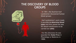
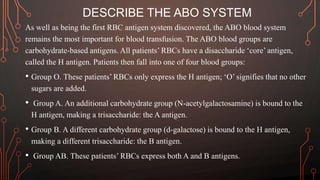
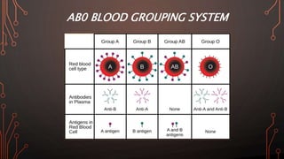
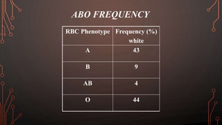
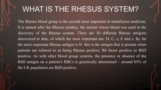
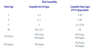
1. ABO blood groups and RH
Ahmed Maki Radeef (M.Sc. Anesthesia Technology )
2. THE DISCOVERY OF BLOOD
GROUPS
In 1901, the Austrian Karl
Landsteiner discovered human
blood groups
Karl Landsteiner's work made
it possible to determine blood
types and thus paved the way
for blood transfusions to be
carried out safely
For this discovery he was
awarded the Nobel Prize in
Physiology or Medicine in
1930.
3. WHAT ARE RED BLOOD CELL ANTIGENS?
RBCs can be thought of as ‘bags of Hb’. However, the composition
of the ‘bag’ itself differs between patients. The RBCs have surface
antigens that act as markers, identifying the RBC to the immune
system. RBC surface antigens may be polypeptides,
polysaccharides and glycoproteins. Which specific antigens are
expressed is determined genetically. There are at least 30 different
RBC antigen systems, the most important of which are:
• The ABO blood group system.
• The Rhesus blood group system.
4. DESCRIBE THE ABO SYSTEM
As well as being the first RBC antigen system discovered, the ABO blood system
remains the most important for blood transfusion. The ABO blood groups are
carbohydrate-based antigens. All patients’ RBCs have a disaccharide ‘core’ antigen,
called the H antigen. Patients then fall into one of four blood groups:
• Group O. These patients’ RBCs only express the H antigen; ‘O’ signifies that no other
sugars are added.
• Group A. An additional carbohydrate group (N-acetylgalactosamine) is bound to the
H antigen, making a trisaccharide: the A antigen.
• Group B. A different carbohydrate group (d-galactose) is bound to the H antigen,
making a different trisaccharide: the B antigen.
• Group AB. These patients’ RBCs express both A and B antigens.
5. In the UK, the most common blood groups are O (44%) and A (42%).
Group B makes up 10%, while group AB is found in only 4% of
patients.
16. ABO ANTIBODY
• Not detectable in newborn
• After 3-6 mo. Papered
• Depend on age
• Depend on disease
CML (Chronic myeloid leukemia)
Hypogammaglobinemia
Agammaglubinemia
BMT (Bone marrow transplant)
17. ABO ANTIBODY FACTS
Complement can be activated with ABO antibodies
(mostly IgM, some IgG)
Anti-A, Anti-B, Anti-A,B
Clinically Significant
Yes
Abs class
IgM, less IgG
18. WHAT IS THE RHESUS SYSTEM?
The Rhesus blood group is the second most important in transfusion medicine.
It is named after the Rhesus monkey, the animal whose blood was used in the
discovery of the Rhesus system. There are 50 different Rhesus antigens
discovered to date, of which the most important are: D, C, c, E and e. By far
the most important Rhesus antigen is D this is the antigen that is present when
patients are referred to as being Rhesus positive, Rh factor positive or RhD
positive. As with other blood group systems, the presence or absence of the
RhD antigen on a patient’s RBCs is genetically determined – around 85% of
the UK population are RhD positive.
19. WHY DOES THE IMMUNE SYSTEM DEVELOP
ANTIBODIES TO RED BLOOD CELL ANTIGENS?
The immune system develops antibodies to fragments of foreign
material presented by antigen-presenting cells. Patients may develop
antibodies to non-self RBC antigens for two reasons:
• Exposure to foreign RBCs; for example, following a blood transfusion
or placental abruption.
• Exposure to environmental antigens (food, bacteria, etc.) that happen
to have a chemical structure similar to that of a non-self RBC antigen
results in the production of antibodies which also cross-react with non-
self RBCs.
20. WHAT IS MEANT BY THE TERMS ‘ALLOGENIC’
AND ‘AUTOLOGOUS’ BLOOD TRANSFUSION?
Allogenic blood transfusion is where donor blood, usually packed RBCs,
is given intravenously to a recipient. In contrast, autologous blood
transfusion is where blood is taken from a patient and re-infused back
to the same patient when required (for example, intraoperative cell
salvage, preoperative autologous blood donation, acute normovolaemic
haemodilution).
25. WHAT IS MEANT BY THE TERM
‘HAEMOLYTIC TRANSFUSION REACTION’?
Early allogenic blood transfusions were fraught with complications:
many patients died after receiving incompatible blood. It was not until
the ABO blood group system was discovered that the reasons for these
deaths became clear.
A haemolytic transfusion reaction will occur if the recipient’s plasma
contains antibodies that are reactive against the donor’s RBC antigens.
The recipient’s antibodies coat the donor RBCs: the antibody– antigen
complex activates complement, leading to haemolysis of donor RBCs.
26. HAEMOLYTIC TRANSFUSION REACTIONS ARE
TWO TYPES
Immediate haemolytic transfusion reaction.
ABO incompatibility causes rapid intravascular haemolysis, the severity
depending on the antibody titre. Urticaria, flushing, chest pain,
dyspnoea, jaundice, tachycardia, shock, haemoglobinuria and DIC may
occur. Transfusion of RhD-incompatible blood tends to result in
extravascular haemolysis, which is usually less severe than
intravascular haemolysis.
27. Delayed haemolytic transfusion reaction. Minor
RhD antigens and the minor blood group systems may cause a delayed
haemolytic transfusion reaction, occurring 7–21 days following
transfusion. Delayed transfusion reactions are difficult to prevent.
Following a prior exposure to a blood antigen, patients develop a low
titre of antibody – too low for laboratory detection. When incompatible
blood is transfused, a secondary immune response occurs: it takes time
for new IgG antibodies to be produced, leading to a delay before
haemolysis is evident.
28. WHAT IS MEANT BY THE TERMS ‘UNIVERSAL
DONOR’AND ‘UNIVERSAL RECIPIENT’?
There are two groups of patients of particular importance in transfusion
medicine – the universal donor and the universal recipient:
The universal donor is a blood group O RhD negative patient. This is
because:
Group O RBCs only express the core H antigen, so are safe for donation
to recipients with anti-A or anti-B antibodies.
RhD-negative RBCs are safe to transfuse in recipients with anti-RhD
antibodies.
29. In an emergency situation, where there is insufficient time to establish the
blood type of the recipient (for example, trauma, ruptured aortic aneurysm,
obstetric haemorrhage), it is considered safe to transfuse with O negative
blood. However, there may still be minor blood group antigen–antibody
incompatibility reactions; a full cross-match is required to ensure blood is
fully compatible.
30. The universal recipient is more of academic than clinical interest. Blood group
AB RhD-positive patients can receive donor blood irrespective of its
ABO and Rh status:
Their plasma has neither anti-A nor anti-B antibodies, because their RBCs
contain both antigens.
Similarly, their plasma does not contain anti- RhD antibodies, because their
RBCs express the RhD antigen.
It is important to note that the minor blood group antigens are not necessarily
compatible; a crossmatch is still required.
31. WHAT IS A ‘CROSS-MATCH’?
Cross-match is a compatibility test between donor and recipient blood. There
are two types:
Major cross-match. The recipient’s serum is mixed with donor RBCs – this
is a test of compatibility between the recipient’s antibodies and donor
antigens. If donor RBCs clump together, the blood is incompatible.
Minor cross-match. The recipient’s RBCs are mixed with the donor’s
plasma. Minor crossmatch is no longer routinely performed because RBCs
are now transfused as packed cells, and thus contain an insignificant amount
of donor plasma.
32. MASSIVE TRANSFUSION
Massive transfusion is the transfusion of a greater volume of stored blood than
a patient’s circulating volume in a 24 h period, or transfusion of more than half
a patient’s circulating volume in a 4 h period (note: there are many other
similar definitions in circulation). Massive transfusion carries all the risks of a
single unit transfusion outlined above, but also has additional risks:
• Hypothermia.
• Dilutional coagulopathy.
• Hypocalcaemia.
• Hyperkalaemia.
• Acidosis.
33. HYPOTHERMIA
Transfusion of multiple units of refrigerated packed cells leads to a fall in core
body temperature through conduction. Hypothermia is associated with
coagulopathy, cardiac arrhythmias, and reduced tissue oxygenation (as the
oxyhaemoglobin dissociation curve is shifted to the left), and a worse overall
outcome. The risk of hypothermia can be reduced by warming all transfused
blood and fluids, as well as active warming of the patient; for example, by
using a forced-air warmer.
34. DILUTIONAL COAGULOPATHY
Multiple transfusions of packed RBCs along with crystalloid administration
leads to dilution of plasma constituents. Of particular importance are clotting
factors and platelets, because haemostasis will become impossible, leading to
further haemorrhage and a requirement for more blood products. Aggressive
and pre-emptive replacement of clotting factors and platelets with FFP,
cryoprecipitate and platelet concentrate is required.
35. HYPOCALCAEMIA
Packed cells contain only a small amount of the anticoagulant citrate; FFP and
platelet concentrate contain much higher citrate concentrations. The role of
citrate is the chelation of Ca2+, preventing the coagulation of stored blood
products. In massive transfusion, there is sufficient intravascular citrate to
cause a severe hypocalcaemia. Without Ca2+ replacement, this can lead to
hypotension and ECG changes (bradycardia, flat ST segments, prolonged QT
interval). Hypocalcaemia should be treated with 10 mL of 10% calcium
chloride.
36. Supplemental calcium may be needed when
1. The rate of blood infusion is more rapid than 50 ml/min
2. Hypothermia or liver disease interferes with the metabolism of citrate
3. The patient is a neonate.
Patients undergoing liver transplantation are the most likely to experience
citrate intoxication, and these patients may require calcium administration
during a massive transfusion of stored blood.
37. HYPERKALAEMIA
when RBCs are stored, K+ seeps out of the erythrocytes. When blood is
transfused, K+ tends to diffuse back into the RBCs, but this process is hindered
if the patient is acidotic or hypothermic, leading to hyperkalaemia.
38. ACIDOSIS
A unit of packed RBCs has a lower pH (around 6.8) than plasma, mainly as a
result of lactate accumulation due to the anaerobic metabolism of erythrocytes
during storage. Massive transfusion may therefore worsen acidosis, though the
additional lactate is usually rapidly cleared by the liver.
39. DECREASED 2,3-DIPHOSPHOGLYCERATE
Storage of blood is associated with a progressive decrease in concentrations of
2,3-DPG in erythrocytes, which results in increased affinity of hemoglobin for
oxygen (decreased P50 values). Conceivably, this increased affinity could
make less oxygen available for tissues and jeopardize tissue oxygen delivery.
There is speculation that fresh blood (with more oxygen available for tissues)
should be used for critically ill patients. Despite these observations, the clinical
significance of the 2,3-DPG oxygen affinity changes remains unconfirmed.
45. We are have tow type of bleeding:
Internal bleeding
External bleeding
46. Trauma
Clotting disorders
Rupture of blood vessels
Fractures (injury to near by vessels)
Can result in rapid progression to
hypovolemic shock & death!
48. How you can know there is internal bleedi
Vomiting bright red blood (haematemesis)
Bleeding form any body orifice
Dark, tarry stools (melena)
Tender, rigid, or distended abdomen
Pain, discoloration, swelling
tenderness at injury site
49. Managing Internal Bleeding
ABC’s
High concentration oxygen
Assist ventilations
Control external bleeding
Stabilize fractures
Transport rapidly to appropriate facility
52. RBC transfusion can be lifesaving. During the past
two decades, however, safety concerns have
emerged, with suggestions that morbidity and
mortality may be increased in patients who receive
blood transfusions. Therefore, the decision to
transfuse should be individualized, based on a
rational approach and taking into account physiologic
variables in addition to the hemoglobin value. This
strategy, along with the use of alternatives whenever
possible to limit bleeding, should limit unnecessary
exposure to RBCs.