Introduction
• Hemostasis isthe normal physiologic mechanism for
keeping the blood in fluid state in vascular system and
for prevention of hemorrhage by complex interaction of
blood vessel walls, platelets, and plasma proteins.
• Following injury, initially vessel wall and platelets
interact to control hemorrhage by forming a platelet
plug at the site of injury; this is called as primary
hemostasis.
• This is followed by activation of coagulation factors by a
series of enzymatic reactions to form a stable fibrin clot
(platelet plug enmeshed by fibrin); this is secondary
hemostasis.
3.
Blood Vessel Wall
•Transient constriction of blood vessels occurs at the site
of injury which helps to control blood loss.
• Endothelial cells synthesize von Willebrand factor
(vWF), tissue factor, and platelet activating factor,
which promote hemostasis.
• Also, following injury, subendothelial collagen is
exposed which provides site for attachment of platelets
(adhesion). Endothelial cells synthesize prostacycline
(inhibits platelet aggregation), protein S (a cofactor for
protein C, which is an inhibitor of coagulation), and
tissue plasminogen activator (activates fibrinolysis).
4.
Platelets
• Platelets areproduced by cytoplasmic
fragmentation of megakaryocytes in bone marrow.
Life-span of platelets is about 7-10 days. Normal
platelet count in peripheral blood is 1.5-4.0 lac/µl.
About 2/3rd of platelets in the body are circulating
in peripheral blood, while 1/3rd are pooled in
spleen.
• Main functions of platelets in hemostasis are
adhesion, release reaction, and aggregation.
6.
• Plasma proteins,which regulate hemostasis,
are coagulation factors, coagulation inhibitors,
and proteins of fibrinolytic system.
• When activated, coagulation factors interact
with each other in a sequential manner to
ultimately form a fibrin clot and arrest
bleeding.
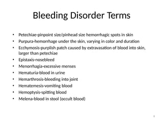
Bleeding Disorder Terms
8
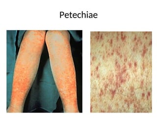
•Petechiae-pinpoint size/pinhead size hemorrhagic spots in skin
• Purpura-hemorrhage under the skin, varying in color and duration
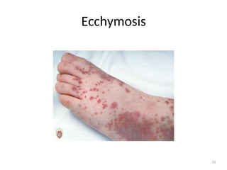
• Ecchymosis-purplish patch caused by extravasation of blood into skin,
larger than petechiae
• Epistaxis-nosebleed
• Menorrhagia-excessive menses
• Hematuria-blood in urine
• Hemarthrosis-bleeding into joint
• Hematemesis-vomiting blood
• Hemoptysis-spitting blood
• Melena-blood in stool (occult blood)
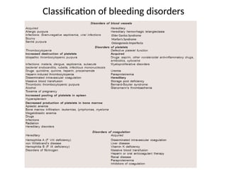
Bleeding due tovascular defects
12
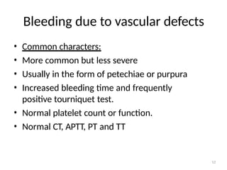
• Common characters:
• More common but less severe
• Usually in the form of petechiae or purpura
• Increased bleeding time and frequently
positive tourniquet test.
• Normal platelet count or function.
• Normal CT, APTT, PT and TT
13.
Tourniquet test
• Microvascularfragility may be demonstrated by a
positive "tourniquet test“.
• This test is performed by inflating a blood
pressure cuff on the arm to midway between
systolic and diastolic blood pressures for five
minutes. The pressure is released for at least one
minute and the skin below the cuff is examined
for petechiae. A finding of 10 or more petechiae
in a one square inch area is considered positive.
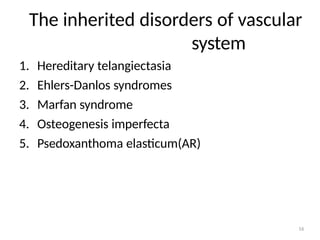
INHERITED DISORDERS
22
1. HEREDITARYTELANGIECTASIA
• AD trait
• Characterised by the presence of flat ,red
or purple lesions on mucous membranes
• Due to the lack of elastic fibers in the
vessel wall
• Epistaxis is the most common manifestation
2.EHLERS –DANLOS SYNDROME
24
•Autosomal dominant trait, EDS IV (vascular
type) is associated with mutations in COL3A1
located on long arm of Chromosome 2.
• Defect in collagen synthesis resulting in weak
subendothelial connective tissue and produce
bleeding.
• Extreme fragility of vessels, Easy bruising,
rupture of large vessels, petechiae, GI bleeding
are common.
3.MARFAN SYNDROME
26
• Marfansyndrome is an autosomal dominant
disorder that has been linked to the FBN1 gene
on chromosome 15. FBN1 encodes a protein
called fibrillin.
• Characteristic defects are long
extremities,spidery fingers,dislocation of lens,
and easy bruising.
• Bruising may be caused by abnormalities of the
vessels.
4.OSTEOGENESIS IMPERFECTA
28
• Autosomaldominant trait .
• Disorder of the genes for type 1 procollagens
which cause patchy ,defective bone matrix.
• Easy and spontaneous bruising,epistaxis,and
intracranial hemorrhage are the bleeding
symptoms.
29.
5.PSEUDOXANTHOMA ELASTICUM
29
• Autosomalrecessive manner.
• PXE is caused by mutations in the ABCC6
(ATP-binding cassette subfamily C member
6) gene, located on short-arm of
human chromosome 16.
• Due to the presence of abnormal elastic
tissue in the skin and all arteries.
• Easy bruising,petechiae and purpura are
common.
30.
ACQUIRED DISORDERS
30
• 1.SENILEPURPURA
• Usually occur in old age.
• A deficient supportive sub-endothelial
connective tissue leads to easy rupture of
vessels and bleeding occurs.
• Common sites of bleeding are extensor
surface of forearm and hand.
2.CUSHING SYNDROME
32
Relatedto altered connective tissue support of
the blood vessel wall.
It may due to abnormalities of
mucopolysaccharides in the supporting tissue
33.
3.SCURVY
• Caused deficientVitamin C
• Vitamin C is required for vessel collagen
integrity.
• Acts as “cement” holding endothelial
cells together.
• Lack of Vitamin C prevents proper
collagen formation.
• Result: bleed and vessel fragility.
• Symptoms include gum bleeding, petechiae
and bleeding into tissues and muscles.
• Treated with Vitamin C.
33
34.
4.PARAPROTEIN DISORDERS
34
Paraproteins aremonoclonal Immunoglobulins.
• Occurs due to the deposition of protein in the
vascular wall.
• Symptoms (hemostasis) include
purpura, bleeding and thrombosis.
• Bleeding symptoms include
epistaxis,petechiae,and hemorrhage into
organs.
35.
5.AMYLOIDOSIS
35
• Due tothe deposition of amyloid in the skin
and vascular walls.
• Leads to fragility of the vessel and
to bruising.
• Bleeding into visceral organs can occur
• Thrombosis is common.
36.
ALLERGIC PURPURA
36
• Usuallyoccur in children.
• The skin lesions begin as urticaria,change to
pink, then to red and hemorrhagic.
37.
INFECTIONS
37
• Purpura associatedwith infections can caused
by damage to the blood vessels
(nonthrombocytopenic purpura)
• It is related to nonspecific immune
complex formed by the antigenic agent and
its corresponding antibody.
• This complex attach to either the endothelial
cells or to the underlying subendothelial
structures ,results in inflammation and vasculitis.
38.
DRUG INDUCED
38
• Aspirin,quinineand warfarins
• Cause vasculitis with the appearance of
ecchymoses in the absence of
thrombocytopenia.
39.
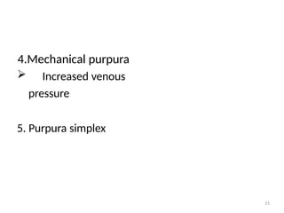
PURPURA SIMPLEX
39
• Easybruising syndrome
• Commonly seen in young woman
• Spontaneous small ecchymoses appear mainly
on the skin of the thighs or upper arms-devil’s
pinches.