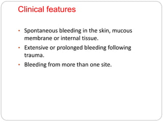
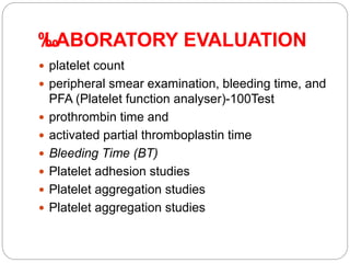
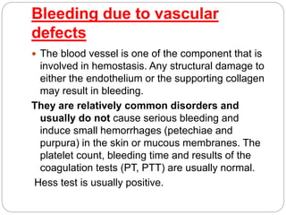
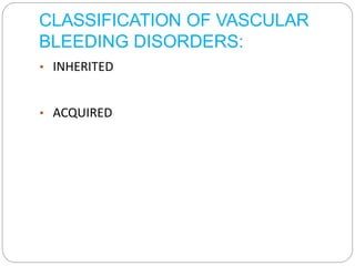
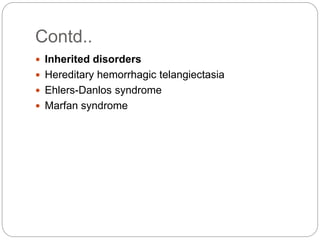
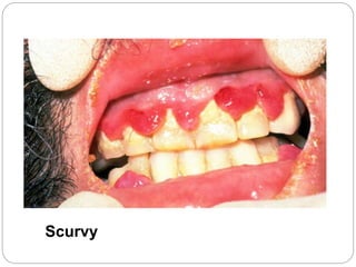
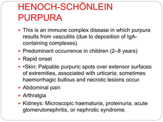
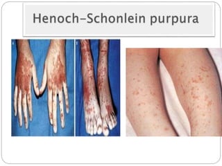
By :
Mohamed Ahmed
Outline
 Introduction to bleeding disorders
 Bleeding due to vascular causes
 Bleeding disorders is a general term for a group of
conditions (acquired or inherited) in which there is an
abnormal tendency to bleeding /hemorrhage due to
failure of hemostasis.
 The bleeding may be heavy and prolonged which may
occur after an insignificant injury or spontaneously.
 The pattern of bleeding is relatively predictable
depending on the aetiology.
 Vascular and platelet disorders tend to be associated
with bleeding from mucous membranes and into the
skin.
 whereas in coagulation disorders the bleeding is often
into joints or soft tissue
 Petechiae : are small (1 to 2 mm in diameter),
red to purple hemorrhagic spots in the skin,
mucous membranes or serosal surfaces. They
are most commonly found with low platelet counts
(thrombocytopenia) or defective platelet function.
 Purpura: it means purple. They are slightly larger
(> 3 mm) than petechiae. The causes are
thrombocytopenia, increased vascular fragility
and vasculitis.
 Ecchymoses: They are larger (>1 to 2 cm) and
result from blood escaping through endothelium
into intact subcutaneous tissue.
DISORDERS OF HEMOSTASIS
 Normal hemostasis requires a well controlled
collaboration among the five components of
hemostasis namely:
vascular endothelium, platelets, coagulation
system, coagulation regulatory system and
fibrinolytic system.
Clinical features
• Spontaneous bleeding in the skin, mucous
membrane or internal tissue.
• Extensive or prolonged bleeding following
trauma.
• Bleeding from more than one site.
Causes of bleeding disorders
• 1.Due to vascular defects
• 2.Due to platelet defects
• 3.Due to coagulation defects
‰
‰
LABORATORY EVALUATION
 platelet count
 peripheral smear examination, bleeding time, and
PFA (Platelet function analyser)-100Test
 prothrombin time and
 activated partial thromboplastin time
 Bleeding Time (BT)
 Platelet adhesion studies
 Platelet aggregation studies
Bleeding due to vascular
defects
 The blood vessel is one of the component that is
involved in hemostasis. Any structural damage to
either the endothelium or the supporting collagen
may result in bleeding.
They are relatively common disorders and
usually do not cause serious bleeding and
induce small hemorrhages (petechiae and
purpura) in the skin or mucous membranes. The
platelet count, bleeding time and results of the
coagulation tests (PT, PTT) are usually normal.
Hess test is usually positive.
CLASSIFICATION OF VASCULAR
BLEEDING DISORDERS:
• INHERITED
• ACQUIRED
Bleeding due to vascular
defects
 Acquired disorders
 1. Due to decreased amount of connective tissue
 Senile purpura
 Scurvy
 Cushing syndrome and steroid therapy
 2. Due to vasculitis
 Henoch-Schönlein purpura
 Infections
 Drug reactions
 3. Associated with plasma cell neoplasms
 Amyloidosis
 4. Miscellaneous
 Simple easy bruising
Contd..
 Inherited disorders
 Hereditary hemorrhagic telangiectasia
 Ehlers-Danlos syndrome
 Marfan syndrome
1-Acquired disorders
 Senile Purpura: It is age-related atrophy of
supporting connective tissue of blood vessels.
 Scurvy: Lack of vitamin C (scurvy) is associated
with microvascular bleeding resulting from
impaired formation of collagens.
 Cushing syndrome and steroid therapy: In
Cushing syndrome, protein-wasting due to
excessive corticosteroid production causes loss
of perivascular supporting tissue.
 Simple easy bruising is a common benign
disorder which occurs in otherwise healthy
women, especially those of child‐bearing age.
Senile Purpura
Scurvy
HENOCH-SCHÖNLEIN
PURPURA
 This is an immune complex disease in which purpura
results from vasculitis (due to deposition of IgA-
containing complexes).
 Predominant occurrence in children (2–8 years)
 Rapid onset
 •Skin: Palpable purpuric spots over extensor surfaces
of extremities, associated with urticaria; sometimes
haemorrhagic bullous and necrotic lesions occur.
 Abdominal pain
 Arthralgia
 Kidneys: Microscopic haematuria, proteinuria, acute
glomerulonephritis, or nephrotic syndrome.
 The disease is self-limited but recurrent episodes
are common. Chronic glomerulonephritis, and
CNS bleeding are rare complications.
 Diagnosis is based on typical clinical features and
exclusion of haematological causes of purpura.
 Glucocorticoids are sometimes administered for
symptomatic relief.
Infections
 Meningococcemia, septicemia, infective
endocarditis and several of the rickettsioses.
 Mechanisms include invasion of vessel, septic
emboli, immune complex vasculitis or result of
disseminated intravascular coagulation (DIC).
 Amyloidosis
Petechiae in Vasculitis
(Rocky Mountain Spotted Fever)
2- Inherited Disorders
 Hereditary Hemorrhagic Telangiectasia
(Weber-Osler-Rendu syndrome)
• It is an autosomal dominant disorder of vascular
malformation characterized by dilated, tortuous,
thin walled blood vessels of the skin and mucous
membranes.
Clinical picture include epistaxis, bleeding from
tongue mouth and GIT.
Repeated bleeding may result in iron deficiency
anemia.
 Ehlers-Danlos Syndrome: It is a congenital
disorder of defective collagen synthesis
characterized by joint hyper mobility skin laxity
and easy bruising.
 The capillaries are poorly supported by
subendothelial collagen and result in bleeding
mainly in the form of ecchymoses.
Diagnosis of vascular purpuras
 Superficial, mild bleeding from skin and mucous
membranes
 Bleeding from skin more in dependent portions of
body
 Screening tests of haemostasis often normal
 Associated clinical features usually characteristic
Vascular purpuras

Vascular purpuras

  • 1.
  • 2.
    Outline  Introduction tobleeding disorders  Bleeding due to vascular causes
  • 3.
     Bleeding disordersis a general term for a group of conditions (acquired or inherited) in which there is an abnormal tendency to bleeding /hemorrhage due to failure of hemostasis.  The bleeding may be heavy and prolonged which may occur after an insignificant injury or spontaneously.  The pattern of bleeding is relatively predictable depending on the aetiology.  Vascular and platelet disorders tend to be associated with bleeding from mucous membranes and into the skin.  whereas in coagulation disorders the bleeding is often into joints or soft tissue
  • 6.
     Petechiae :are small (1 to 2 mm in diameter), red to purple hemorrhagic spots in the skin, mucous membranes or serosal surfaces. They are most commonly found with low platelet counts (thrombocytopenia) or defective platelet function.  Purpura: it means purple. They are slightly larger (> 3 mm) than petechiae. The causes are thrombocytopenia, increased vascular fragility and vasculitis.
  • 7.
     Ecchymoses: Theyare larger (>1 to 2 cm) and result from blood escaping through endothelium into intact subcutaneous tissue.
  • 10.
    DISORDERS OF HEMOSTASIS Normal hemostasis requires a well controlled collaboration among the five components of hemostasis namely: vascular endothelium, platelets, coagulation system, coagulation regulatory system and fibrinolytic system.
  • 12.
    Clinical features • Spontaneousbleeding in the skin, mucous membrane or internal tissue. • Extensive or prolonged bleeding following trauma. • Bleeding from more than one site.
  • 13.
    Causes of bleedingdisorders • 1.Due to vascular defects • 2.Due to platelet defects • 3.Due to coagulation defects
  • 14.
    ‰ ‰ LABORATORY EVALUATION  plateletcount  peripheral smear examination, bleeding time, and PFA (Platelet function analyser)-100Test  prothrombin time and  activated partial thromboplastin time  Bleeding Time (BT)  Platelet adhesion studies  Platelet aggregation studies  Platelet aggregation studies
  • 15.
    Bleeding due tovascular defects  The blood vessel is one of the component that is involved in hemostasis. Any structural damage to either the endothelium or the supporting collagen may result in bleeding. They are relatively common disorders and usually do not cause serious bleeding and induce small hemorrhages (petechiae and purpura) in the skin or mucous membranes. The platelet count, bleeding time and results of the coagulation tests (PT, PTT) are usually normal. Hess test is usually positive.
  • 16.
    CLASSIFICATION OF VASCULAR BLEEDINGDISORDERS: • INHERITED • ACQUIRED
  • 17.
    Bleeding due tovascular defects  Acquired disorders  1. Due to decreased amount of connective tissue  Senile purpura  Scurvy  Cushing syndrome and steroid therapy  2. Due to vasculitis  Henoch-Schönlein purpura  Infections  Drug reactions  3. Associated with plasma cell neoplasms  Amyloidosis  4. Miscellaneous  Simple easy bruising
  • 18.
    Contd..  Inherited disorders Hereditary hemorrhagic telangiectasia  Ehlers-Danlos syndrome  Marfan syndrome
  • 19.
    1-Acquired disorders  SenilePurpura: It is age-related atrophy of supporting connective tissue of blood vessels.  Scurvy: Lack of vitamin C (scurvy) is associated with microvascular bleeding resulting from impaired formation of collagens.  Cushing syndrome and steroid therapy: In Cushing syndrome, protein-wasting due to excessive corticosteroid production causes loss of perivascular supporting tissue.  Simple easy bruising is a common benign disorder which occurs in otherwise healthy women, especially those of child‐bearing age.
  • 20.
  • 21.
  • 23.
    HENOCH-SCHÖNLEIN PURPURA  This isan immune complex disease in which purpura results from vasculitis (due to deposition of IgA- containing complexes).  Predominant occurrence in children (2–8 years)  Rapid onset  •Skin: Palpable purpuric spots over extensor surfaces of extremities, associated with urticaria; sometimes haemorrhagic bullous and necrotic lesions occur.  Abdominal pain  Arthralgia  Kidneys: Microscopic haematuria, proteinuria, acute glomerulonephritis, or nephrotic syndrome.
  • 26.
     The diseaseis self-limited but recurrent episodes are common. Chronic glomerulonephritis, and CNS bleeding are rare complications.  Diagnosis is based on typical clinical features and exclusion of haematological causes of purpura.  Glucocorticoids are sometimes administered for symptomatic relief.
  • 27.
    Infections  Meningococcemia, septicemia,infective endocarditis and several of the rickettsioses.  Mechanisms include invasion of vessel, septic emboli, immune complex vasculitis or result of disseminated intravascular coagulation (DIC).  Amyloidosis
  • 28.
    Petechiae in Vasculitis (RockyMountain Spotted Fever)
  • 29.
    2- Inherited Disorders Hereditary Hemorrhagic Telangiectasia (Weber-Osler-Rendu syndrome) • It is an autosomal dominant disorder of vascular malformation characterized by dilated, tortuous, thin walled blood vessels of the skin and mucous membranes. Clinical picture include epistaxis, bleeding from tongue mouth and GIT. Repeated bleeding may result in iron deficiency anemia.
  • 31.
     Ehlers-Danlos Syndrome:It is a congenital disorder of defective collagen synthesis characterized by joint hyper mobility skin laxity and easy bruising.  The capillaries are poorly supported by subendothelial collagen and result in bleeding mainly in the form of ecchymoses.
  • 33.
    Diagnosis of vascularpurpuras  Superficial, mild bleeding from skin and mucous membranes  Bleeding from skin more in dependent portions of body  Screening tests of haemostasis often normal  Associated clinical features usually characteristic