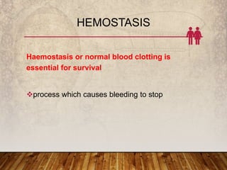
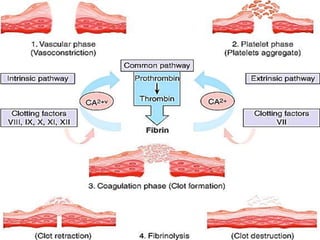
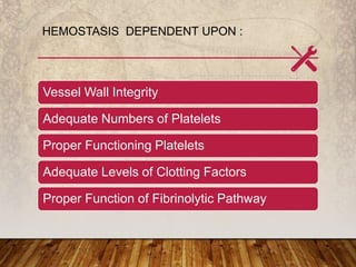
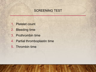
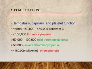
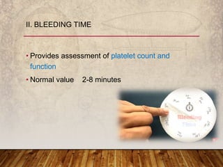
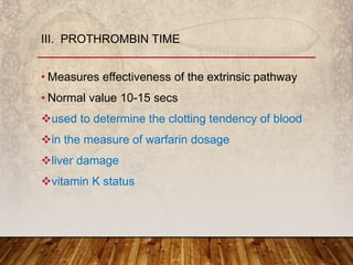
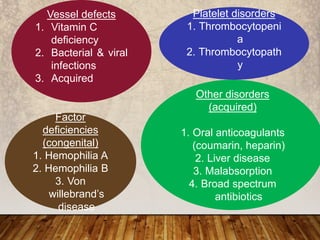
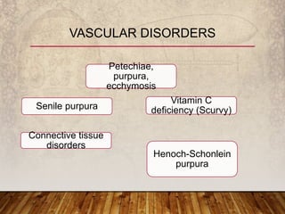
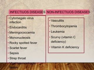
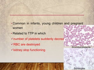
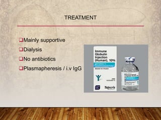
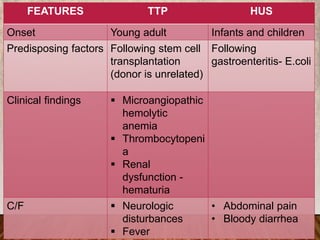
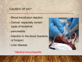
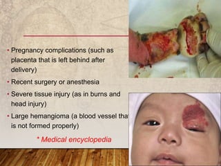
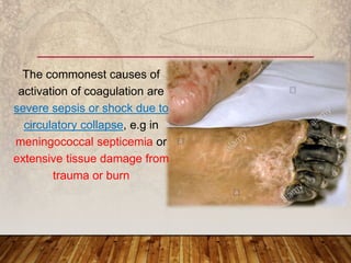
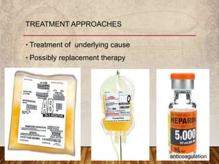
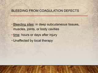
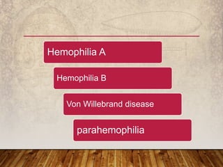
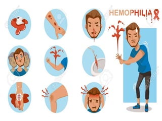
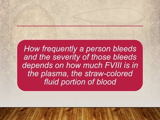
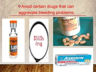
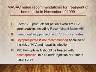
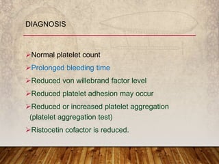
BLEEDING DISORDERS
AND THEIR
MANAGEMENT
Dr. Eaketha Lokesh
PG 1ST Year
Dept: of Prosthodontics
KMCT Dental College
CONTENTS
• Introduction
• Bleeding disorders
• History
• Hemostasis
• Screening test
• INR
• Vessel defects
CONTENTS
• Introduction
• Bleeding disorders
• History
• Hemostasis
• Screening test
• INR
• Vessel defects
• Platelet disorders
• Coagulation disorders
• WBDR
• Recent advancements
• Summary
• References
• Vascular damage results in initiation of clotting
with the goal of producing a localized platelet or
fibrin plug to prevent blood loss
• This action is followed by processes that lead to
clot containment, would healing, clot dissolution,
and tissue regeneration and remodeling.
INTRODUCTION
BLEEDING DISORDERS???
• caused by the abnormalities of hemostasis and /
or coagulation
• characterized by local or extensive skin or
mucocutaneous hemorrhage derived from
capillary bleeding is usually spontaneous or from
slight trauma
• 1800 - U.S., the transmission of hemophilia from
mothers to sons.
• 1803, a Philadelphia physician named Dr. John
Conrad Otto wrote an account of "a hemorrhagic
disposition existing in certain families."
HISTORY
• The word "Hemophilia" first appeared - University
of Zurich in 1828
• 1960s - clotting factors were identified and named.
• 1950s , early 1960s - hemophilia and other
bleeding problems were treated with whole blood
or fresh plasma.
There wasn't enough of the factor VIII or IX
proteins in these blood products to stop serious
internal bleeding.
• 1960 - cryoprecipitate by Dr. Judith
Pool
• 1960s , early 1970s - concentrates
containing factor VIII and IX (freeze-
dried powdered concentrates)
• 1990s - modern treatment-safer factor
concentrates
• Vessel constricts
• Circulating platelets adhere to the vessel
• An intricate series of enzymatic reactions
involving coagulation proteins
• produces fibrin to form a stable haemostatic plug
BLOOD VESSEL INJURY TRIGGERS THE
FOLLOWING SEQUENCE:
HEMOSTASIS
Haemostasis or normal blood clotting is
essential for survival
process which causes bleeding to stop
Vascular phase
Platelet phase
Coagulation phase
Fibrinolytic phase
• Primary hemostasis -platelet plug formation at
sites of injury
• Secondary hemostasis - plasma coagulation
system reaction resulting in fibrin formation
• Primary and secondary hemostasis are closely
linked
• When a blood vessel is damaged, results in
vasoconstriction
VASCULAR PHASE
• Platelets adhere to the damaged surface and form
a temporary plug
PLATELET PHASE
• Through two separate pathways the conversion of
fibrinogen to fibrin is complete
COAGULATION PHASE
• Anticlotting mechanisms are activated to allow clot
disintegration and repair of the damaged vessel
FIBRINOLYTIC PHASE
HEMOSTASIS DEPENDENT UPON :
Vessel Wall Integrity
Adequate Numbers of Platelets
Proper Functioning Platelets
Adequate Levels of Clotting Factors
Proper Function of Fibrinolytic Pathway
1. Platelet count
2. Bleeding time
3. Prothrombin time
4. Partial thromboplastin time
5. Thrombin time
SCREENING TEST
• Hemostasis, capillary and platelet function
• Normal 150,000 - 450,000 cells/mm 3
• < 150,000 thrombocytopenia
50,000 - 100,000 mild thrombocytopenia
50,000- severe thrombocytopenia
• ˃ 450,000 cells/mm3 thrombocytosis
1. PLATELET COUNT
• Provides assessment of platelet count and
function
• Normal value 2-8 minutes
II. BLEEDING TIME
• Measures effectiveness of the extrinsic pathway
• Normal value 10-15 secs
used to determine the clotting tendency of blood
in the measure of warfarin dosage
liver damage
vitamin K status
III. PROTHROMBIN TIME
• PT measures factors II , V, VII, X and fibrinogen .
• used in conjunction with the activated partial
thromboplastin time (aPTT) which measures the
intrinsic pathway .
• Measures effectiveness of the intrinsic pathway
• Normal value 25-40 secs
IV. PARTIAL THROMBOPLASTIN TIME
• Time for thrombin to convert fibrinogen to fibrin
• A measure of fibrinolytic pathway
• Normal value 9-13 secs
V. THROMBIN TIME
• Preferred test of choice for patients taking vitamin
K antagonists (VKA).
• To assess the risk of bleeding or the coagulation
status of the patients.
Patients taking oral anticoagulants are required to
monitor INR to adjust the VKA doses
because these vary between patients.
INTERNATIONAL NORMALIZED RATIO (INR)
• The INR is derived from prothrombin time (PT)
• Standardized for the potency of the thromboplastin
reagent
• Developed by the World Health Organization
(WHO) using the following formula:
INR = Patient PT ÷ Control PT
• Dimensionless
• Ranges from a score of 0.9-1.3
Tripathi et al . Clinical evaluation of whole blood prothrombin time (PT) and
international normalized ratio (INR) using a Laser Speckle Rheology sensor. Sci
• For normal patients who are not on anticoagulation, the
INR is usually 1.0 regardless of the ISI or the particular
performing laboratory
• High INR level (INR=5) high chance of
bleeding
• INR=0.5 high chance of having a clot
Tripathi et al . Clinical evaluation of whole blood prothrombin time (PT) and international
normalized ratio (INR) using a Laser Speckle Rheology sensor. Sci Rep. 2017 Aug
WHAT CAUSES
BLEEDING
DISORDERS???
1. Vessel defects
2. Platelet disorders
3. Factor deficiencies
4. Other disorders
CAUSES OF BLEEDING DISORDERS
(National Hemophilia Association)
Vessel defects
1. Vitamin C
deficiency
2. Bacterial & viral
infections
3. Acquired
Platelet disorders
1. Thrombocytopeni
a
2. Thrombocytopath
y
Factor
deficiencies
(congenital)
1. Hemophilia A
2. Hemophilia B
3. Von
willebrand’s
disease
Other disorders
(acquired)
1. Oral anticoagulants
(coumarin, heparin)
2. Liver disease
3. Malabsorption
4. Broad spectrum
antibiotics
VASCULAR DISORDERS
Petechiae,
purpura,
ecchymosis
Senile purpura
Vitamin C
deficiency (Scurvy)
Connective tissue
disorders
Henoch-Schonlein
purpura
Tiny blood vessels
(capillaries) link the
smallest parts of your
arteries to the smallest
parts of your veins.
• Small (1–2 mm) red or purple
spot on the skin, caused by a
minor bleed from
broken capillary blood vessels
• Petechiae appear when
capillaries bleed, leaking blood
into the skin
1. PETECHIAE
• Cytomegalo virus
infection
• Endocarditis
• Meningococcemia
• Mononucleosis
• Rocky spotted fever
• Scarlet fever
• Sepsis
• Strep throat
• Viral haemorrhagic
fevers
• Vasculitis
• Thrombocytopenia
• Leukemia
• Scurvy (vitamin C
deficiency)
• Vitamin K deficiency
INFECTIUOS DISEASES NON-INFECTIUOS DISEASES
• the appearance of red or purple discolorations on
the skin that do not blanch on applying pressure
• They are caused by bleeding underneath the skin
2. PURPURA
The medical term for a
subcutaneous hematoma
larger than 1 centimeter,
commonly called a BRUISE
both in the skin as well as in
a mucous membrane
3. ECCHYMOSIS
In the purpuric disorders, petechiae commonly are
associated with multiple superficial ecchymoses,
which usually develop without perceptible trauma
but seldom spread into deeper tissues.
Small isolated ecchymoses are commonly noted
in apparently normal women, especially on the
legs, and in small children.
1-2 mm
≥ 3 mm
˃ 1-2 cm
• Bleeding into joint spaces.
• Most seen in hemophilia A or hemophilia B
• Rare in disorders of the vessels and platelets or in
acquired coagulation disorders.
4. HEMARTHROSIS
• common, benign condition
• characterised by the recurrent formation of
purple ecchymoses (bruises) on the extensor
surfaces of forearms following minor trauma
5. SENILE PURPURA
• It is also known as Bateman purpura, after
British dermatology pioneer Thomas Bateman,
who first described it in 1818;
• and actinic purpura, because of its association
with sun damage.
(Senile Purpura. Ken et al. DermNet NZ 2014)
RISK FACTORS
oral or topical corticosteroids
 Characterised by irregularly-shaped macules, 1 –
4 cm in diameter, that are dark purple with well-
defined margins.
The lesions do not undergo the colour changes of
a bruise and take up to three weeks to resolve.
Surrounding skin is typically thin, inelastic and
pigmented in association with others signs of skin
ageing and sun damage.
Extensor surface of forearms and dorsal aspect of
hands
Infrequently, they also occur on necks and faces
• benign and self-resolving
• Patients should be educated on sun
protection measures, includes
sunscreen application and sun-protective clothing
to protect their skin from further photodamage
MANAGEMENT
• Vitamin C (ascorbic acid)
• Humans can not synthesize vitamin C due to
deficiency of l-gulonolactone oxidase (rich in citrus
food)
• Deficiency- results in SCURVY
6. VITAMIN C DEFICIENCY
• Elderly patients seem to require vitamin C in large
quantities.
• They feel better and are able to do more work
when vitamin C is added to their diet.
• Vitamin C is nontoxic even in massive doses.
National Academy of Sciences (RDA)
Recommended Dietary Allowances in 2000 for vitamin
C
75+age male → 50mg
 Female → 30mg
Young individuals (male = female)→ 40-30mg
proline
5-hydroxyproline
lysine
5-
hydroxylysine
Vitamin C
Hydroxylases
Mirhadi SA et al., 1990
AMINOACIDS IN COLLAGEN
The protective role of vitamin C can help our
skin and gums from pinpoint hemorrhage
(scurvy disease).
Cardiovascular diseases, cancers, and joint
diseases are all associated with vitamin c
deficiency and can be partly prevented by
optimal intake of vitamin c.
Kurl S et al, 2002
spongy and swollen gumsloose teeth
Cork screw hair pattern with tiny
bleeding points around orifice of a hair
follicle
Woody legs with large
spontaneous bruises in
lower extremities.
delayed wound healing
Vitamin C achieves much of its
protective effect by functioning as an
antioxidant and preventing oxygen-
based damage to our cells.
Structures that contain fat (like the
lipoprotein molecules that carry fat
around our body) are particularly
dependent on vitamin C for protection
Reaven PD, Witztum JL:
1996
• disorder that causes inflammation
and bleeding in the small blood
vessels of skin, joints, intestines and
kidneys.
• The most striking feature - purplish
rash, typically on the lower legs and
buttocks
• Children: ages of 2 and 6
7. HENOCH-SCHONLEIN PURPURA
Rarely serious kidney
damage can occur
PLATELET
DISORDERS
• Bleeding sites localized to superficial sites such as
the skin and mucous membranes
• Immediately after trauma or surgery
• Readily controlled by local measures
BLEEDING FROM A PLATELET
DISORDER
QUANTITATIVE
Abnormal distribution
Dilution effect
Decreased production
Increased destruction
QUALITATIVE
Inherited disorders
(rare)
Acquired disorders
Medications
Chronic renal failure
Cardiopulmonary
bypass
PLATELET DISORDERS
• Defined as reduced in the platelet count ˂150,
000µL
spontaneous bleeding
prolonged bleeding time
normal PT and PTT
1. THROMBOCYTOPENIA
THE RISK OF BLEEDING DEPENDS ON THE
LEVEL OF THE PLATELET COUNT:
Mild
thrombocytopeni
a (platelet ˂150
000 cells/µL)
Moderate
thrombocytopeni
a (platelet 20 000
- 50 000 cells/µL)
Severe
thrombocytopeni
a (platelet ˂ 20
000 cells/µL)
ETIOLOGY
Decreased
production of
platelets
Increased
platelets
destruction
Dilutional
Sequestration
Congenital
(Fanconianae
mia, Wiskot-
Aldrich
Syndrome
Acquired
(aplastic
anaemia,
marrow
infiltration, drug
induced )
Immunologic
destruction (ITP,
SLE , Alloimmune
neonatal
thrombocytopenia)
Non-
immunologic
destruction
(HUS, TTP,
DIC, CHD)
Hypersplenis
m
Massive
blood
transfusion
• Bruising
• Petechiae
• Purpura
• Mucosal bleeding (epistaxis and gum bleeding)
• Major haemorrhage (severe GI bleeding,
intracranial bleeding or haematuria is less
common)
• Normal platelet count may present in platelet
dysfunction
SIGN AND SYMPTOM
• IMMUNE THROMBOCYTOPENIA
• may occur when the immune system mistakenly
attacks platelets
• In children, it may follow a viral infection
• In adults - chronic
• Low platelet count
• Easy bruising
IDIOPATHIC THROMBOCYTOPENIC PURPURA
(ITP)
• There are two clinical subtypes of ITP:
• Acute ITP
• Chronic ITP (starts after the disease has been
present for & ˃6 months)
Increased destruction of platelets – Thrombocytopenia
Phagocytosis of antibody-coated platelets by the
reticuloendothelial system.
Autoantibodies (IgG or IgM) directed against platelet
membrane antigens (especially glycoprotein complex
IIb/IIIa).
Inappropriate immune recovery follows an acute viral
infection in children.
PATHOGENESIS OF ITP
• acute ITP - sudden onset
• chronic ITP - insidious onset
• Petechiae or purpura feet, legs, arms, and
buttocks.
• Mucosal bleeding.
• Palatal petechiae, epistaxis, hematuria,
menorrhagia, GI bleeding.
• Rarely, intracranial hemorrhage may occur in long
standing severe thrombocytopenia.
CLINICAL MANIFESTATIONS
• Not all children with acute ITP need
hospitalization.
• Treatment is indicated if there is:
Life threatening bleeding episode (e.g. intra
cranial haemorrhage) regardless of platelet count.
Platelet count ˂20,000/mm³ with mucosal
bleeding.
 Platelet count ˂10,000/mm³with any bleeding.
MANAGEMENT OF ITP
Choice of treatment:
• Oral prednisolone - 4 mg/kg/day for 7 days,
taper and discontinue at 21 days.
• IV Methylprednisolone - 30 mg/kg/day for 3 days.
• IV Immunoglobulin - 0.8 g/kg/dose for 1 day OR
250 mg/kg for 2 days.
• IV Anti-Rh(D) immunoglobulin - (50 – 75 µ/kg) in
Rhesus positive patients – may cause haemolytic
anaemia
• Splenectomy is only for life threatening in acute
ITP
• For chronic ITP:
 Repeated treatment with IV immunoglobulin or IV
anti-D or high dose pulse steroids are effective in
delaying the need for splenectomy
Splenectomy is effective in inducing remission in
70-80% of childhood chronic ITP
• Intracranial hemorrhage - 50% mortality rate
• Risk of ICH highest in:
Platelet count ˂ 20 000/mm³
History of head trauma
Uses of aspirin (inhibitor of platelet aggregation)
Presence of cerebral arteriovenous malformation
• 50% of all ICH occurs after 1 month of
presentation, 30% after 6 months
COMPLICATION
• clinical syndrome characterized by progressive
renal failure that is associated with
microangiopathic hemolytic
anemia and thrombocytopenia.
• HUS is the most common cause of acute kidney
injury in children
HEMOLYTIC UREMIC SYNDROME
(HUS)
• Common in infants, young children and pregnant
women
• Related to TTP in which
number of platelets suddenly decreases
RBC are destroyed
kidney stop functioning
Schiocytes
thrombocytopenia
• HUS is rare, but can occur with certain bacterial
infection (E.coli or shigelladysenteriae)
with the use some drugs (quinine, cyclosporine,
mitomycin C)
Toxin producing organism such as E.coli cause
endothelial damage that activates localized
clotting, leading to platelet aggregation and
consumption
Mainly supportive
Dialysis
No antibiotics
Plasmapheresis / i.v IgG
TREATMENT
• Resembles hemolytic uremic syndrome but occur
more commonly in adults than in children
• Spontaneous aggregation of platelets and
activation of coagulation in small blood vessels
• In TTP, platelet consumption, precipitated by a
congenital or acquired deficiency of
metalloproteinase that cleaves vWF
THROMBOTIC THROMBOCYTOPENIC
PURPURA (TTP)
FEATURES TTP HUS
Onset Young adult Infants and children
Predisposing factors Following stem cell
transplantation
(donor is unrelated)
Following
gastroenteritis- E.coli
Clinical findings  Microangiopathic
hemolytic
anemia
 Thrombocytopeni
a
 Renal
dysfunction -
hematuria
C/F  Neurologic
disturbances
 Fever
• Abdominal pain
• Bloody diarrhea
• Progressive, malignant disease of the blood
forming organs
• Marked by distorted proliferation and
development of leukocytes and their precursors
in the blood and the bone marrow
MARROW INFILTRATION
(LEUKEMIA)
• Overproduction of these white cells(immature or
abnormal forms) → suppresses the production of
normal WBC, RBC and platelets
• Lead to increase susceptibility to infection ,
anemia and bleeding
• Result from infiltration of bone marrow or other organs with
leukemic blast cells
CLINICAL PRESENTATION
• Progress rapidly in some children
• Blood count is abnormal,
• low hemoglobin
• thrombocytopenia
• evidence of circulating blast cells in most children
Bone marrow examination to confirm the
diagnosis
• a condition in which blood clots form throughout
the body, blocking small blood vessels
• Serious medical condition
• Occurs as secondary complication of variety
diseases.
DISSEMINATED INTRAVASCULAR
COAGULATION (DIC)
Caused by the systemic
activation of coagulation
pathways → formation of
thrombi throughout the
microcirculation and
widespread thromboses.
• May be acute or chronic.
• Initiated through the TISSUE FACTOR PATHWAY
• Consumption of platelets and coagulation factors
• Secondarily activation of fibrinolysis
• As consequence, there is depletion of the
elements required for hemostasis ( consumptive
coagulopathy)
• Blood transfusion reaction
• Cancer, especially certain
types of leukemia
• pancreatitis
• Infection in the blood (bacteria
or fungus)
• Liver disease
CAUSES OF DIC*
* Medical encyclopedia
• Pregnancy complications (such as
placenta that is left behind after
delivery)
• Recent surgery or anesthesia
• Severe tissue injury (as in burns and
head injury)
• Large hemangioma (a blood vessel that
is not formed properly)
* Medical encyclopedia
The commonest causes of
activation of coagulation are
severe sepsis or shock due to
circulatory collapse, e.g in
meningococcal septicemia or
extensive tissue damage from
trauma or burn
INFECTIOUS
 Meningococcemia
 Other gram –vebact (Salmonella, E.coli,
Haemophilus)
 Virus (CMV, herpes, hemorrhagic
fevers)
 Rickettsia, Malaria and fungus
TISSUE INJURY
 CNS trauma
 crush injury
 Multiple fracture with fat emboli
 Profound shock of asphyxia
Hypothermia/hyperthermia
 Massive burns
MALIGNANCY
 Acute promyelocytic leukemia
 Acute monoblastic or myelocytic
leukemia widespread malignancies
(neuroblastoma)
HEREDITARY THROMBOTIC
DISORDERS
 Antithrombin III def.
 Homozygous protein C def.
MICROANGIOPATHIC DISORDER
 Severe TTP/ hemolytic uremic
syndrome
 Giant hemangioma
VENOM/ TOXIN
 Snake bites
 Insect bites
GIT DISORDER
 Fulminant hepatitis
 severe IBD
 Reye syndrome
NEWBORN
 Maternal toxemia
 G. B strep. Infection
 Abruptioplacentae
 Congenital viral disease (CMV,
herpes)
MISCELLANEOUS
 Severe acute graft rejection
 Acute hemolytic transfusion reaction
 Heparin induced thrombosis
• Treatment of underlying cause
• Possibly replacement therapy
TREATMENT APPROACHES
anticoagulation
• Immediate correction of the cause is the priority
(eg, broad-spectrum antibiotic treatment of
suspected gram-negative sepsis, evacuation of
the uterus in abruptio placentae).
• If treatment is effective, disseminated
intravascular coagulation should subside quickly.
• any of several blood disorders characterized by
dysfunctional platelets (thrombocytes), which
result in prolonged bleeding time, defective clot
formation, and a tendency to hemorrhage.
• Denotes abnormal platelet function
• Platelet count normal
• Thrombocytopathy may be congenital or acquired
2. THROMBOCYTOPATHY
INHERITED
• Von willebrand disease
• Thrombasthenia
• Bernard-soulier
syndrome
• Down syndrome
• Wiskott-aldrich
syndrome (an immune
disorder).
ACQUIRED
• Leukemia
• Pernicious anemia
• Scurvy
• uremia
THROMBOCYTOPATHY
https://www.britannica.com/editor/The-Editors-of-Encyclopaedia-
Britannica/4419
• Platelet transfusion to control bleeding
• Curing of the underlying disease usually results in
improved platelet function
MANAGEMENT
BLEEDING DISORDERS
AND THEIR
MANAGEMENT
Eaketha Lokesh
PG 1ST Year
CONTENTS- I
• Introduction
• Bleeding disorders
• History
• Hemostasis
• Screening test
• INR
• Vessel defects
• Platelet disorders
• Coagulation disorders
• Prosthetic considerationssssssss
• WBDR
• Recent advancements
• Summary
• References
CONTENTS- II
COAGULATION
DISORDERS
• Bleeding sites: in deep subcutaneous tissues,
muscles, joints, or body cavities
• time: hours or days after injury
• Unaffected by local therapy
BLEEDING FROM COAGULATION DEFECTS
Hemophilia A
Hemophilia B
Von Willebrand disease
parahemophilia
• HEMOPHILIA ----------"Royal disease"
• Hemophilia gene was passed from Queen
Victoria, who became queen of England in 1837,
to the ruling families of Russia, Spain, and
Germany.
HEMOPHILIA
 Factor VIII (FVIII) deficiency
CLASSIC HEMOPHILIA
A genetic disorder caused by missing or defective
factor VIII, a clotting protein.
Although it is passed down from parents to
children, about 1/3 of cases are caused by a
spontaneous mutation, a change in a gene
HEMOPHILIA A
Incidence:
approximately 1 in 5,000 live births
(US Centers for Disease Control and Prevention)
X-linked recessive
• Normal plasma levels of FVIII range from 50% to
150%
• Levels below 50%, or half of what is needed to
form a clot, determine a person’s symptoms.
Mild hemophilia A- 6% up to 49% of FVIII in the
blood
Moderate hemophilia A- 1% up to 5% of FVIII in
the blood
Severe hemophilia A - <1% of FVIII in the blood
How frequently a person bleeds
and the severity of those bleeds
depends on how much FVIII is in
the plasma, the straw-colored
fluid portion of blood
MANAGEMENT
Avoid certain drugs that can
aggravate bleeding problems:
I. Factor VIII products for patients who are HIV
seronegative, including Recombinant factor VIII
II. Immunoaffinity purified factor VIII concentrates
III. Cryoprecipitate is not recommended because of
the risk of HIV and hepatitis infection.
IV. Mild hemophilia A should be treated with
desmopressin, in a DDAVP injection or Stimate
nasal spray
MASAC made recommendations for treatment of
hemophilia in November of 1999
COMPLICATIONS
 Chronic joint deformities
 Intracerebral
hemorrhage
• CHRISTMAS DISEASE
• deficiency in clotting factor IX
• Hemophilia B is four times less common than
hemophilia A
• X-linked recessive
• Incidence: 1 in 35000 men
HEMOPHILIA B
• Severe (factor levels less than 1%) represent
approximately 60% of cases
• Moderate (factor levels of 1-5%) represent
approximately 15% of cases
• Mild (factor levels of 6%-30%) represent
approximately 25% of cases
• Nose bleeds
• Bruising
• Spontaneous bleeding
• Bleeding into joints and associated pain and
swelling
• Gastrointestinal tract and Urinary tract
hemorrhage
• Blood in the urine or stool
• Prolonged bleeding from cuts, tooth extraction,
surgery, following circumcision
SYMPTOMS
• Infusing the missing clotting factor
• To prevent a bleeding crisis- taught to administer
factor IX concentrates at home at the first signs of
bleeding
• Factor IX concentrate may be given prior to dental
extractions and surgery to prevent bleeding
TREATMENT
Milder forms of
hemophilia need to
have dental or other
surgery, the drug
desmopressin acetate
(DDAVP) may be
given to improve
clotting temporarily so
that transfusions can
be avoided.
Recombinant factor IX products for patients who
are HIV seronegative
Patients who are HIV-seropositive should also be
treated with high purity products such as
immunoaffinity purified and recombinant factor VIII
products.
MASAC MADE RECOMMENDATIONS FOR
TREATMENT OF HEMOPHILIA B IN NOVEMBER OF
1999
For patients with inhibitors to
factors VIII and IX, there is
Recombinant Factor VIIa
(NovoSeven)
Produced by baby hamster
kidney cells, no human
albumin or other proteins are
used in its production, reducing
virus risk.
There is also Porcine factor VIII (Hyate C) and
activated prothrombin complex concentrates
Not currently available
hereditary
deficiency or
abnormality of the
von Willebrand
factor in the blood,
a protein that
affects platelet
function
VON WILLEBRAND DISEASE
• Type I:
Most common and mildest form of von Willebrand
disease.
Levels of von Willebrand factor are lower than
normal, reduced levels of factor VIII.
CLASSIFICATIONS
• Type II:
• Von Willebrand factor itself has an abnormality.
• Depending on the abnormality, they may be
classified as:
Type II a - the level of von Willebrand factor is
reduced, as is the ability of platelets to clump
together.
Type II b -although the factor itself is defective,
the ability of platelets to clump together is actually
increased.
• Type III:
Severe von Willebrand disease.
Total absence of von Willebrand factor, and
 factor VIII levels are often less than 10%
• Pseudo (or platelet-type) von Willebrand disease:
Resembles Type IIb von Willebrand disease,
but the defects appears to be in the platelets,
rather than the von Willebrand factor.
Inheritance Pattern occurs in men and women
equally.
Types I and II - inherited as a "dominant" pattern.
Type III von Willebrand disease - inherited in a
"recessive" pattern
Normal platelet count
Prolonged bleeding time
Reduced von willebrand factor level
Reduced platelet adhesion may occur
Reduced or increased platelet aggregation
(platelet aggregation test)
Ristocetin cofactor is reduced.
DIAGNOSIS
Stimate, desmopressin acetate (DDAVP),nasal
spray or injection
Viral-inactivated factor VIII preparations rich in
von Willebrand factor, such as Alphanate,
Humate-P and Koate DVI, are recommended
Cryoprecipitate is not recommended except in
life-threatening emergencies because of the risk of
HIV and hepatitis infection
MASAC MADE RECOMMENDATIONS FOR
TREATMENT OF VON WILLEBRAND DISEASE IN
NOVEMBER OF 1999
After surgery, hemorrhaging may occur.
The condition is worsened by the use of aspirin
and other nonsteroidal anti-inflammatory drugs.
 Women may have risks during pregnancy and
childbirth.
PROSTHETIC
CONSIDERATIONS
IN BLEEDING
DISORDERS
• Good thorough medical
history- family history ,
personal history, medications,
spontaneous bleeding
• Physical examination
• Screening clinical lab tests
• Excessive bleeding following
surgical proceedure
DENTAL EVALUATION
Aspirin
Anticoagulant
Alcohol
Antibiotics
anticancer
REVIEW OF MEDICATIONS
5
A
1. Removable prosthodontics dentures
donot usually involve a considerable risk
of bleeding.
2. Trauma should be minimized by careful
post-insertion adjustments.
3. Oral tissues should be handled delicately
during the various clinical stages of
prosthesis fabrication to reduce the risk
of eccymosis
• Only global registry collecting standardized clinical
data on people with hemophilia from around the
world
WBDR
• New clotting products and drugs - desmopressin
acetate also known as DDAVP.
• Synthetic (not derived from plasma) clotting
products that take advantage of recombinant
technologies.
• Better screening methods to detect and remove
viruses and other agents from factor concentrates
and blood products
RECENT ADVANCEMENTS
• Improved surgical options
• Advanced genetic testing methods
• Medically supervised home-infusion therapy
• Prophylactic treatment
SUMMARY
• National hemophilia foundation
• The worldwide incidence of hemophilia is
estimated at more than 400,000 people. 108 Kar,
A., Phadnis, S., Dharmarajan, S., & Nakade, J.
(2014). Epidemiology & social costs of
haemophilia in India. The Indian Journal of
Medical Research, 140(1), 19–31.
REFERENCES
• Davidson’s “principles and practice of medicine-
19th edition;- Hanslet, Chilvers, Boon, Colledge,
Hunters.
• Medical emergencies in the dental practice –
malammed; 5th edition
• Bailey and Love’s Short practice of surgery-23rd
edition; Russel, Williams, Bulstrode;
• Complications in Oral and Maxillofacial surgery-
1st edition; Kaban, Pogrel, Perrot.
• Concise Medical Physiology;-5th edition;
Chaudhari
• Systemic disease in dental treatment-1st edition;-
Michael.J.Tullman, Spencer.W.Redding.
• Clinical hematology-7th edition;-
Maxwell.M.Wintrobe.
Bleeding disorders and their management

Bleeding disorders and their management

  • 2.
    BLEEDING DISORDERS AND THEIR MANAGEMENT Dr.Eaketha Lokesh PG 1ST Year Dept: of Prosthodontics KMCT Dental College
  • 3.
    CONTENTS • Introduction • Bleedingdisorders • History • Hemostasis • Screening test • INR • Vessel defects
  • 4.
    CONTENTS • Introduction • Bleedingdisorders • History • Hemostasis • Screening test • INR • Vessel defects
  • 5.
    • Platelet disorders •Coagulation disorders • WBDR • Recent advancements • Summary • References
  • 6.
    • Vascular damageresults in initiation of clotting with the goal of producing a localized platelet or fibrin plug to prevent blood loss • This action is followed by processes that lead to clot containment, would healing, clot dissolution, and tissue regeneration and remodeling. INTRODUCTION
  • 7.
    BLEEDING DISORDERS??? • causedby the abnormalities of hemostasis and / or coagulation • characterized by local or extensive skin or mucocutaneous hemorrhage derived from capillary bleeding is usually spontaneous or from slight trauma
  • 8.
    • 1800 -U.S., the transmission of hemophilia from mothers to sons. • 1803, a Philadelphia physician named Dr. John Conrad Otto wrote an account of "a hemorrhagic disposition existing in certain families." HISTORY
  • 9.
    • The word"Hemophilia" first appeared - University of Zurich in 1828 • 1960s - clotting factors were identified and named.
  • 10.
    • 1950s ,early 1960s - hemophilia and other bleeding problems were treated with whole blood or fresh plasma. There wasn't enough of the factor VIII or IX proteins in these blood products to stop serious internal bleeding.
  • 11.
    • 1960 -cryoprecipitate by Dr. Judith Pool • 1960s , early 1970s - concentrates containing factor VIII and IX (freeze- dried powdered concentrates) • 1990s - modern treatment-safer factor concentrates
  • 12.
    • Vessel constricts •Circulating platelets adhere to the vessel • An intricate series of enzymatic reactions involving coagulation proteins • produces fibrin to form a stable haemostatic plug BLOOD VESSEL INJURY TRIGGERS THE FOLLOWING SEQUENCE:
  • 14.
    HEMOSTASIS Haemostasis or normalblood clotting is essential for survival process which causes bleeding to stop
  • 15.
  • 16.
    • Primary hemostasis-platelet plug formation at sites of injury • Secondary hemostasis - plasma coagulation system reaction resulting in fibrin formation • Primary and secondary hemostasis are closely linked
  • 17.
    • When ablood vessel is damaged, results in vasoconstriction VASCULAR PHASE
  • 18.
    • Platelets adhereto the damaged surface and form a temporary plug PLATELET PHASE
  • 19.
    • Through twoseparate pathways the conversion of fibrinogen to fibrin is complete COAGULATION PHASE
  • 21.
    • Anticlotting mechanismsare activated to allow clot disintegration and repair of the damaged vessel FIBRINOLYTIC PHASE
  • 22.
    HEMOSTASIS DEPENDENT UPON: Vessel Wall Integrity Adequate Numbers of Platelets Proper Functioning Platelets Adequate Levels of Clotting Factors Proper Function of Fibrinolytic Pathway
  • 23.
    1. Platelet count 2.Bleeding time 3. Prothrombin time 4. Partial thromboplastin time 5. Thrombin time SCREENING TEST
  • 24.
    • Hemostasis, capillaryand platelet function • Normal 150,000 - 450,000 cells/mm 3 • < 150,000 thrombocytopenia 50,000 - 100,000 mild thrombocytopenia 50,000- severe thrombocytopenia • ˃ 450,000 cells/mm3 thrombocytosis 1. PLATELET COUNT
  • 25.
    • Provides assessmentof platelet count and function • Normal value 2-8 minutes II. BLEEDING TIME
  • 26.
    • Measures effectivenessof the extrinsic pathway • Normal value 10-15 secs used to determine the clotting tendency of blood in the measure of warfarin dosage liver damage vitamin K status III. PROTHROMBIN TIME
  • 27.
    • PT measuresfactors II , V, VII, X and fibrinogen . • used in conjunction with the activated partial thromboplastin time (aPTT) which measures the intrinsic pathway .
  • 28.
    • Measures effectivenessof the intrinsic pathway • Normal value 25-40 secs IV. PARTIAL THROMBOPLASTIN TIME
  • 29.
    • Time forthrombin to convert fibrinogen to fibrin • A measure of fibrinolytic pathway • Normal value 9-13 secs V. THROMBIN TIME
  • 30.
    • Preferred testof choice for patients taking vitamin K antagonists (VKA). • To assess the risk of bleeding or the coagulation status of the patients. Patients taking oral anticoagulants are required to monitor INR to adjust the VKA doses because these vary between patients. INTERNATIONAL NORMALIZED RATIO (INR)
  • 31.
    • The INRis derived from prothrombin time (PT) • Standardized for the potency of the thromboplastin reagent • Developed by the World Health Organization (WHO) using the following formula: INR = Patient PT ÷ Control PT • Dimensionless • Ranges from a score of 0.9-1.3 Tripathi et al . Clinical evaluation of whole blood prothrombin time (PT) and international normalized ratio (INR) using a Laser Speckle Rheology sensor. Sci
  • 32.
    • For normalpatients who are not on anticoagulation, the INR is usually 1.0 regardless of the ISI or the particular performing laboratory • High INR level (INR=5) high chance of bleeding • INR=0.5 high chance of having a clot Tripathi et al . Clinical evaluation of whole blood prothrombin time (PT) and international normalized ratio (INR) using a Laser Speckle Rheology sensor. Sci Rep. 2017 Aug
  • 33.
  • 34.
    1. Vessel defects 2.Platelet disorders 3. Factor deficiencies 4. Other disorders CAUSES OF BLEEDING DISORDERS (National Hemophilia Association)
  • 35.
    Vessel defects 1. VitaminC deficiency 2. Bacterial & viral infections 3. Acquired Platelet disorders 1. Thrombocytopeni a 2. Thrombocytopath y Factor deficiencies (congenital) 1. Hemophilia A 2. Hemophilia B 3. Von willebrand’s disease Other disorders (acquired) 1. Oral anticoagulants (coumarin, heparin) 2. Liver disease 3. Malabsorption 4. Broad spectrum antibiotics
  • 36.
    VASCULAR DISORDERS Petechiae, purpura, ecchymosis Senile purpura VitaminC deficiency (Scurvy) Connective tissue disorders Henoch-Schonlein purpura
  • 37.
    Tiny blood vessels (capillaries)link the smallest parts of your arteries to the smallest parts of your veins.
  • 38.
    • Small (1–2mm) red or purple spot on the skin, caused by a minor bleed from broken capillary blood vessels • Petechiae appear when capillaries bleed, leaking blood into the skin 1. PETECHIAE
  • 40.
    • Cytomegalo virus infection •Endocarditis • Meningococcemia • Mononucleosis • Rocky spotted fever • Scarlet fever • Sepsis • Strep throat • Viral haemorrhagic fevers • Vasculitis • Thrombocytopenia • Leukemia • Scurvy (vitamin C deficiency) • Vitamin K deficiency INFECTIUOS DISEASES NON-INFECTIUOS DISEASES
  • 41.
    • the appearanceof red or purple discolorations on the skin that do not blanch on applying pressure • They are caused by bleeding underneath the skin 2. PURPURA
  • 42.
    The medical termfor a subcutaneous hematoma larger than 1 centimeter, commonly called a BRUISE both in the skin as well as in a mucous membrane 3. ECCHYMOSIS
  • 43.
    In the purpuricdisorders, petechiae commonly are associated with multiple superficial ecchymoses, which usually develop without perceptible trauma but seldom spread into deeper tissues. Small isolated ecchymoses are commonly noted in apparently normal women, especially on the legs, and in small children.
  • 44.
    1-2 mm ≥ 3mm ˃ 1-2 cm
  • 45.
    • Bleeding intojoint spaces. • Most seen in hemophilia A or hemophilia B • Rare in disorders of the vessels and platelets or in acquired coagulation disorders. 4. HEMARTHROSIS
  • 46.
    • common, benigncondition • characterised by the recurrent formation of purple ecchymoses (bruises) on the extensor surfaces of forearms following minor trauma 5. SENILE PURPURA
  • 47.
    • It isalso known as Bateman purpura, after British dermatology pioneer Thomas Bateman, who first described it in 1818; • and actinic purpura, because of its association with sun damage. (Senile Purpura. Ken et al. DermNet NZ 2014)
  • 48.
    RISK FACTORS oral ortopical corticosteroids
  • 49.
     Characterised byirregularly-shaped macules, 1 – 4 cm in diameter, that are dark purple with well- defined margins. The lesions do not undergo the colour changes of a bruise and take up to three weeks to resolve.
  • 50.
    Surrounding skin istypically thin, inelastic and pigmented in association with others signs of skin ageing and sun damage. Extensor surface of forearms and dorsal aspect of hands Infrequently, they also occur on necks and faces
  • 51.
    • benign andself-resolving • Patients should be educated on sun protection measures, includes sunscreen application and sun-protective clothing to protect their skin from further photodamage MANAGEMENT
  • 52.
    • Vitamin C(ascorbic acid) • Humans can not synthesize vitamin C due to deficiency of l-gulonolactone oxidase (rich in citrus food) • Deficiency- results in SCURVY 6. VITAMIN C DEFICIENCY
  • 53.
    • Elderly patientsseem to require vitamin C in large quantities. • They feel better and are able to do more work when vitamin C is added to their diet. • Vitamin C is nontoxic even in massive doses.
  • 54.
    National Academy ofSciences (RDA) Recommended Dietary Allowances in 2000 for vitamin C 75+age male → 50mg  Female → 30mg Young individuals (male = female)→ 40-30mg
  • 55.
  • 56.
    The protective roleof vitamin C can help our skin and gums from pinpoint hemorrhage (scurvy disease). Cardiovascular diseases, cancers, and joint diseases are all associated with vitamin c deficiency and can be partly prevented by optimal intake of vitamin c. Kurl S et al, 2002
  • 57.
    spongy and swollengumsloose teeth Cork screw hair pattern with tiny bleeding points around orifice of a hair follicle Woody legs with large spontaneous bruises in lower extremities. delayed wound healing
  • 58.
    Vitamin C achievesmuch of its protective effect by functioning as an antioxidant and preventing oxygen- based damage to our cells. Structures that contain fat (like the lipoprotein molecules that carry fat around our body) are particularly dependent on vitamin C for protection Reaven PD, Witztum JL: 1996
  • 59.
    • disorder thatcauses inflammation and bleeding in the small blood vessels of skin, joints, intestines and kidneys. • The most striking feature - purplish rash, typically on the lower legs and buttocks • Children: ages of 2 and 6 7. HENOCH-SCHONLEIN PURPURA
  • 60.
  • 61.
  • 62.
    • Bleeding siteslocalized to superficial sites such as the skin and mucous membranes • Immediately after trauma or surgery • Readily controlled by local measures BLEEDING FROM A PLATELET DISORDER
  • 63.
    QUANTITATIVE Abnormal distribution Dilution effect Decreasedproduction Increased destruction QUALITATIVE Inherited disorders (rare) Acquired disorders Medications Chronic renal failure Cardiopulmonary bypass PLATELET DISORDERS
  • 64.
    • Defined asreduced in the platelet count ˂150, 000µL spontaneous bleeding prolonged bleeding time normal PT and PTT 1. THROMBOCYTOPENIA
  • 65.
    THE RISK OFBLEEDING DEPENDS ON THE LEVEL OF THE PLATELET COUNT: Mild thrombocytopeni a (platelet ˂150 000 cells/µL) Moderate thrombocytopeni a (platelet 20 000 - 50 000 cells/µL) Severe thrombocytopeni a (platelet ˂ 20 000 cells/µL)
  • 66.
    ETIOLOGY Decreased production of platelets Increased platelets destruction Dilutional Sequestration Congenital (Fanconianae mia, Wiskot- Aldrich Syndrome Acquired (aplastic anaemia, marrow infiltration,drug induced ) Immunologic destruction (ITP, SLE , Alloimmune neonatal thrombocytopenia) Non- immunologic destruction (HUS, TTP, DIC, CHD) Hypersplenis m Massive blood transfusion
  • 67.
    • Bruising • Petechiae •Purpura • Mucosal bleeding (epistaxis and gum bleeding) • Major haemorrhage (severe GI bleeding, intracranial bleeding or haematuria is less common) • Normal platelet count may present in platelet dysfunction SIGN AND SYMPTOM
  • 68.
    • IMMUNE THROMBOCYTOPENIA •may occur when the immune system mistakenly attacks platelets • In children, it may follow a viral infection • In adults - chronic • Low platelet count • Easy bruising IDIOPATHIC THROMBOCYTOPENIC PURPURA (ITP)
  • 69.
    • There aretwo clinical subtypes of ITP: • Acute ITP • Chronic ITP (starts after the disease has been present for & ˃6 months)
  • 70.
    Increased destruction ofplatelets – Thrombocytopenia Phagocytosis of antibody-coated platelets by the reticuloendothelial system. Autoantibodies (IgG or IgM) directed against platelet membrane antigens (especially glycoprotein complex IIb/IIIa). Inappropriate immune recovery follows an acute viral infection in children. PATHOGENESIS OF ITP
  • 71.
    • acute ITP- sudden onset • chronic ITP - insidious onset • Petechiae or purpura feet, legs, arms, and buttocks. • Mucosal bleeding. • Palatal petechiae, epistaxis, hematuria, menorrhagia, GI bleeding. • Rarely, intracranial hemorrhage may occur in long standing severe thrombocytopenia. CLINICAL MANIFESTATIONS
  • 72.
    • Not allchildren with acute ITP need hospitalization. • Treatment is indicated if there is: Life threatening bleeding episode (e.g. intra cranial haemorrhage) regardless of platelet count. Platelet count ˂20,000/mm³ with mucosal bleeding.  Platelet count ˂10,000/mm³with any bleeding. MANAGEMENT OF ITP
  • 73.
    Choice of treatment: •Oral prednisolone - 4 mg/kg/day for 7 days, taper and discontinue at 21 days. • IV Methylprednisolone - 30 mg/kg/day for 3 days. • IV Immunoglobulin - 0.8 g/kg/dose for 1 day OR 250 mg/kg for 2 days. • IV Anti-Rh(D) immunoglobulin - (50 – 75 µ/kg) in Rhesus positive patients – may cause haemolytic anaemia
  • 74.
    • Splenectomy isonly for life threatening in acute ITP • For chronic ITP:  Repeated treatment with IV immunoglobulin or IV anti-D or high dose pulse steroids are effective in delaying the need for splenectomy Splenectomy is effective in inducing remission in 70-80% of childhood chronic ITP
  • 75.
    • Intracranial hemorrhage- 50% mortality rate • Risk of ICH highest in: Platelet count ˂ 20 000/mm³ History of head trauma Uses of aspirin (inhibitor of platelet aggregation) Presence of cerebral arteriovenous malformation • 50% of all ICH occurs after 1 month of presentation, 30% after 6 months COMPLICATION
  • 76.
    • clinical syndromecharacterized by progressive renal failure that is associated with microangiopathic hemolytic anemia and thrombocytopenia. • HUS is the most common cause of acute kidney injury in children HEMOLYTIC UREMIC SYNDROME (HUS)
  • 77.
    • Common ininfants, young children and pregnant women • Related to TTP in which number of platelets suddenly decreases RBC are destroyed kidney stop functioning Schiocytes thrombocytopenia
  • 78.
    • HUS israre, but can occur with certain bacterial infection (E.coli or shigelladysenteriae) with the use some drugs (quinine, cyclosporine, mitomycin C) Toxin producing organism such as E.coli cause endothelial damage that activates localized clotting, leading to platelet aggregation and consumption
  • 79.
  • 80.
    • Resembles hemolyticuremic syndrome but occur more commonly in adults than in children • Spontaneous aggregation of platelets and activation of coagulation in small blood vessels • In TTP, platelet consumption, precipitated by a congenital or acquired deficiency of metalloproteinase that cleaves vWF THROMBOTIC THROMBOCYTOPENIC PURPURA (TTP)
  • 81.
    FEATURES TTP HUS OnsetYoung adult Infants and children Predisposing factors Following stem cell transplantation (donor is unrelated) Following gastroenteritis- E.coli Clinical findings  Microangiopathic hemolytic anemia  Thrombocytopeni a  Renal dysfunction - hematuria C/F  Neurologic disturbances  Fever • Abdominal pain • Bloody diarrhea
  • 82.
    • Progressive, malignantdisease of the blood forming organs • Marked by distorted proliferation and development of leukocytes and their precursors in the blood and the bone marrow MARROW INFILTRATION (LEUKEMIA)
  • 83.
    • Overproduction ofthese white cells(immature or abnormal forms) → suppresses the production of normal WBC, RBC and platelets • Lead to increase susceptibility to infection , anemia and bleeding
  • 84.
    • Result frominfiltration of bone marrow or other organs with leukemic blast cells CLINICAL PRESENTATION
  • 86.
    • Progress rapidlyin some children • Blood count is abnormal, • low hemoglobin • thrombocytopenia • evidence of circulating blast cells in most children Bone marrow examination to confirm the diagnosis
  • 87.
    • a conditionin which blood clots form throughout the body, blocking small blood vessels • Serious medical condition • Occurs as secondary complication of variety diseases. DISSEMINATED INTRAVASCULAR COAGULATION (DIC)
  • 88.
    Caused by thesystemic activation of coagulation pathways → formation of thrombi throughout the microcirculation and widespread thromboses.
  • 89.
    • May beacute or chronic. • Initiated through the TISSUE FACTOR PATHWAY • Consumption of platelets and coagulation factors • Secondarily activation of fibrinolysis • As consequence, there is depletion of the elements required for hemostasis ( consumptive coagulopathy)
  • 90.
    • Blood transfusionreaction • Cancer, especially certain types of leukemia • pancreatitis • Infection in the blood (bacteria or fungus) • Liver disease CAUSES OF DIC* * Medical encyclopedia
  • 91.
    • Pregnancy complications(such as placenta that is left behind after delivery) • Recent surgery or anesthesia • Severe tissue injury (as in burns and head injury) • Large hemangioma (a blood vessel that is not formed properly) * Medical encyclopedia
  • 92.
    The commonest causesof activation of coagulation are severe sepsis or shock due to circulatory collapse, e.g in meningococcal septicemia or extensive tissue damage from trauma or burn
  • 93.
    INFECTIOUS  Meningococcemia  Othergram –vebact (Salmonella, E.coli, Haemophilus)  Virus (CMV, herpes, hemorrhagic fevers)  Rickettsia, Malaria and fungus TISSUE INJURY  CNS trauma  crush injury  Multiple fracture with fat emboli  Profound shock of asphyxia Hypothermia/hyperthermia  Massive burns MALIGNANCY  Acute promyelocytic leukemia  Acute monoblastic or myelocytic leukemia widespread malignancies (neuroblastoma) HEREDITARY THROMBOTIC DISORDERS  Antithrombin III def.  Homozygous protein C def. MICROANGIOPATHIC DISORDER  Severe TTP/ hemolytic uremic syndrome  Giant hemangioma VENOM/ TOXIN  Snake bites  Insect bites GIT DISORDER  Fulminant hepatitis  severe IBD  Reye syndrome NEWBORN  Maternal toxemia  G. B strep. Infection  Abruptioplacentae  Congenital viral disease (CMV, herpes) MISCELLANEOUS  Severe acute graft rejection  Acute hemolytic transfusion reaction  Heparin induced thrombosis
  • 95.
    • Treatment ofunderlying cause • Possibly replacement therapy TREATMENT APPROACHES anticoagulation
  • 96.
    • Immediate correctionof the cause is the priority (eg, broad-spectrum antibiotic treatment of suspected gram-negative sepsis, evacuation of the uterus in abruptio placentae). • If treatment is effective, disseminated intravascular coagulation should subside quickly.
  • 97.
    • any ofseveral blood disorders characterized by dysfunctional platelets (thrombocytes), which result in prolonged bleeding time, defective clot formation, and a tendency to hemorrhage. • Denotes abnormal platelet function • Platelet count normal • Thrombocytopathy may be congenital or acquired 2. THROMBOCYTOPATHY
  • 98.
    INHERITED • Von willebranddisease • Thrombasthenia • Bernard-soulier syndrome • Down syndrome • Wiskott-aldrich syndrome (an immune disorder). ACQUIRED • Leukemia • Pernicious anemia • Scurvy • uremia THROMBOCYTOPATHY https://www.britannica.com/editor/The-Editors-of-Encyclopaedia- Britannica/4419
  • 99.
    • Platelet transfusionto control bleeding • Curing of the underlying disease usually results in improved platelet function MANAGEMENT
  • 101.
  • 102.
    CONTENTS- I • Introduction •Bleeding disorders • History • Hemostasis • Screening test • INR • Vessel defects • Platelet disorders
  • 103.
    • Coagulation disorders •Prosthetic considerationssssssss • WBDR • Recent advancements • Summary • References CONTENTS- II
  • 104.
  • 105.
    • Bleeding sites:in deep subcutaneous tissues, muscles, joints, or body cavities • time: hours or days after injury • Unaffected by local therapy BLEEDING FROM COAGULATION DEFECTS
  • 106.
    Hemophilia A Hemophilia B VonWillebrand disease parahemophilia
  • 107.
    • HEMOPHILIA ----------"Royaldisease" • Hemophilia gene was passed from Queen Victoria, who became queen of England in 1837, to the ruling families of Russia, Spain, and Germany. HEMOPHILIA
  • 109.
     Factor VIII(FVIII) deficiency CLASSIC HEMOPHILIA A genetic disorder caused by missing or defective factor VIII, a clotting protein. Although it is passed down from parents to children, about 1/3 of cases are caused by a spontaneous mutation, a change in a gene HEMOPHILIA A
  • 111.
    Incidence: approximately 1 in5,000 live births (US Centers for Disease Control and Prevention) X-linked recessive
  • 112.
    • Normal plasmalevels of FVIII range from 50% to 150% • Levels below 50%, or half of what is needed to form a clot, determine a person’s symptoms. Mild hemophilia A- 6% up to 49% of FVIII in the blood Moderate hemophilia A- 1% up to 5% of FVIII in the blood Severe hemophilia A - <1% of FVIII in the blood
  • 114.
    How frequently aperson bleeds and the severity of those bleeds depends on how much FVIII is in the plasma, the straw-colored fluid portion of blood
  • 115.
  • 116.
    Avoid certain drugsthat can aggravate bleeding problems:
  • 117.
    I. Factor VIIIproducts for patients who are HIV seronegative, including Recombinant factor VIII II. Immunoaffinity purified factor VIII concentrates III. Cryoprecipitate is not recommended because of the risk of HIV and hepatitis infection. IV. Mild hemophilia A should be treated with desmopressin, in a DDAVP injection or Stimate nasal spray MASAC made recommendations for treatment of hemophilia in November of 1999
  • 118.
    COMPLICATIONS  Chronic jointdeformities  Intracerebral hemorrhage
  • 119.
    • CHRISTMAS DISEASE •deficiency in clotting factor IX • Hemophilia B is four times less common than hemophilia A • X-linked recessive • Incidence: 1 in 35000 men HEMOPHILIA B
  • 120.
    • Severe (factorlevels less than 1%) represent approximately 60% of cases • Moderate (factor levels of 1-5%) represent approximately 15% of cases • Mild (factor levels of 6%-30%) represent approximately 25% of cases
  • 122.
    • Nose bleeds •Bruising • Spontaneous bleeding • Bleeding into joints and associated pain and swelling • Gastrointestinal tract and Urinary tract hemorrhage • Blood in the urine or stool • Prolonged bleeding from cuts, tooth extraction, surgery, following circumcision SYMPTOMS
  • 123.
    • Infusing themissing clotting factor • To prevent a bleeding crisis- taught to administer factor IX concentrates at home at the first signs of bleeding • Factor IX concentrate may be given prior to dental extractions and surgery to prevent bleeding TREATMENT
  • 124.
    Milder forms of hemophilianeed to have dental or other surgery, the drug desmopressin acetate (DDAVP) may be given to improve clotting temporarily so that transfusions can be avoided.
  • 125.
    Recombinant factor IXproducts for patients who are HIV seronegative Patients who are HIV-seropositive should also be treated with high purity products such as immunoaffinity purified and recombinant factor VIII products. MASAC MADE RECOMMENDATIONS FOR TREATMENT OF HEMOPHILIA B IN NOVEMBER OF 1999
  • 126.
    For patients withinhibitors to factors VIII and IX, there is Recombinant Factor VIIa (NovoSeven) Produced by baby hamster kidney cells, no human albumin or other proteins are used in its production, reducing virus risk.
  • 127.
    There is alsoPorcine factor VIII (Hyate C) and activated prothrombin complex concentrates Not currently available
  • 128.
    hereditary deficiency or abnormality ofthe von Willebrand factor in the blood, a protein that affects platelet function VON WILLEBRAND DISEASE
  • 129.
    • Type I: Mostcommon and mildest form of von Willebrand disease. Levels of von Willebrand factor are lower than normal, reduced levels of factor VIII. CLASSIFICATIONS
  • 130.
    • Type II: •Von Willebrand factor itself has an abnormality. • Depending on the abnormality, they may be classified as: Type II a - the level of von Willebrand factor is reduced, as is the ability of platelets to clump together. Type II b -although the factor itself is defective, the ability of platelets to clump together is actually increased.
  • 131.
    • Type III: Severevon Willebrand disease. Total absence of von Willebrand factor, and  factor VIII levels are often less than 10%
  • 132.
    • Pseudo (orplatelet-type) von Willebrand disease: Resembles Type IIb von Willebrand disease, but the defects appears to be in the platelets, rather than the von Willebrand factor.
  • 133.
    Inheritance Pattern occursin men and women equally. Types I and II - inherited as a "dominant" pattern. Type III von Willebrand disease - inherited in a "recessive" pattern
  • 134.
    Normal platelet count Prolongedbleeding time Reduced von willebrand factor level Reduced platelet adhesion may occur Reduced or increased platelet aggregation (platelet aggregation test) Ristocetin cofactor is reduced. DIAGNOSIS
  • 135.
    Stimate, desmopressin acetate(DDAVP),nasal spray or injection Viral-inactivated factor VIII preparations rich in von Willebrand factor, such as Alphanate, Humate-P and Koate DVI, are recommended Cryoprecipitate is not recommended except in life-threatening emergencies because of the risk of HIV and hepatitis infection MASAC MADE RECOMMENDATIONS FOR TREATMENT OF VON WILLEBRAND DISEASE IN NOVEMBER OF 1999
  • 136.
    After surgery, hemorrhagingmay occur. The condition is worsened by the use of aspirin and other nonsteroidal anti-inflammatory drugs.  Women may have risks during pregnancy and childbirth.
  • 137.
  • 138.
    • Good thoroughmedical history- family history , personal history, medications, spontaneous bleeding • Physical examination • Screening clinical lab tests • Excessive bleeding following surgical proceedure DENTAL EVALUATION
  • 139.
  • 140.
    1. Removable prosthodonticsdentures donot usually involve a considerable risk of bleeding. 2. Trauma should be minimized by careful post-insertion adjustments. 3. Oral tissues should be handled delicately during the various clinical stages of prosthesis fabrication to reduce the risk of eccymosis
  • 141.
    • Only globalregistry collecting standardized clinical data on people with hemophilia from around the world WBDR
  • 142.
    • New clottingproducts and drugs - desmopressin acetate also known as DDAVP. • Synthetic (not derived from plasma) clotting products that take advantage of recombinant technologies. • Better screening methods to detect and remove viruses and other agents from factor concentrates and blood products RECENT ADVANCEMENTS
  • 143.
    • Improved surgicaloptions • Advanced genetic testing methods • Medically supervised home-infusion therapy • Prophylactic treatment
  • 144.
  • 145.
    • National hemophiliafoundation • The worldwide incidence of hemophilia is estimated at more than 400,000 people. 108 Kar, A., Phadnis, S., Dharmarajan, S., & Nakade, J. (2014). Epidemiology & social costs of haemophilia in India. The Indian Journal of Medical Research, 140(1), 19–31. REFERENCES
  • 146.
    • Davidson’s “principlesand practice of medicine- 19th edition;- Hanslet, Chilvers, Boon, Colledge, Hunters. • Medical emergencies in the dental practice – malammed; 5th edition • Bailey and Love’s Short practice of surgery-23rd edition; Russel, Williams, Bulstrode;
  • 147.
    • Complications inOral and Maxillofacial surgery- 1st edition; Kaban, Pogrel, Perrot. • Concise Medical Physiology;-5th edition; Chaudhari • Systemic disease in dental treatment-1st edition;- Michael.J.Tullman, Spencer.W.Redding. • Clinical hematology-7th edition;- Maxwell.M.Wintrobe.

Editor's Notes

  • #19 Damage to blood vessel walls exposes subendothelium proteins, most notably von Willebrand factor (vWF), present under the endothelium . vWF is a protein secreted by healthy endothelium, forming a layer between the endothelium and underlying basement membrane . 9. When the endothelium is damaged, the normally-isolated, underlying vWF is exposed to blood and recruits Factor VIII , collagen , and other clotting factors. Circulating platelets bind to collagen with surface collagen-specific glycoprotein Ia/IIa receptors. Platelet activation 10. This adhesion is strengthened further by additional circulating proteins vWF), which forms additional links between the platelets glycoprotein Ib/IX/V and the collagen fibrils. These adhesions activate the platelets
  • #71 Auto immune blood disorder
  • #79 (nonimmune, Coombs-negative)
  • #89 Mostly presents insidiously over several weeks with some or all of following signs and symptoms: Malaise Infections Pallor Abnormal bruising Hepatosplenomegaly Lymphadenopathy Bone pain
  • #101 CRYOPRECIPITATE, FRESH FROZEN PLASMA, HEPARIN AS ANTICOAGULANT