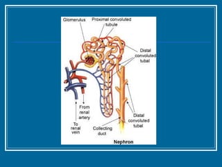
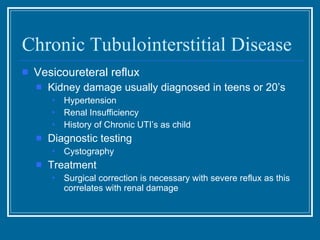
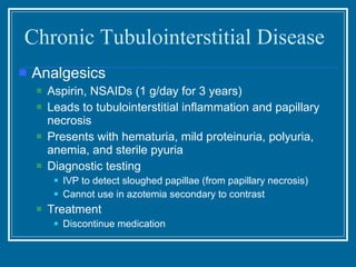
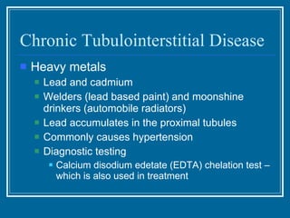
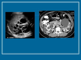
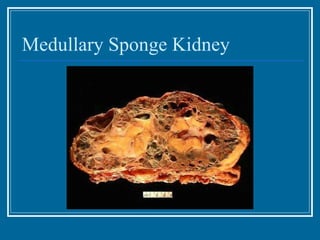
Non-Glomerular Diseases Clinical Medicine I Patrick Carter MPAS, PA-C February 25, 2011
Objectives Define tubulo-interstitial nephropathy  Differentiate between acute interstitial nephropathy and chronic interstitial nephropathy based upon:  Etiology – main causes both drug and non-drug  Clinical features  Laboratory findings Treatment Prognosis
Objectives Define polycystic kidney disease (PKD)  Review the prevalence of PKD and most common long term prognosis Review the diagnosis of PKD including: Etiology Clinical features Laboratory findings and imaging findings Use of gene therapy Treatment Prognosis Define medullary sponge kidney identifying presenting signs and symptoms, laboratory findings, and treatment
 
Tubulointerstitial Diseases May be acute or chronic Acute Most commonly associated with toxins and ischemia Characterized by interstitial edema, infiltration with PMN’s, and patchy areas of tubular cell necrosis Chronic Interstitial fibrosis and more widespread tubular abnormalities – atrophy, luminal dilatation, and thickening of the tubule basement membrane Mononuclear cell predominance
Acute Tubulointerstitial Disease Causes Drugs Antibiotics, NSAIDs, Diuretics, PPIs, H2 receptor blockers Infection  Bacteria – strep, staph, salmonella Viruses – EBV, CMV, HIV Ischemia Renal Artery Stenosis
Chronic Tubulointerstitial Disease Characterized by Decreased urinary concentrating ability Reduced GFR Small contracted kidneys Four main causes Obstructive uropathy – most common cause Vesicoureteral reflux – 2 nd  most common cause Analgesics – NSAID’s, ASA, High Dose Tylenol Heavy metals – Chronic Lead & Cadmium exposure
Chronic Tubulointerstitial Disease Obstructive uropathy Prolonged obstruction of the urinary tract Urinary output alternates between Polyuria and Oliguria Azotemia Hypertension Major causes are prostatic disease, bilateral ureteral calculi, and cancer } Due to increased renin-angiotensin production
Chronic Tubulointerstitial Disease Obstructive Uropathy Urinalysis -  Often negative but can show Hematuria Pyuria Bacteriuria CT scanning and MRI are best diagnostic tests Treatment Treat underlying obstruction
Chronic Tubulointerstitial Disease Vesicoureteral reflux Second most common cause Reflux is primarily a disorder of childhood Due to incompetent vesicoureteral sphincter Reflux of urine causes inflammatory response and fibrosis
Chronic Tubulointerstitial Disease Vesicoureteral reflux Kidney damage usually diagnosed in teens or 20’s Hypertension Renal Insufficiency History of Chronic UTI’s as child Diagnostic testing Cystography Treatment Surgical correction is necessary with severe reflux as this correlates with renal damage
Chronic Tubulointerstitial Disease Analgesics Aspirin, NSAIDs (1 g/day for 3 years) Leads to tubulointerstitial inflammation and papillary necrosis Presents with hematuria, mild proteinuria, polyuria, anemia, and sterile pyuria Diagnostic testing IVP to detect sloughed papillae (from papillary necrosis) Cannot use in azotemia secondary to contrast Treatment Discontinue medication
Chronic Tubulointerstitial Disease Heavy metals Lead and cadmium Welders (lead based paint) and moonshine drinkers (automobile radiators) Lead accumulates in the proximal tubules Commonly causes hypertension Diagnostic testing Calcium disodium edetate (EDTA) chelation test – which is also used in treatment
Chronic Tubulointerstitial Disease Signs and symptoms Polyuria Due to inability to concentrate the urine Dehydration  Laboratory findings Urinalysis is nonspecific Mild proteinuria Hyperkalemia Hyperchloremic renal tubular acidosis Reduced ammonia production Inability to acidify distal tubules Proximal tubule bicarbonate wasting
Chronic Tubulointerstitial Disease Prognosis Depends on the disorder responsible The degree of interstitial fibrosis can help predict recovery of renal function Once there is loss of parenchyma, progression to ESRD can’t be prevented
Polycystic Kidney Disease Key Features Common hereditary disease, affecting 500,000 individuals in United States End-stage renal disease develops in 50% of patients by age 60 10% of dialysis patients Family history is positive in 75%
Polycystic Kidney Disease Key Features Autosomal dominant Two genes account for this disorder  ADPKD1  on the short arm of chromosome 16 (85% of patients) ADPKD2  on chromosome 4 Seen primarily in adults Progressive expansion of numerous fluid-filled cysts results in massive enlargement of the kidneys
Polycystic Kidney Disease Signs and symptoms Often asymptomatic until their 50s  Abdominal or flank pain Abdominal mass HTN (50%) Fever (infection) Hematuria UTI **Combination of HTN and an abdominal mass suggests the disease
Polycystic Kidney Disease Extrarenal manifestations Likely due to abnormality in collagen and extracellular matrix SAH, cerebral hemorrhage - aneurysms in the circle of Willis in 10–15% Aortic aneurysms Aortic valve abnormalities – MVP, aortic regurg. Hepatic cysts Colonic diverticulae Abdominal and inguinal hernias
Polycystic Kidney Disease
Polycystic Kidney Disease Labs Urinalysis May be normal  Or show hematuria and mild proteinuria CBC Hgb and Hct often normal – erythropoietin produced by the cysts
Polycystic Kidney Disease Diagnosis Renal ultrasound  Diagnostic criteria 2 or > cysts in one kidney and at least 1 cyst in the contralateral kidney in < 30 years 2 or > cysts in each kidney age 30-59 4 or > cysts in subjects >60 years (increased frequency of benign simple cysts) CT scan or T2-weighted MRI is more sensitive in subjects <30 years of age Cerebral arteriography screening if family history of aneurysms
 
Polycystic Kidney Disease Treatment Largely supportive HTN Aggressive Treatment (<130 / 85 is recommended) Multidrug approach including agents to inhibit RAS Cyst rupture  Bed rest Analgesics, not NSAIDs Cyst pain  Drainage by percutaneous aspiration Sclerotherapy with alcohol Cyst infection Antibiotics: fluoroquinolones, trimethoprim-sulfamethoxazole
Polycystic Kidney Disease Prognosis Renal function declines progressively over the course of 10-20 years after diagnosis Risk factors for progressive kidney disease include – younger age at diagnosis, AA race, male gender, HTN,  ADPKD1  mutation Can undergo kidney transplantation
Medullary Sponge Kidney Relatively common and benign disorder Autosomal dominant mutations in the  MCKD1  or  MCKD2  genes  Present at birth but not usually diagnosed until the 40s Kidneys have irregular enlargement of the medullary and interpapillary collecting ducts Medullary cysts causing a “Swiss cheese” appearance
Medullary Sponge Kidney
Medullary Sponge Kidney Signs/symptoms/clinical findings Polyuria - Decreased urinary concentrating ability Gross or microscopic hematuria Recurrent UTI’s Nephrolithiasis Diagnosis IVP – striation in the papillary portions of the kidney U/S or CT – hyperdense papillae with medullary cysts
Medullary Sponge Kidney Treatment No known therapy Adequate fluid intake to prevent stone formation
Any Questions?

Non Glomerular Disease

  • 1.
    Non-Glomerular Diseases ClinicalMedicine I Patrick Carter MPAS, PA-C February 25, 2011
  • 2.
    Objectives Define tubulo-interstitialnephropathy Differentiate between acute interstitial nephropathy and chronic interstitial nephropathy based upon: Etiology – main causes both drug and non-drug Clinical features Laboratory findings Treatment Prognosis
  • 3.
    Objectives Define polycystickidney disease (PKD) Review the prevalence of PKD and most common long term prognosis Review the diagnosis of PKD including: Etiology Clinical features Laboratory findings and imaging findings Use of gene therapy Treatment Prognosis Define medullary sponge kidney identifying presenting signs and symptoms, laboratory findings, and treatment
  • 4.
  • 5.
    Tubulointerstitial Diseases Maybe acute or chronic Acute Most commonly associated with toxins and ischemia Characterized by interstitial edema, infiltration with PMN’s, and patchy areas of tubular cell necrosis Chronic Interstitial fibrosis and more widespread tubular abnormalities – atrophy, luminal dilatation, and thickening of the tubule basement membrane Mononuclear cell predominance
  • 6.
    Acute Tubulointerstitial DiseaseCauses Drugs Antibiotics, NSAIDs, Diuretics, PPIs, H2 receptor blockers Infection Bacteria – strep, staph, salmonella Viruses – EBV, CMV, HIV Ischemia Renal Artery Stenosis
  • 7.
    Chronic Tubulointerstitial DiseaseCharacterized by Decreased urinary concentrating ability Reduced GFR Small contracted kidneys Four main causes Obstructive uropathy – most common cause Vesicoureteral reflux – 2 nd most common cause Analgesics – NSAID’s, ASA, High Dose Tylenol Heavy metals – Chronic Lead & Cadmium exposure
  • 8.
    Chronic Tubulointerstitial DiseaseObstructive uropathy Prolonged obstruction of the urinary tract Urinary output alternates between Polyuria and Oliguria Azotemia Hypertension Major causes are prostatic disease, bilateral ureteral calculi, and cancer } Due to increased renin-angiotensin production
  • 9.
    Chronic Tubulointerstitial DiseaseObstructive Uropathy Urinalysis - Often negative but can show Hematuria Pyuria Bacteriuria CT scanning and MRI are best diagnostic tests Treatment Treat underlying obstruction
  • 10.
    Chronic Tubulointerstitial DiseaseVesicoureteral reflux Second most common cause Reflux is primarily a disorder of childhood Due to incompetent vesicoureteral sphincter Reflux of urine causes inflammatory response and fibrosis
  • 11.
    Chronic Tubulointerstitial DiseaseVesicoureteral reflux Kidney damage usually diagnosed in teens or 20’s Hypertension Renal Insufficiency History of Chronic UTI’s as child Diagnostic testing Cystography Treatment Surgical correction is necessary with severe reflux as this correlates with renal damage
  • 12.
    Chronic Tubulointerstitial DiseaseAnalgesics Aspirin, NSAIDs (1 g/day for 3 years) Leads to tubulointerstitial inflammation and papillary necrosis Presents with hematuria, mild proteinuria, polyuria, anemia, and sterile pyuria Diagnostic testing IVP to detect sloughed papillae (from papillary necrosis) Cannot use in azotemia secondary to contrast Treatment Discontinue medication
  • 13.
    Chronic Tubulointerstitial DiseaseHeavy metals Lead and cadmium Welders (lead based paint) and moonshine drinkers (automobile radiators) Lead accumulates in the proximal tubules Commonly causes hypertension Diagnostic testing Calcium disodium edetate (EDTA) chelation test – which is also used in treatment
  • 14.
    Chronic Tubulointerstitial DiseaseSigns and symptoms Polyuria Due to inability to concentrate the urine Dehydration Laboratory findings Urinalysis is nonspecific Mild proteinuria Hyperkalemia Hyperchloremic renal tubular acidosis Reduced ammonia production Inability to acidify distal tubules Proximal tubule bicarbonate wasting
  • 15.
    Chronic Tubulointerstitial DiseasePrognosis Depends on the disorder responsible The degree of interstitial fibrosis can help predict recovery of renal function Once there is loss of parenchyma, progression to ESRD can’t be prevented
  • 16.
    Polycystic Kidney DiseaseKey Features Common hereditary disease, affecting 500,000 individuals in United States End-stage renal disease develops in 50% of patients by age 60 10% of dialysis patients Family history is positive in 75%
  • 17.
    Polycystic Kidney DiseaseKey Features Autosomal dominant Two genes account for this disorder ADPKD1 on the short arm of chromosome 16 (85% of patients) ADPKD2 on chromosome 4 Seen primarily in adults Progressive expansion of numerous fluid-filled cysts results in massive enlargement of the kidneys
  • 18.
    Polycystic Kidney DiseaseSigns and symptoms Often asymptomatic until their 50s Abdominal or flank pain Abdominal mass HTN (50%) Fever (infection) Hematuria UTI **Combination of HTN and an abdominal mass suggests the disease
  • 19.
    Polycystic Kidney DiseaseExtrarenal manifestations Likely due to abnormality in collagen and extracellular matrix SAH, cerebral hemorrhage - aneurysms in the circle of Willis in 10–15% Aortic aneurysms Aortic valve abnormalities – MVP, aortic regurg. Hepatic cysts Colonic diverticulae Abdominal and inguinal hernias
  • 20.
  • 21.
    Polycystic Kidney DiseaseLabs Urinalysis May be normal Or show hematuria and mild proteinuria CBC Hgb and Hct often normal – erythropoietin produced by the cysts
  • 22.
    Polycystic Kidney DiseaseDiagnosis Renal ultrasound Diagnostic criteria 2 or > cysts in one kidney and at least 1 cyst in the contralateral kidney in < 30 years 2 or > cysts in each kidney age 30-59 4 or > cysts in subjects >60 years (increased frequency of benign simple cysts) CT scan or T2-weighted MRI is more sensitive in subjects <30 years of age Cerebral arteriography screening if family history of aneurysms
  • 23.
  • 24.
    Polycystic Kidney DiseaseTreatment Largely supportive HTN Aggressive Treatment (<130 / 85 is recommended) Multidrug approach including agents to inhibit RAS Cyst rupture Bed rest Analgesics, not NSAIDs Cyst pain Drainage by percutaneous aspiration Sclerotherapy with alcohol Cyst infection Antibiotics: fluoroquinolones, trimethoprim-sulfamethoxazole
  • 25.
    Polycystic Kidney DiseasePrognosis Renal function declines progressively over the course of 10-20 years after diagnosis Risk factors for progressive kidney disease include – younger age at diagnosis, AA race, male gender, HTN, ADPKD1 mutation Can undergo kidney transplantation
  • 26.
    Medullary Sponge KidneyRelatively common and benign disorder Autosomal dominant mutations in the MCKD1 or MCKD2 genes Present at birth but not usually diagnosed until the 40s Kidneys have irregular enlargement of the medullary and interpapillary collecting ducts Medullary cysts causing a “Swiss cheese” appearance
  • 27.
  • 28.
    Medullary Sponge KidneySigns/symptoms/clinical findings Polyuria - Decreased urinary concentrating ability Gross or microscopic hematuria Recurrent UTI’s Nephrolithiasis Diagnosis IVP – striation in the papillary portions of the kidney U/S or CT – hyperdense papillae with medullary cysts
  • 29.
    Medullary Sponge KidneyTreatment No known therapy Adequate fluid intake to prevent stone formation
  • 30.

Editor's Notes

  • #9 Cancer of the cervix, colon and bladder