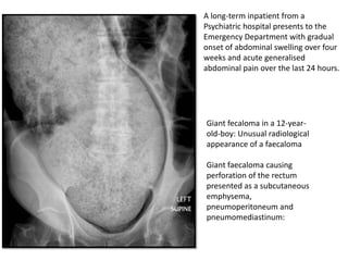
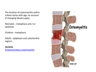
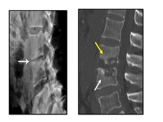
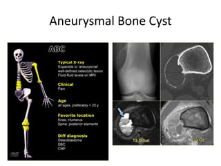
DR.MITUSHA VERMA
DEPT.OF RADIODIAGNOSIS
Beyond Chest in Radiographs….
Basis of X-rays…
Key points
• An X-ray image is a map of
X-ray attenuation
• Attenuation of X-rays is
variable depending on
density and thickness of
tissues
• Describing X-ray
abnormalities in terms of
density may help in
determining the tissue
involved
Plain Abdominal Radiographs
RCR guidelines for the use of plain abdominal radiography
Acute abdominal pain: if perforation or obstruction suspected
Acute small or large bowel obstruction
Inflammatory bowel disease of the colon: acute exacerbation
Palpable mass (indicated in specific circumstances)
Constipation (indicated in specific circumstances)
Acute and chronic pancreatitis
Suspected ureteric colic/stones
Renal failure
Haematuria
Foreign body in pharynx/upper oesophagus
Smooth and small foreign body, eg, coin
Sharp/poisonous foreign body
Blunt or stab abdominal injury
Perforation
Key points
• Bowel perforation is a surgical emergency
• An ERECT chest X-ray should be requested if
perforation is suspected
• Be familiar with Rigler's sign
Rigler's/double wall sign - example
The double wall (Rigler's) sign is visible
Gas separates bowel segments and forms
sharp angles and triangles (*)
Football sign –
A large volume of free gas has risen to the
front of the peritoneal cavity resulting in a
large round black area - 'football sign‘
Liver edge -
Gas may be seen outlining soft tissues structures such as the
falciform ligament, or the liver edge
Free gas mimics
Normal stomach bubble - erect chest X-ray
Round/ovoid - 'bubble' shape
Thick upper wall
Fluid level or food contents
Chilaiditi's phenomenon -
Gas forms a near crescent shape under the
right hemidiaphragm
There is however a thick hemidiaphragm
(partly consisting of bowel wall)
Gas can be seen to lie within bowel
Importantly, this patient with hyperexpanded
lungs, due to emphysema, did not have acute
abdominal pain
False Rigler's/double wall sign
Gas seen on both sides of the bowel wall is
contained within adjacent bowel
There are no black triangles or sharp angles on
the outside of the bowel wall
False football sign - example
1 - Perirenal fat (retroperitoneal)
2 - Peritoneal fat (next to the liver)
3 - Abdominal wall fat (separating muscles of
the abdominal wall)
Small Bowel Obstruction
Key points
Dilated small bowel >3cm is considered abnormal
Small bowel obstruction and ileus can have similar
appearances
Large Bowel Obstruction
Key points
Dilatation of the caecum >9cm is abnormal
Dilatation of any other part of the colon
>6cm is abnormal
Abdominal X-ray may demonstrate the level
of obstruction
Abdominal X-ray cannot reliably differentiate
mechanical obstruction from pseudo-
obstruction
Volvulus
Sigmoid volvulus - coffee bean sign
Sigmoid volvulus classically results in the
formation of a loop of sigmoid colon, which is
twisted at the root of the sigmoid mesentery,
which lies in the left iliac fossa (LIF).
The loop of dilated bowel usually points
upwards towards the diaphragm
Bowel wall inflammation
Mucosal thickening - 'thumbprinting'
Lead pipe colon
This patient with ulcerative colitis has a featureless segment of transverse colon with shows
loss of the normal haustral markings.
Toxic megacolon
The colon is very dilated in this patient with
acute abdominal pain, sepsis, and a known
history of ulcerative colitis. The clinical features
and X-ray appearances are consistent with
toxic megacolon.
There is evidence of bowel wall oedema with
'thumbprinting', and pseudopolyps or 'mucosal
islands'.
A long-term inpatient from a
Psychiatric hospital presents to the
Emergency Department with gradual
onset of abdominal swelling over four
weeks and acute generalised
abdominal pain over the last 24 hours.
Giant fecaloma in a 12-year-
old-boy: Unusual radiological
appearance of a faecaloma
Giant faecaloma causing
perforation of the rectum
presented as a subcutaneous
emphysema,
pneumoperitoneum and
pneumomediastinum:
Key Points…
Suspected bowel obstruction or perforation are the main
indications for abdominal X-ray
An ERECT chest X-ray should be requested if perforation is
suspected
The pattern of bowel dilatation may help determine a level of
obstruction
Occasionally features of inflammatory bowel disease are
demonstrated on abdominal x-rays
BONES….
Osteomyelitis
Plain film
Earliest in adjacent soft tissues +/- muscle outlines with swelling
and loss/blurring of normal fat planes.
 An effusion may be seen in an adjacent joint.
Osteomyelitis must extend at least 1 cm and compromise 30 to
50% of bone mineral content to produce noticeable changes in plain
radiographs.
OM may not be obvious until 5 to 7 days in children and 10 to 14
days in adults.
Osteomyelitis refers to bony inflammation that is almost always due
to infection, typically bacterial
 Regional osteopaenia
 Periosteal reaction / periosteal thickening -
variable, and may appear aggressive including
formation of a Codman's triangle
 Focal bony lysis
 Endosteal scalloping
 Loss of bony trabecular architecture
 New bone apposition
 Eventual peripheral sclerosis
The location of osteomyelitis within
a bone varies with age, on account
of changing blood supply:
Neonates - metaphysis and / or
epiphysis
Children - metaphysis
Adults - epiphyses and subchondral
regions
Variants
Emphysematous osteomyelitis
Tumours
Aneurysmal Bone Cyst
Enchondroma
The differential diagnosis based on the
radiograph is:
 fibrous dysplasia,
 enchondroma,
 eosinophilic granuloma
 hemangioma.
The coronal T1-WI after Gd with fatsat
shows a lobulated lesion with peripheral
enhancement consistent with
the diagnosis of an enchondroma.
Eosinophilic granuloma
Ewings Sarcoma
Typical presentation: ill-defined osteolytic
lesion with a moth-eaten or permeative type
of bone destruction, irregular cortical
destruction and aggressive periostitis in the
lower extremity of a child.
Plain radiographs usually illustrate the
malignant nature.
Based on the age, the location and the
radiographic appearance the diagnosis of
Ewing sarcoma can be made in over 70% of
cases.
In long bones, the tumor is most commonly
located centrally in the meta- or diaphysis
MR imaging reveals the soft tissue extension.
Giant Cell Tumour
ARTHRITIS….
Soft-tissue swelling and early
erosions in the proximal
interphalangeal joints
Prominent juxta-articular osteopenia
in all interphalangeal joints
RHEUMATOIDARTHRITIS
Subluxation in the metacarpophalangeal joints,
with ulnar deviation
Marked ankylosis
Partial collapse of fused carpal bones with
subluxation at the radiocarpal joint
Concentric joint-space loss. Subchondral erosions and sclerosis of the
femoral head
Septic Arthritis…
The earliest plain film radiographic findings of septic arthritis are
soft tissue swelling around the joint and a widened joint space from
joint effusion.
Osteonecrosis and complete collapse of the femoral head are present
GOUT
Sclerosis and joint-space narrowing are seen
in the first metatarsophalangeal joint, as well
as in the fourth interphalangeal joint
Extensive bony erosions are noted throughout
the carpal bones. Urate depositions may be
present in the periarticular areas.
ANKYLOSING SPONDYLITIS
Bilateral sacroiliac joint erosions and iliac side
subchondral sclerosis
Complete fusion of both sacroiliac jointsNormal SI Joint.
Bamboo spine. Frontal radiograph shows
complete fusion of the vertebral bodies.
Extensive facet joint ankylosis and posterior
ligamentous ossification produce the trolley
track appearance
Vertebral fusion. Lateral radiograph shows
solid ankylosis of all cervical facet joints from
C2 downwards. Extensive anterior and
posterior syndesmophytes are noted.
Destruction of intervetebral disc and
adjacent vertebral body
-Early course there will be narrowing of
disc space + erosion of adjoining surface
of vertebral body.
-Later, bone destruction may lead to
collapse of the vertebral body, forming
the gibbus (sharp angulations)
-Paravetebral abscess may present
-Bony fusion of vertebral bodies across
obliterated disc space when healing
occurs
POTT’S SPINE
Thank you…

Beyond chest in radiographs

  • 1.
  • 2.
    Basis of X-rays… Keypoints • An X-ray image is a map of X-ray attenuation • Attenuation of X-rays is variable depending on density and thickness of tissues • Describing X-ray abnormalities in terms of density may help in determining the tissue involved
  • 3.
    Plain Abdominal Radiographs RCRguidelines for the use of plain abdominal radiography Acute abdominal pain: if perforation or obstruction suspected Acute small or large bowel obstruction Inflammatory bowel disease of the colon: acute exacerbation Palpable mass (indicated in specific circumstances) Constipation (indicated in specific circumstances) Acute and chronic pancreatitis Suspected ureteric colic/stones Renal failure Haematuria Foreign body in pharynx/upper oesophagus Smooth and small foreign body, eg, coin Sharp/poisonous foreign body Blunt or stab abdominal injury
  • 4.
    Perforation Key points • Bowelperforation is a surgical emergency • An ERECT chest X-ray should be requested if perforation is suspected • Be familiar with Rigler's sign
  • 5.
    Rigler's/double wall sign- example The double wall (Rigler's) sign is visible Gas separates bowel segments and forms sharp angles and triangles (*)
  • 6.
    Football sign – Alarge volume of free gas has risen to the front of the peritoneal cavity resulting in a large round black area - 'football sign‘ Liver edge - Gas may be seen outlining soft tissues structures such as the falciform ligament, or the liver edge
  • 7.
    Free gas mimics Normalstomach bubble - erect chest X-ray Round/ovoid - 'bubble' shape Thick upper wall Fluid level or food contents Chilaiditi's phenomenon - Gas forms a near crescent shape under the right hemidiaphragm There is however a thick hemidiaphragm (partly consisting of bowel wall) Gas can be seen to lie within bowel Importantly, this patient with hyperexpanded lungs, due to emphysema, did not have acute abdominal pain
  • 8.
    False Rigler's/double wallsign Gas seen on both sides of the bowel wall is contained within adjacent bowel There are no black triangles or sharp angles on the outside of the bowel wall False football sign - example 1 - Perirenal fat (retroperitoneal) 2 - Peritoneal fat (next to the liver) 3 - Abdominal wall fat (separating muscles of the abdominal wall)
  • 9.
    Small Bowel Obstruction Keypoints Dilated small bowel >3cm is considered abnormal Small bowel obstruction and ileus can have similar appearances
  • 10.
    Large Bowel Obstruction Keypoints Dilatation of the caecum >9cm is abnormal Dilatation of any other part of the colon >6cm is abnormal Abdominal X-ray may demonstrate the level of obstruction Abdominal X-ray cannot reliably differentiate mechanical obstruction from pseudo- obstruction
  • 11.
    Volvulus Sigmoid volvulus -coffee bean sign Sigmoid volvulus classically results in the formation of a loop of sigmoid colon, which is twisted at the root of the sigmoid mesentery, which lies in the left iliac fossa (LIF). The loop of dilated bowel usually points upwards towards the diaphragm
  • 12.
    Bowel wall inflammation Mucosalthickening - 'thumbprinting'
  • 13.
    Lead pipe colon Thispatient with ulcerative colitis has a featureless segment of transverse colon with shows loss of the normal haustral markings.
  • 14.
    Toxic megacolon The colonis very dilated in this patient with acute abdominal pain, sepsis, and a known history of ulcerative colitis. The clinical features and X-ray appearances are consistent with toxic megacolon. There is evidence of bowel wall oedema with 'thumbprinting', and pseudopolyps or 'mucosal islands'.
  • 15.
    A long-term inpatientfrom a Psychiatric hospital presents to the Emergency Department with gradual onset of abdominal swelling over four weeks and acute generalised abdominal pain over the last 24 hours. Giant fecaloma in a 12-year- old-boy: Unusual radiological appearance of a faecaloma Giant faecaloma causing perforation of the rectum presented as a subcutaneous emphysema, pneumoperitoneum and pneumomediastinum:
  • 16.
    Key Points… Suspected bowelobstruction or perforation are the main indications for abdominal X-ray An ERECT chest X-ray should be requested if perforation is suspected The pattern of bowel dilatation may help determine a level of obstruction Occasionally features of inflammatory bowel disease are demonstrated on abdominal x-rays
  • 17.
  • 18.
    Osteomyelitis Plain film Earliest inadjacent soft tissues +/- muscle outlines with swelling and loss/blurring of normal fat planes.  An effusion may be seen in an adjacent joint. Osteomyelitis must extend at least 1 cm and compromise 30 to 50% of bone mineral content to produce noticeable changes in plain radiographs. OM may not be obvious until 5 to 7 days in children and 10 to 14 days in adults. Osteomyelitis refers to bony inflammation that is almost always due to infection, typically bacterial
  • 19.
     Regional osteopaenia Periosteal reaction / periosteal thickening - variable, and may appear aggressive including formation of a Codman's triangle  Focal bony lysis  Endosteal scalloping  Loss of bony trabecular architecture  New bone apposition  Eventual peripheral sclerosis
  • 20.
    The location ofosteomyelitis within a bone varies with age, on account of changing blood supply: Neonates - metaphysis and / or epiphysis Children - metaphysis Adults - epiphyses and subchondral regions Variants Emphysematous osteomyelitis
  • 22.
  • 23.
  • 24.
  • 25.
    The differential diagnosisbased on the radiograph is:  fibrous dysplasia,  enchondroma,  eosinophilic granuloma  hemangioma. The coronal T1-WI after Gd with fatsat shows a lobulated lesion with peripheral enhancement consistent with the diagnosis of an enchondroma.
  • 26.
  • 27.
    Ewings Sarcoma Typical presentation:ill-defined osteolytic lesion with a moth-eaten or permeative type of bone destruction, irregular cortical destruction and aggressive periostitis in the lower extremity of a child. Plain radiographs usually illustrate the malignant nature. Based on the age, the location and the radiographic appearance the diagnosis of Ewing sarcoma can be made in over 70% of cases. In long bones, the tumor is most commonly located centrally in the meta- or diaphysis MR imaging reveals the soft tissue extension.
  • 28.
  • 29.
  • 30.
    Soft-tissue swelling andearly erosions in the proximal interphalangeal joints Prominent juxta-articular osteopenia in all interphalangeal joints RHEUMATOIDARTHRITIS
  • 31.
    Subluxation in themetacarpophalangeal joints, with ulnar deviation Marked ankylosis Partial collapse of fused carpal bones with subluxation at the radiocarpal joint
  • 32.
    Concentric joint-space loss.Subchondral erosions and sclerosis of the femoral head Septic Arthritis… The earliest plain film radiographic findings of septic arthritis are soft tissue swelling around the joint and a widened joint space from joint effusion.
  • 33.
    Osteonecrosis and completecollapse of the femoral head are present
  • 34.
    GOUT Sclerosis and joint-spacenarrowing are seen in the first metatarsophalangeal joint, as well as in the fourth interphalangeal joint Extensive bony erosions are noted throughout the carpal bones. Urate depositions may be present in the periarticular areas.
  • 35.
    ANKYLOSING SPONDYLITIS Bilateral sacroiliacjoint erosions and iliac side subchondral sclerosis Complete fusion of both sacroiliac jointsNormal SI Joint.
  • 36.
    Bamboo spine. Frontalradiograph shows complete fusion of the vertebral bodies. Extensive facet joint ankylosis and posterior ligamentous ossification produce the trolley track appearance Vertebral fusion. Lateral radiograph shows solid ankylosis of all cervical facet joints from C2 downwards. Extensive anterior and posterior syndesmophytes are noted.
  • 37.
    Destruction of intervetebraldisc and adjacent vertebral body -Early course there will be narrowing of disc space + erosion of adjoining surface of vertebral body. -Later, bone destruction may lead to collapse of the vertebral body, forming the gibbus (sharp angulations) -Paravetebral abscess may present -Bony fusion of vertebral bodies across obliterated disc space when healing occurs POTT’S SPINE
  • 41.