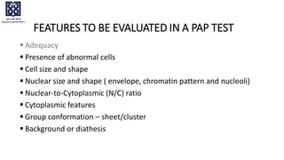
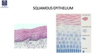
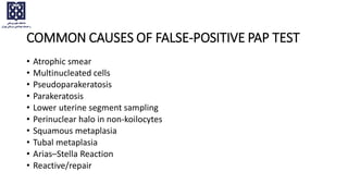
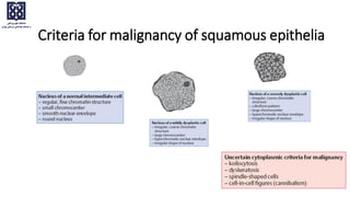
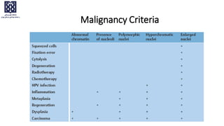
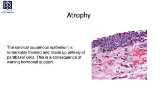
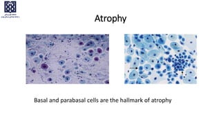
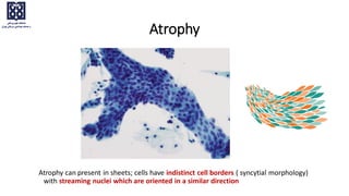
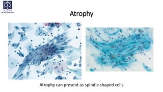
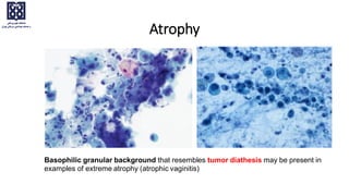
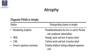
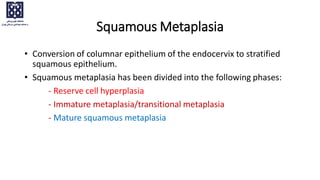
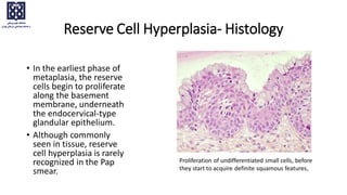
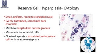
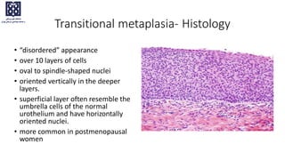
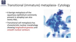
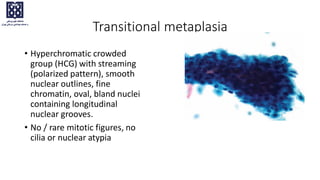
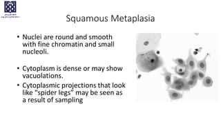
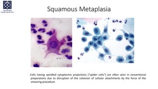
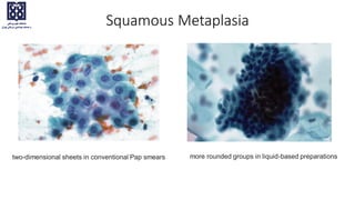
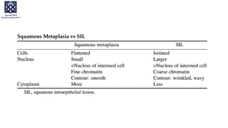
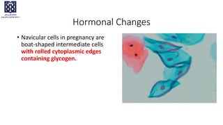
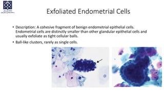
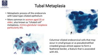
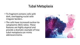
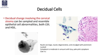
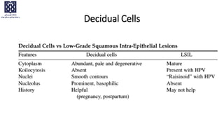
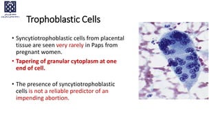
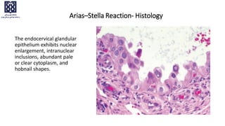
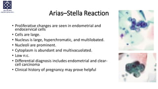
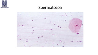
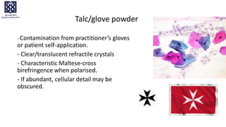
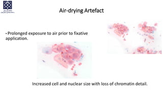
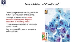
NEGATIVE FOR INTRAEPITHELIAL LESION AND MALIGNANCY
NON-NEOPLASTIC CELLULAR VARIATIONS
Dr Fereshteh Ameli
FEATURES TO BE EVALUATED IN A PAP TEST
 Adequacy
 Presence of abnormal cells
 Cell size and shape
 Nuclear size and shape ( envelope, chromatin pattern and nucleoli)
 Nuclear-to-Cytoplasmic (N/C) ratio
 Cytoplasmic features
 Group conformation – sheet/cluster
 Background or diathesis
SQUAMOUS EPITHELIUM
COMMON CAUSES OF FALSE-POSITIVE PAP TEST
• Atrophic smear
• Multinucleated cells
• Pseudoparakeratosis
• Parakeratosis
• Lower uterine segment sampling
• Perinuclear halo in non-koilocytes
• Squamous metaplasia
• Tubal metaplasia
• Arias–Stella Reaction
• Reactive/repair
Criteria for malignancy of squamous epithelia
Malignancy Criteria
Reliability of the criteria
• None of the criteria for malignancy is absolutely reliable, since they
occasionally also occur with certain benign processes.
• The more criteria are detected simultaneously in the cytological
smear, the more likely they are to indicate the existence of a
malignant lesion.
Atrophy
The cervical squamous epithelium is
remarkably thinned and made up entirely of
parabasal cells. This is a consequence of
waning hormonal support.
Atrophic Smear Pattern
• Menopause
• Postpartum and lactation (glycogeneated parabasal cells)
• Depo-Provera or other progestational drugs
• Bilateral oophorectomy
• Pituitary dysfunction
• Ovarian insufficiency
• Sever hypothyroidism
• Androgens (exogenous,tumor)
• Turner’s syndrome
Basal and parabasal cells are the hallmark of atrophy
Atrophy
Atrophy can present in sheets; cells have indistinct cell borders ( syncytial morphology)
with streaming nuclei which are oriented in a similar direction
Atrophy
Atrophy
Atrophy can present as spindle shaped cells
Basophilic granular background that resembles tumor diathesis may be present in
examples of extreme atrophy (atrophic vaginitis)
Atrophy
Atrophy
Pseudoparakeratosis in atrophic vaginitis
Multinucleated histiocytic giant cells are a nonspecific
finding & are often seen in postmenopausal and postpartum
specimens.
Atrophy
DIFFERENTIAL DIAGNOSIS OF GIANT MULTINUCLEATED CELLS
• Atrophy
• Folic acid deficiency
• Tissue repair
• Viral infection
• Granuloma
• Radiation
• Syncytiotrophoblast
• Squamous carcinoma
Dark blue blobs (thought to represent either condensed mucus or
degenerated bare nuclei ) are seen in some atrophic smears.
Atrophy
Atrophy
Keratotic cellular changes: Hyperkeratosis
• Hyperkeratosis is a benign
response of stratified squamous
epithelium due to chronic
mucosal irritation, as in uterine
prolapse.
• The keratotic surface looks white,
which is termed leukoplakia
Anucleate, mature, polygonal squamous cells appear as
isolated cells or plaques of tightly adherent cells with ghostlike
“nuclear holes”.
• Keratosis may cover a lesion, thereby masking it. Thus, the underlying
abnormality may not be sampled when taking the Pap smear, which
only scrapes the mucosal surface.
• When extensive hyperkeratosis is present, an underlying neoplastic or
nonneoplastic process may be associated and should be considered.
• Patients with extensive clusters should be followed more closely to
rule out a hidden underlying dysplasia or cancer
Keratotic cellular changes: Hyperkeratosis
• A large fragment of anucleated
squames is unusual and urges
careful search for a keratinizing
neoplasm.
• If none is found, the
hyperkeratosis may be due to
uterine prolapse alone, or with use
of a pessary.
• Clinical correlation is needed to
define the reason for the keratosis.
Keratotic cellular changes: Hyperkeratosis
Keratotic cellular changes: Parakeratosis
• Parakeratosis, a benign reactive change
also caused by chronic irritation, is
characterized by small, heavily
keratinized squamous cells with dense
orangeophilic cytoplasm and small,
pyknotic nuclei.
• If all cells have the same small-size
nuclei, and are in an ordered pattern,
the diagnosis is benign.
• If atypical nuclear changes are
present, an atypical squamous cell
(ASC-US/ASCH) or SIL interpretation
should be considered.
Keratotic cellular changes: Parakeratosis
Nuclei within it are small and bland
Lower uterine segment sampling
• Aggressive sampling may result in the
presence of cells from the lower uterine
segment in cervicovaginal samples.
• Cells from the lower uterine segment
generally present as a tissue fragment
rather than individual cells or small groups
of cells.
• Biphasic tissue fragments with densely
packed spindle cells and vascular spaces.
Capillaries are seen traversing the stroma.
Lower uterine segment sampling
• Conversion of columnar epithelium of the endocervix to stratified
squamous epithelium.
• Squamous metaplasia has been divided into the following phases:
- Reserve cell hyperplasia
- Immature metaplasia/transitional metaplasia
- Mature squamous metaplasia
Squamous Metaplasia
• In the earliest phase of
metaplasia, the reserve
cells begin to proliferate
along the basement
membrane, underneath
the endocervical-type
glandular epithelium.
• Although commonly
seen in tissue, reserve
cell hyperplasia is rarely
recognized in the Pap
smear.
Reserve Cell Hyperplasia- Histology
Proliferation of undifferentiated small cells, before
they start to acquire definite squamous features,
Reserve Cell Hyperplasia- Cytology
• Small, uniform, round to elongated nuclei
• Evenly distributed, sometimes dark
chromatin
• May have longitudinal nuclear grooves
• May mimic endometrial cells.
• Clue to diagnosis is associated endocervical
cells or immature metaplasia.
Transitional metaplasia- Histology
• “disordered” appearance
• over 10 layers of cells
• oval to spindle-shaped nuclei
• oriented vertically in the deeper
layers.
• superficial layer often resemble the
umbrella cells of the normal
urothelium and have horizontally
oriented nuclei.
• more common in postmenopausal
women
• A benign metaplasia of the
squamous epithelium commonly
present in atrophy) can also
mimic HSIL.
• Transitional cell metaplasia has
characteristic nuclear morphology
showing longitudinal grooves and
smooth nuclear contours
Transitional (immature) metaplasia- Cytology
Transitional metaplasia
• Hyperchromatic crowded
group (HCG) with streaming
(polarized pattern), smooth
nuclear outlines, fine
chromatin, oval, bland nuclei
containing longitudinal
nuclear grooves.
• No / rare mitotic figures, no
cilia or nuclear atypia
Squamous Metaplasia
• Nuclei are round and smooth
with fine chromatin and small
nucleoli.
• Cytoplasm is dense or may show
vacuolations.
• Cytoplasmic projections that look
like “spider legs” may be seen as
a result of sampling
Cells having spindled cytoplasmic projections (“spider cells”) are often seen in conventional
preparations due to disruption of the cohesion of cellular attachments by the force of the
smearing procedure.
Squamous Metaplasia
more rounded groups in liquid-based preparations
two-dimensional sheets in conventional Pap smears
Squamous Metaplasia
Hormonal Changes
• Navicular cells in pregnancy are
boat-shaped intermediate cells
with rolled cytoplasmic edges
containing glycogen.
Exfoliated Endometrial Cells
• Description: A cohesive fragment of benign endometrial epithelial cells.
Endometrial cells are distinctly smaller than other glandular epithelial cells and
usually exfoliate as tight cellular balls.
• Ball-like clusters, rarely as single cells.
Significance of endometrial cells depends on:
• Age of patient.
• Last menstrual period.
• Hormone therapy status
• Abnormal uterine bleeding
• IUD use.
• Recent instrumentation
• Presence of endometriosis
Endometrial Cells vs Endocervical Cells
• Endometrial cells are packed together with scant cytoplasm.
• Endocervical cells form looser clusters and are more abundant
cytoplasm
• Endometrial cells in women 45 years or older may be a/w benign
endometrium, hormonal alterations, and, less commonly,
endometrial or uterine abnormalities. Endometrial evaluation is
recommended in postmenopausal women.
• Woman’s LMP is provided: Endometrial cells correlate with the
menstrual history provided.
Exfoliated Endometrial Cells
Tubal Metaplasia
• Metaplastic process of the endocervix
with tubal-type ciliated epithelium
• More common in women aged 35 or
older; also known as “ciliated cell”
metaplasia, mimics glandular neoplasia
particularly AIS.
Columnar ciliated endocervical cells that may
occur in small groups or as pseudostratified
crowded groups almost appear to form a
feathered border, a feature that is associated
with AIS.
• Tis fragment contains cells with
dark, overlapping nuclei with
irregular borders.
• The cells have increased nuclear-to-
cytoplasmic (N/C) ratios. These
features are highly atypical and
provide a dramatic example of how
tubal metaplasia can mimic
adenocarcinoma.
Tubal Metaplasia
Decidual Cells
• Decidual change involving the cervical
stroma can be sampled and resemble
epithelial cell abnormalities, both LSIL
and HSIL.
Nuclei are large, round, degenerative, and smudged with prominent
nucleoli
Cytoplasm is moderate in amount with lacy, pale-pink cytoplasm
Low n:c
Decidual Cells
Trophoblastic Cells
• Syncytiotrophoblastic cells from placental
tissue are seen very rarely in Paps from
pregnant women.
• Tapering of granular cytoplasm at one
end of cell.
• The presence of syncytiotrophoblastic
cells is not a reliable predictor of an
impending abortion.
The endocervical glandular
epithelium exhibits nuclear
enlargement, intranuclear
inclusions, abundant pale
or clear cytoplasm, and
hobnail shapes.
Arias–Stella Reaction- Histology
Arias–Stella Reaction
• Proliferative changes are seen in endometrial and
endocervical cells
• Cells are large.
• Nucleus is large, hyperchromatic, and multilobated.
• Nucleoli are prominent.
• Cytoplasm is abundant and multivacuolated.
• Low n:c.
• Differential diagnosis includes endometrial and clear-
cell carcinoma
• Clinical history of pregnancy may prove helpful
Miscellaneous components of smears
Spermatozoa
Talc/glove powder
- Contamination from practitioner’s gloves
or patient self-application.
- Clear/translucent refractile crystals
- Characteristic Maltese-cross
birefringence when polarised.
- If abundant, cellular detail may be
obscured.
Air-drying Artefact
• Prolonged exposure to air prior to fixative
application.
Increased cell and nuclear size with loss of chromatin detail.
Brown Artefact – “Corn Flakes”
• Air trapping between surface grooves of
mature squamous cells and coverslip.
• Thought to be caused by a delay
between last the xylene stage and
application of mounting media.
• Brown granular cytoplasmic staining in
squamous cells
-Can be removed by reverse processing
and re-staining.
Lubricant
• Contamination from lubricant used
when inserting speculum.
• Intense blue/purple staining material,
which can obscure cellular material.
Pap smaer cytology

Pap smaer cytology

  • 2.
    NEGATIVE FOR INTRAEPITHELIALLESION AND MALIGNANCY NON-NEOPLASTIC CELLULAR VARIATIONS Dr Fereshteh Ameli
  • 3.
    FEATURES TO BEEVALUATED IN A PAP TEST  Adequacy  Presence of abnormal cells  Cell size and shape  Nuclear size and shape ( envelope, chromatin pattern and nucleoli)  Nuclear-to-Cytoplasmic (N/C) ratio  Cytoplasmic features  Group conformation – sheet/cluster  Background or diathesis
  • 4.
  • 5.
    COMMON CAUSES OFFALSE-POSITIVE PAP TEST • Atrophic smear • Multinucleated cells • Pseudoparakeratosis • Parakeratosis • Lower uterine segment sampling • Perinuclear halo in non-koilocytes • Squamous metaplasia • Tubal metaplasia • Arias–Stella Reaction • Reactive/repair
  • 6.
    Criteria for malignancyof squamous epithelia
  • 7.
  • 8.
    Reliability of thecriteria • None of the criteria for malignancy is absolutely reliable, since they occasionally also occur with certain benign processes. • The more criteria are detected simultaneously in the cytological smear, the more likely they are to indicate the existence of a malignant lesion.
  • 9.
    Atrophy The cervical squamousepithelium is remarkably thinned and made up entirely of parabasal cells. This is a consequence of waning hormonal support.
  • 10.
    Atrophic Smear Pattern •Menopause • Postpartum and lactation (glycogeneated parabasal cells) • Depo-Provera or other progestational drugs • Bilateral oophorectomy • Pituitary dysfunction • Ovarian insufficiency • Sever hypothyroidism • Androgens (exogenous,tumor) • Turner’s syndrome
  • 11.
    Basal and parabasalcells are the hallmark of atrophy Atrophy
  • 12.
    Atrophy can presentin sheets; cells have indistinct cell borders ( syncytial morphology) with streaming nuclei which are oriented in a similar direction Atrophy
  • 13.
    Atrophy Atrophy can presentas spindle shaped cells
  • 14.
    Basophilic granular backgroundthat resembles tumor diathesis may be present in examples of extreme atrophy (atrophic vaginitis) Atrophy
  • 15.
  • 16.
    Multinucleated histiocytic giantcells are a nonspecific finding & are often seen in postmenopausal and postpartum specimens. Atrophy
  • 17.
    DIFFERENTIAL DIAGNOSIS OFGIANT MULTINUCLEATED CELLS • Atrophy • Folic acid deficiency • Tissue repair • Viral infection • Granuloma • Radiation • Syncytiotrophoblast • Squamous carcinoma
  • 18.
    Dark blue blobs(thought to represent either condensed mucus or degenerated bare nuclei ) are seen in some atrophic smears. Atrophy
  • 19.
  • 20.
    Keratotic cellular changes:Hyperkeratosis • Hyperkeratosis is a benign response of stratified squamous epithelium due to chronic mucosal irritation, as in uterine prolapse. • The keratotic surface looks white, which is termed leukoplakia Anucleate, mature, polygonal squamous cells appear as isolated cells or plaques of tightly adherent cells with ghostlike “nuclear holes”.
  • 21.
    • Keratosis maycover a lesion, thereby masking it. Thus, the underlying abnormality may not be sampled when taking the Pap smear, which only scrapes the mucosal surface. • When extensive hyperkeratosis is present, an underlying neoplastic or nonneoplastic process may be associated and should be considered. • Patients with extensive clusters should be followed more closely to rule out a hidden underlying dysplasia or cancer Keratotic cellular changes: Hyperkeratosis
  • 22.
    • A largefragment of anucleated squames is unusual and urges careful search for a keratinizing neoplasm. • If none is found, the hyperkeratosis may be due to uterine prolapse alone, or with use of a pessary. • Clinical correlation is needed to define the reason for the keratosis. Keratotic cellular changes: Hyperkeratosis
  • 23.
    Keratotic cellular changes:Parakeratosis • Parakeratosis, a benign reactive change also caused by chronic irritation, is characterized by small, heavily keratinized squamous cells with dense orangeophilic cytoplasm and small, pyknotic nuclei.
  • 24.
    • If allcells have the same small-size nuclei, and are in an ordered pattern, the diagnosis is benign. • If atypical nuclear changes are present, an atypical squamous cell (ASC-US/ASCH) or SIL interpretation should be considered. Keratotic cellular changes: Parakeratosis Nuclei within it are small and bland
  • 25.
    Lower uterine segmentsampling • Aggressive sampling may result in the presence of cells from the lower uterine segment in cervicovaginal samples. • Cells from the lower uterine segment generally present as a tissue fragment rather than individual cells or small groups of cells. • Biphasic tissue fragments with densely packed spindle cells and vascular spaces. Capillaries are seen traversing the stroma. Capillaries are seen traversing the stroma.
  • 26.
  • 27.
    • Conversion ofcolumnar epithelium of the endocervix to stratified squamous epithelium. • Squamous metaplasia has been divided into the following phases: - Reserve cell hyperplasia - Immature metaplasia/transitional metaplasia - Mature squamous metaplasia Squamous Metaplasia
  • 28.
    • In theearliest phase of metaplasia, the reserve cells begin to proliferate along the basement membrane, underneath the endocervical-type glandular epithelium. • Although commonly seen in tissue, reserve cell hyperplasia is rarely recognized in the Pap smear. Reserve Cell Hyperplasia- Histology Proliferation of undifferentiated small cells, before they start to acquire definite squamous features,
  • 29.
    Reserve Cell Hyperplasia-Cytology • Small, uniform, round to elongated nuclei • Evenly distributed, sometimes dark chromatin • May have longitudinal nuclear grooves • May mimic endometrial cells. • Clue to diagnosis is associated endocervical cells or immature metaplasia.
  • 30.
    Transitional metaplasia- Histology •“disordered” appearance • over 10 layers of cells • oval to spindle-shaped nuclei • oriented vertically in the deeper layers. • superficial layer often resemble the umbrella cells of the normal urothelium and have horizontally oriented nuclei. • more common in postmenopausal women
  • 31.
    • A benignmetaplasia of the squamous epithelium commonly present in atrophy) can also mimic HSIL. • Transitional cell metaplasia has characteristic nuclear morphology showing longitudinal grooves and smooth nuclear contours Transitional (immature) metaplasia- Cytology
  • 32.
    Transitional metaplasia • Hyperchromaticcrowded group (HCG) with streaming (polarized pattern), smooth nuclear outlines, fine chromatin, oval, bland nuclei containing longitudinal nuclear grooves. • No / rare mitotic figures, no cilia or nuclear atypia
  • 33.
    Squamous Metaplasia • Nucleiare round and smooth with fine chromatin and small nucleoli. • Cytoplasm is dense or may show vacuolations. • Cytoplasmic projections that look like “spider legs” may be seen as a result of sampling
  • 34.
    Cells having spindledcytoplasmic projections (“spider cells”) are often seen in conventional preparations due to disruption of the cohesion of cellular attachments by the force of the smearing procedure. Squamous Metaplasia
  • 35.
    more rounded groupsin liquid-based preparations two-dimensional sheets in conventional Pap smears Squamous Metaplasia
  • 37.
    Hormonal Changes • Navicularcells in pregnancy are boat-shaped intermediate cells with rolled cytoplasmic edges containing glycogen.
  • 38.
    Exfoliated Endometrial Cells •Description: A cohesive fragment of benign endometrial epithelial cells. Endometrial cells are distinctly smaller than other glandular epithelial cells and usually exfoliate as tight cellular balls. • Ball-like clusters, rarely as single cells.
  • 39.
    Significance of endometrialcells depends on: • Age of patient. • Last menstrual period. • Hormone therapy status • Abnormal uterine bleeding • IUD use. • Recent instrumentation • Presence of endometriosis
  • 40.
    Endometrial Cells vsEndocervical Cells • Endometrial cells are packed together with scant cytoplasm. • Endocervical cells form looser clusters and are more abundant cytoplasm
  • 41.
    • Endometrial cellsin women 45 years or older may be a/w benign endometrium, hormonal alterations, and, less commonly, endometrial or uterine abnormalities. Endometrial evaluation is recommended in postmenopausal women. • Woman’s LMP is provided: Endometrial cells correlate with the menstrual history provided. Exfoliated Endometrial Cells
  • 43.
    Tubal Metaplasia • Metaplasticprocess of the endocervix with tubal-type ciliated epithelium • More common in women aged 35 or older; also known as “ciliated cell” metaplasia, mimics glandular neoplasia particularly AIS. Columnar ciliated endocervical cells that may occur in small groups or as pseudostratified crowded groups almost appear to form a feathered border, a feature that is associated with AIS.
  • 44.
    • Tis fragmentcontains cells with dark, overlapping nuclei with irregular borders. • The cells have increased nuclear-to- cytoplasmic (N/C) ratios. These features are highly atypical and provide a dramatic example of how tubal metaplasia can mimic adenocarcinoma. Tubal Metaplasia
  • 45.
    Decidual Cells • Decidualchange involving the cervical stroma can be sampled and resemble epithelial cell abnormalities, both LSIL and HSIL. Nuclei are large, round, degenerative, and smudged with prominent nucleoli Cytoplasm is moderate in amount with lacy, pale-pink cytoplasm Low n:c
  • 46.
  • 47.
    Trophoblastic Cells • Syncytiotrophoblasticcells from placental tissue are seen very rarely in Paps from pregnant women. • Tapering of granular cytoplasm at one end of cell. • The presence of syncytiotrophoblastic cells is not a reliable predictor of an impending abortion.
  • 48.
    The endocervical glandular epitheliumexhibits nuclear enlargement, intranuclear inclusions, abundant pale or clear cytoplasm, and hobnail shapes. Arias–Stella Reaction- Histology
  • 49.
    Arias–Stella Reaction • Proliferativechanges are seen in endometrial and endocervical cells • Cells are large. • Nucleus is large, hyperchromatic, and multilobated. • Nucleoli are prominent. • Cytoplasm is abundant and multivacuolated. • Low n:c. • Differential diagnosis includes endometrial and clear- cell carcinoma • Clinical history of pregnancy may prove helpful
  • 50.
  • 51.
  • 52.
    Talc/glove powder - Contaminationfrom practitioner’s gloves or patient self-application. - Clear/translucent refractile crystals - Characteristic Maltese-cross birefringence when polarised. - If abundant, cellular detail may be obscured.
  • 53.
    Air-drying Artefact • Prolongedexposure to air prior to fixative application. Increased cell and nuclear size with loss of chromatin detail.
  • 54.
    Brown Artefact –“Corn Flakes” • Air trapping between surface grooves of mature squamous cells and coverslip. • Thought to be caused by a delay between last the xylene stage and application of mounting media. • Brown granular cytoplasmic staining in squamous cells -Can be removed by reverse processing and re-staining.
  • 55.
    Lubricant • Contamination fromlubricant used when inserting speculum. • Intense blue/purple staining material, which can obscure cellular material.