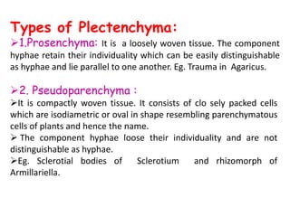
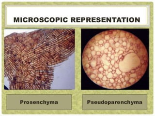
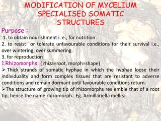
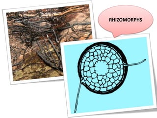
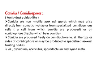
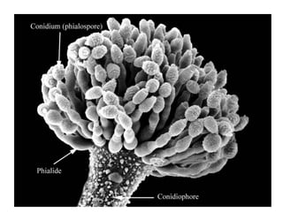
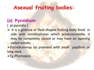
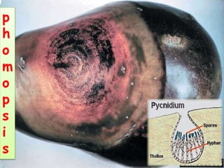
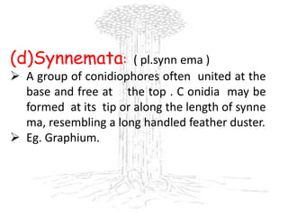
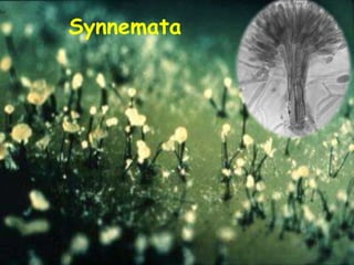
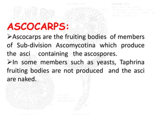
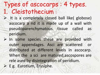
Downloaded 143 times

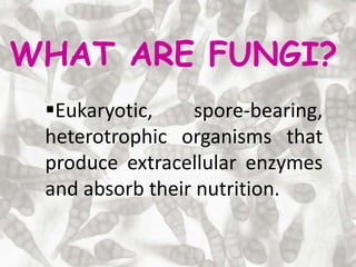
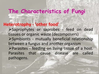
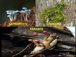
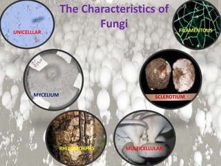
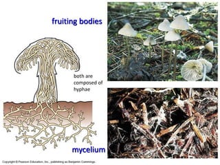
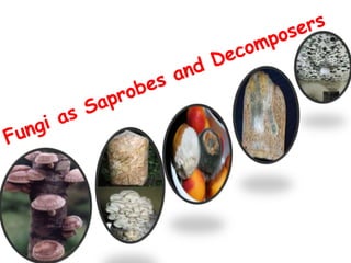
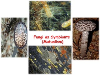
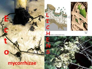
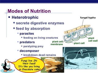
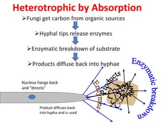
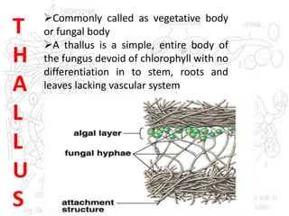
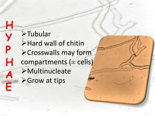
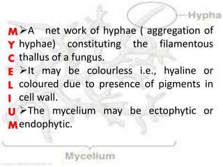
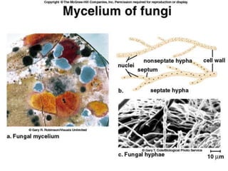
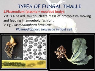
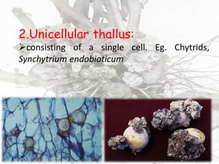
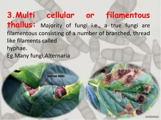
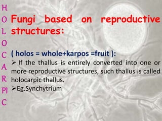
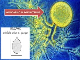
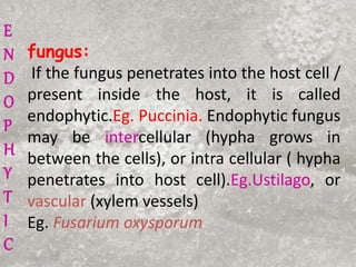
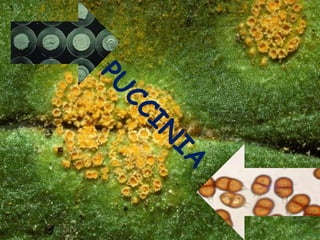
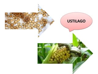
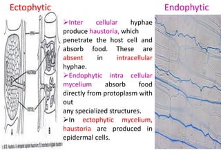
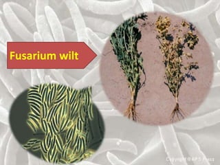
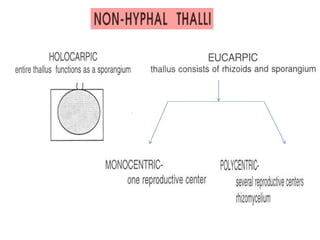
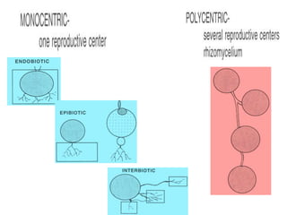
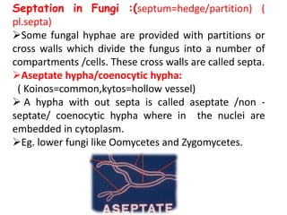

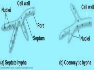
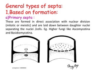
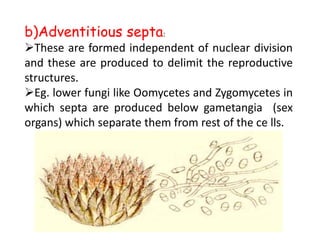
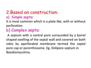
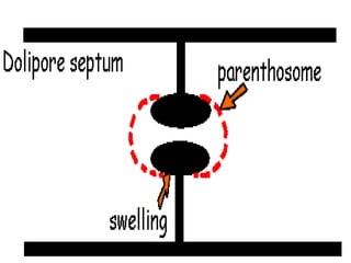
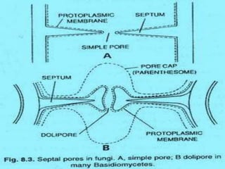
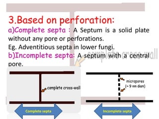
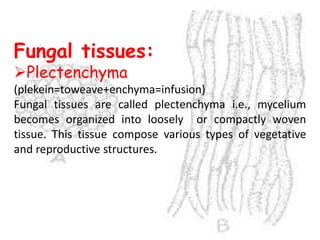
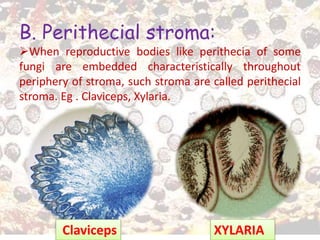
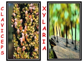
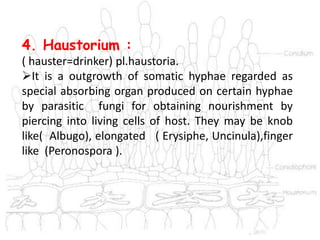
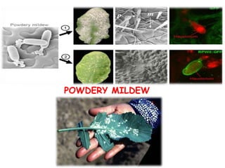
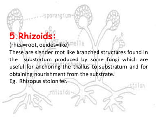
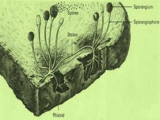
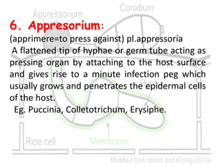
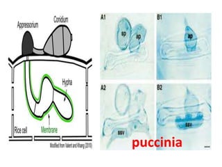
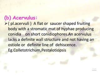
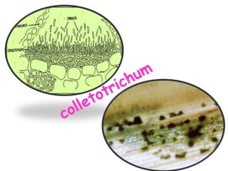
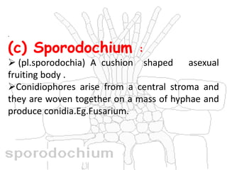
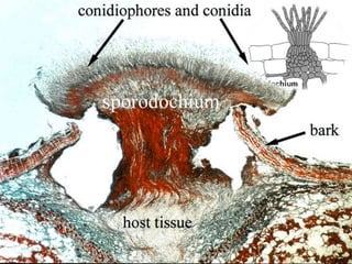


The document discusses various terms used to describe the phenotypic structure of fungi. It defines key terms like mycelium, hyphae, septa, and modifications of mycelium. It describes different types of fungal thalli like holocarpic, eucarpic, ectophytic, endophytic. It also discusses specialized somatic structures produced by fungi for nutrition, survival, and reproduction, including rhizomorphs, sclerotia, stroma, haustoria, and asexual fruiting bodies.


![General characteristics of_fungi[1]](https://cdn.slidesharecdn.com/ss_thumbnails/generalcharacteristicsoffungi1-200527112152-thumbnail.jpg?width=640&height=640&fit=bounds)


![ANIMAL_CELL_,_TISSUE_AND_ORGAN_CULTURE[1].pptx](https://cdn.slidesharecdn.com/ss_thumbnails/animalcelltissueandorganculture1-260204172026-4462b440-thumbnail.jpg?width=640&height=640&fit=bounds)


