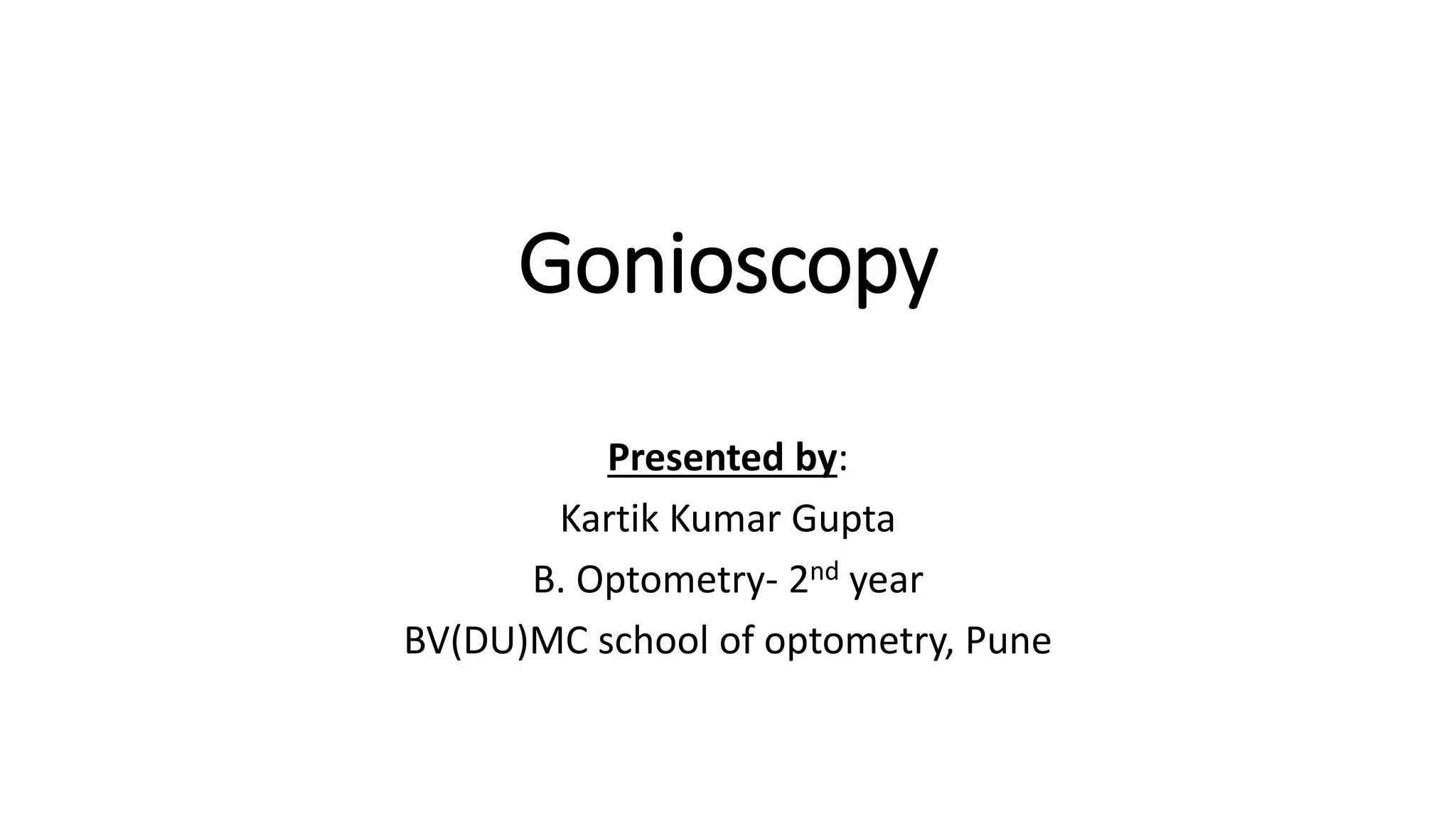
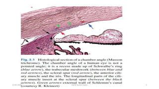
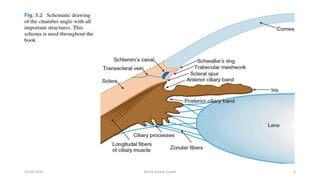
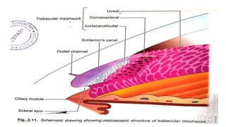
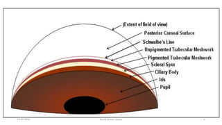
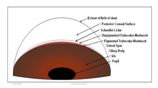
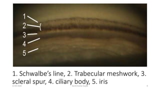
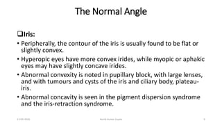
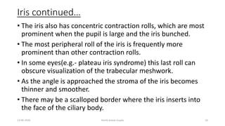
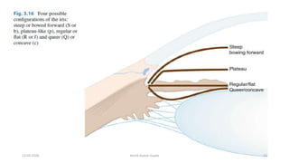
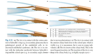
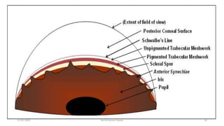
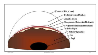
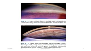
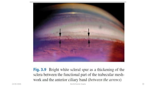
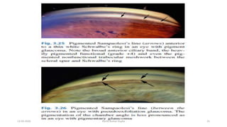
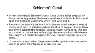
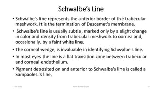
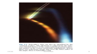
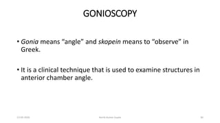
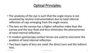
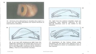
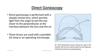
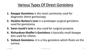
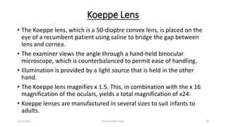
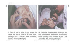
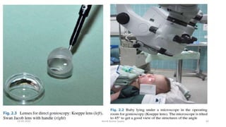
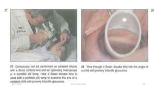
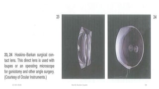
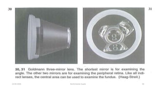
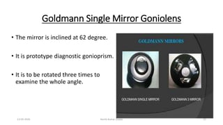
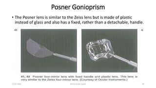
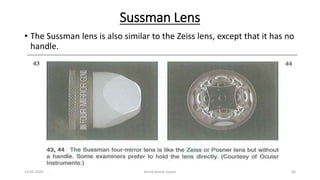
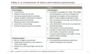
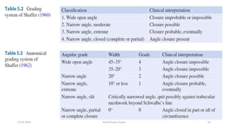
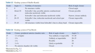
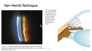
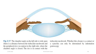
Gonioscopy and optic nerve head evaluationAhmedfaik
this is a simple presentation copy paste from kanski clinical ophthalmology about gonioscopy and optic nerve head changes in glaucoma... hope you get benefit
Gonioscopy and optic nerve head evaluationAhmedfaik
this is a simple presentation copy paste from kanski clinical ophthalmology about gonioscopy and optic nerve head changes in glaucoma... hope you get benefit
Nearsightedness (myopia) is a common vision condition in which near objects appear clear, but objects farther away look blurry. It occurs when the shape of the eye — or the shape of certain parts of the eye — causes light rays to bend (refract) inaccurately. Light rays that should be focused on nerve tissues at the back of the eye (retina) are focused in front of the retina.
Nearsightedness usually develops during childhood and adolescence, and it usually becomes more stable between the ages of 20 and 40. Myopia tends to run in families.
A basic eye exam can confirm nearsightedness. You can compensate for the blurry vision with eyeglasses, contact lenses or refractive surgery.
congenital cataract for undergraduate MBBS Students.
Also covers salient points for PGMEE.
Aetiology, clinical features and management discussed in detail.
Basavarajeeyam is an important text for ayurvedic physician belonging to andhra pradehs. It is a popular compendium in various parts of our country as well as in andhra pradesh. The content of the text was presented in sanskrit and telugu language (Bilingual). One of the most famous book in ayurvedic pharmaceutics and therapeutics. This book contains 25 chapters called as prakaranas. Many rasaoushadis were explained, pioneer of dhatu druti, nadi pareeksha, mutra pareeksha etc. Belongs to the period of 15-16 century. New diseases like upadamsha, phiranga rogas are explained.
Title: Sense of Taste
Presenter: Dr. Faiza, Assistant Professor of Physiology
Qualifications:
MBBS (Best Graduate, AIMC Lahore)
FCPS Physiology
ICMT, CHPE, DHPE (STMU)
MPH (GC University, Faisalabad)
MBA (Virtual University of Pakistan)
Learning Objectives:
Describe the structure and function of taste buds.
Describe the relationship between the taste threshold and taste index of common substances.
Explain the chemical basis and signal transduction of taste perception for each type of primary taste sensation.
Recognize different abnormalities of taste perception and their causes.
Key Topics:
Significance of Taste Sensation:
Differentiation between pleasant and harmful food
Influence on behavior
Selection of food based on metabolic needs
Receptors of Taste:
Taste buds on the tongue
Influence of sense of smell, texture of food, and pain stimulation (e.g., by pepper)
Primary and Secondary Taste Sensations:
Primary taste sensations: Sweet, Sour, Salty, Bitter, Umami
Chemical basis and signal transduction mechanisms for each taste
Taste Threshold and Index:
Taste threshold values for Sweet (sucrose), Salty (NaCl), Sour (HCl), and Bitter (Quinine)
Taste index relationship: Inversely proportional to taste threshold
Taste Blindness:
Inability to taste certain substances, particularly thiourea compounds
Example: Phenylthiocarbamide
Structure and Function of Taste Buds:
Composition: Epithelial cells, Sustentacular/Supporting cells, Taste cells, Basal cells
Features: Taste pores, Taste hairs/microvilli, and Taste nerve fibers
Location of Taste Buds:
Found in papillae of the tongue (Fungiform, Circumvallate, Foliate)
Also present on the palate, tonsillar pillars, epiglottis, and proximal esophagus
Mechanism of Taste Stimulation:
Interaction of taste substances with receptors on microvilli
Signal transduction pathways for Umami, Sweet, Bitter, Sour, and Salty tastes
Taste Sensitivity and Adaptation:
Decrease in sensitivity with age
Rapid adaptation of taste sensation
Role of Saliva in Taste:
Dissolution of tastants to reach receptors
Washing away the stimulus
Taste Preferences and Aversions:
Mechanisms behind taste preference and aversion
Influence of receptors and neural pathways
Impact of Sensory Nerve Damage:
Degeneration of taste buds if the sensory nerve fiber is cut
Abnormalities of Taste Detection:
Conditions: Ageusia, Hypogeusia, Dysgeusia (parageusia)
Causes: Nerve damage, neurological disorders, infections, poor oral hygiene, adverse drug effects, deficiencies, aging, tobacco use, altered neurotransmitter levels
Neurotransmitters and Taste Threshold:
Effects of serotonin (5-HT) and norepinephrine (NE) on taste sensitivity
Supertasters:
25% of the population with heightened sensitivity to taste, especially bitterness
Increased number of fungiform papillae
HOT NEW PRODUCT! BIG SALES FAST SHIPPING NOW FROM CHINA!! EU KU DB BK substit...GL Anaacs
Contact us if you are interested:
Email / Skype : kefaya1771@gmail.com
Threema: PXHY5PDH
New BATCH Ku !!! MUCH IN DEMAND FAST SALE EVERY BATCH HAPPY GOOD EFFECT BIG BATCH !
Contact me on Threema or skype to start big business!!
Hot-sale products:
NEW HOT EUTYLONE WHITE CRYSTAL!!
5cl-adba precursor (semi finished )
5cl-adba raw materials
ADBB precursor (semi finished )
ADBB raw materials
APVP powder
5fadb/4f-adb
Jwh018 / Jwh210
Eutylone crystal
Protonitazene (hydrochloride) CAS: 119276-01-6
Flubrotizolam CAS: 57801-95-3
Metonitazene CAS: 14680-51-4
Payment terms: Western Union,MoneyGram,Bitcoin or USDT.
Deliver Time: Usually 7-15days
Shipping method: FedEx, TNT, DHL,UPS etc.Our deliveries are 100% safe, fast, reliable and discreet.
Samples will be sent for your evaluation!If you are interested in, please contact me, let's talk details.
We specializes in exporting high quality Research chemical, medical intermediate, Pharmaceutical chemicals and so on. Products are exported to USA, Canada, France, Korea, Japan,Russia, Southeast Asia and other countries.
Report Back from SGO 2024: What’s the Latest in Cervical Cancer?bkling
Are you curious about what’s new in cervical cancer research or unsure what the findings mean? Join Dr. Emily Ko, a gynecologic oncologist at Penn Medicine, to learn about the latest updates from the Society of Gynecologic Oncology (SGO) 2024 Annual Meeting on Women’s Cancer. Dr. Ko will discuss what the research presented at the conference means for you and answer your questions about the new developments.
Knee anatomy and clinical tests 2024.pdfvimalpl1234
This includes all relevant anatomy and clinical tests compiled from standard textbooks, Campbell,netter etc..It is comprehensive and best suited for orthopaedicians and orthopaedic residents.
263778731218 Abortion Clinic /Pills In Harare ,sisternakatoto
263778731218 Abortion Clinic /Pills In Harare ,ABORTION WOMEN’S CLINIC +27730423979 IN women clinic we believe that every woman should be able to make choices in her pregnancy. Our job is to provide compassionate care, safety,affordable and confidential services. That’s why we have won the trust from all generations of women all over the world. we use non surgical method(Abortion pills) to terminate…Dr.LISA +27730423979women Clinic is committed to providing the highest quality of obstetrical and gynecological care to women of all ages. Our dedicated staff aim to treat each patient and her health concerns with compassion and respect.Our dedicated group ABORTION WOMEN’S CLINIC +27730423979 IN women clinic we believe that every woman should be able to make choices in her pregnancy. Our job is to provide compassionate care, safety,affordable and confidential services. That’s why we have won the trust from all generations of women all over the world. we use non surgical method(Abortion pills) to terminate…Dr.LISA +27730423979women Clinic is committed to providing the highest quality of obstetrical and gynecological care to women of all ages. Our dedicated staff aim to treat each patient and her health concerns with compassion and respect.Our dedicated group of receptionists, nurses, and physicians have worked together as a teamof receptionists, nurses, and physicians have worked together as a team wwww.lisywomensclinic.co.za/
Flu Vaccine Alert in Bangalore Karnatakaaddon Scans
As flu season approaches, health officials in Bangalore, Karnataka, are urging residents to get their flu vaccinations. The seasonal flu, while common, can lead to severe health complications, particularly for vulnerable populations such as young children, the elderly, and those with underlying health conditions.
Dr. Vidisha Kumari, a leading epidemiologist in Bangalore, emphasizes the importance of getting vaccinated. "The flu vaccine is our best defense against the influenza virus. It not only protects individuals but also helps prevent the spread of the virus in our communities," he says.
This year, the flu season is expected to coincide with a potential increase in other respiratory illnesses. The Karnataka Health Department has launched an awareness campaign highlighting the significance of flu vaccinations. They have set up multiple vaccination centers across Bangalore, making it convenient for residents to receive their shots.
To encourage widespread vaccination, the government is also collaborating with local schools, workplaces, and community centers to facilitate vaccination drives. Special attention is being given to ensuring that the vaccine is accessible to all, including marginalized communities who may have limited access to healthcare.
Residents are reminded that the flu vaccine is safe and effective. Common side effects are mild and may include soreness at the injection site, mild fever, or muscle aches. These side effects are generally short-lived and far less severe than the flu itself.
Healthcare providers are also stressing the importance of continuing COVID-19 precautions. Wearing masks, practicing good hand hygiene, and maintaining social distancing are still crucial, especially in crowded places.
Protect yourself and your loved ones by getting vaccinated. Together, we can help keep Bangalore healthy and safe this flu season. For more information on vaccination centers and schedules, residents can visit the Karnataka Health Department’s official website or follow their social media pages.
Stay informed, stay safe, and get your flu shot today!
NVBDCP.pptx Nation vector borne disease control programSapna Thakur
NVBDCP was launched in 2003-2004 . Vector-Borne Disease: Disease that results from an infection transmitted to humans and other animals by blood-feeding arthropods, such as mosquitoes, ticks, and fleas. Examples of vector-borne diseases include Dengue fever, West Nile Virus, Lyme disease, and malaria.
Ozempic: Preoperative Management of Patients on GLP-1 Receptor Agonists Saeid Safari
Preoperative Management of Patients on GLP-1 Receptor Agonists like Ozempic and Semiglutide
ASA GUIDELINE
NYSORA Guideline
2 Case Reports of Gastric Ultrasound
9. The Normal Angle
Iris:
• Peripherally, the contour of the iris is usually found to be flat or
slightly convex.
• Hyperopic eyes have more convex irides, while myopic or aphakic
eyes may have slightly concave irides.
• Abnormal convexity is noted in pupillary block, with large lenses,
and with tumours and cysts of the iris and ciliary body, plateau-
iris.
• Abnormal concavity is seen in the pigment dispersion syndrome
and the iris-retraction syndrome.
13-03-2020 Kartik Kumar Gupta 9
10. Iris continued…
• The iris also has concentric contraction rolls, which are most
prominent when the pupil is large and the iris bunched.
• The most peripheral roll of the iris is frequently more
prominent than other contraction rolls.
• In some eyes(e.g.- plateau iris syndrome) this last roll can
obscure visualization of the trabecular meshwork.
• As the angle is approached the stroma of the iris becomes
thinner and smoother.
• There may be a scalloped border where the iris inserts into
the face of the ciliary body.
13-03-2020 Kartik Kumar Gupta 10
16. Ciliary Body Band
• The iris usually inserts into the concave face of the ciliary body,
leaving some of the ciliary body visible anterior to the iris.
• The ciliary body band is seen as a light gray to dark brown band
located just anterior to the iris and posterior to the scleral spur.
• This band can be quite wide in myopic or aphakic eyes and
narrow to absent in hyperopic eyes or eyes with anterior
insertions of the iris.
• If the ciliary body band is abnormally deep and not symmetric
with the other eye, the possibility of angle recession,
cyclodialysis, or unilateral high myopia must be considered.
13-03-2020 Kartik Kumar Gupta 16
18. Scleral Spur
• The scleral spur is a ridge of scleral tissue that lies anterior to
the ciliary body band and marks the posterior border of the
trabecular meshwork.
• Appears as a thin band that is usually white or light gray but
which may have a yellowish cast in older individuals.
• Difficult to distinguish from trabecular meshwork in lightly
pigmented eye.
• Although the scleral spur is usually visible, it may be
obscured by iris processes, a high insertion of the iris, iris
bombe, peripheral anterior synechiae, or heavy
pigmentation.
13-03-2020 Kartik Kumar Gupta 18
20. Trabecular Meshwork
• The trabecular meshwork lies between the scleral spur and
Schwalbe's line.
• The meshwork is nonpigmented and smooth in infants but
becomes coarser and more pigmented with advancing age.
• Pigment in the angle is usually heaviest inferiorly owing to
gravitational settling and aqueous circulation.
• With narrow angles there can be more pigment superiorly than
inferiorly as a result of apposition of the iris against trabecular
meshwork.
13-03-2020 Kartik Kumar Gupta 20
21. Trabecular Meshwork cont’d…
• A non-pigmented angle is a pale gray color .
• Trabecular pigmentation usually appears deep within the
posterior trabecular meshwork.
• Sometimes pigment is deposited on the surface of the posterior
trabecular meshwork or over the anterior trabecular meshwork
and Schwalbe’s line.
• Heavy pigmentation may cover all angle structures.
• Increased pigmentation of the angle can be caused by many
pathological processes.
13-03-2020 Kartik Kumar Gupta 21
22. Trabecular meshwork cont’d…
• Heavy angle pigment can accumulate in a line anterior to
Schwalbe's line as a Sampaolesi's line.
• Sampaolesi's Iine is a non-specific finding in heavily pigmented
angles, whether physiologic or pathologic.
• The extra line of pigment can cause confusion in determining the
location of the pigmented trabecular meshwork.
• The corneal wedge can help in locating Schwalbe's line and in
defining whether the pigmentation is in the trabecular meshwork
or anterior to it.
• The anterior border of the trabecular meshwork is usually
smooth, but it can be wavy and irregular.
13-03-2020 Kartik Kumar Gupta 22
26. Schlemm’s Canal
• In most individuals Schlemm's canal is not visible. It lies deep within
the posterior (pigmented)trabecular meshwork, anterior to the scleral
spur, and becomes visible only when filled with blood.
• Blood can occasionally be found in Schlemm's canal in normal eyes. It
may also be seen in situations where the flow of aqueous humor from
Schlemm's canal to the episcleral venous system is impeded. This can
occur when a contact lens with a large diameter (such as a Goldmann
lens) is pressed too firmly against the eye, compressing the episcleral
veins.
• It can also be seen when the pressure in the episcleral venous system
is high or when the intraocular pressure is low.
13-03-2020 Kartik Kumar Gupta 26
27. Schwalbe’s Line
• Schwalbe's line represents the anterior border of the trabecular
meshwork. It is the termination of Descemet's membrane.
• Schwalbe's line is usually subtle, marked only by a slight change
in color and density from trabecular meshwork to cornea and,
occasionally, by a faint white line.
• The corneal wedge, is invaluable in identifying Schwalbe's line.
• In most eyes the line is a flat transition zone between trabecular
and corneal endothelium.
• Pigment deposited on and anterior to Schwalbe's line is called a
Sampaolesi's line,
13-03-2020 Kartik Kumar Gupta 27
30. GONIOSCOPY
• Gonia means “angle” and skopein means to “observe” in
Greek.
• It is a clinical technique that is used to examine structures in
anterior chamber angle.
13-03-2020 Kartik Kumar Gupta 30
31. Optical Principles:
• The anatomy of the eye is such that the angle recess is not
visualized by routine instrumentation due to total internal
reflection of rays emerging from the angle recess.
• The lens on the cornea has a higher refractive index than the
cornea and the tear fluid and thus eliminates the phenomenon
of total internal reflection.
• In modern gonioscopy contact lenses are used to overcome the
problem of total internal reflection.
• Two basic types of lens are used: the direct Lens and the indirect
lens.
13-03-2020 Kartik Kumar Gupta 31
33. Common Indication For Gonioscopy
• Narrow anterior chamber angles by Van Herick (grade II or narrower).
Gonioscopy is the gold standard technique against which screening
tests for narrow angles are compared (e.g. Johnson & Foster 2005).
• Narrow (or closed) angle glaucoma including evaluation and
documentation of peripheral anterior synechiae if present (Johnson &
Foster 2005).
• Primary open angle glaucoma (POAG) and risk factors for POAG (e.g.
elevated intraocular pressure) to confirm ‘primary’ diagnosis.
13-03-2020 Kartik Kumar Gupta 33
34. Common Indication For Gonioscopy Cont’d…
• Secondary open angle glaucoma and risk factors (e.g. chronic
uveitis, pseudoexfoliation, pigment dispersion) to contribute to
determination of disease severity.
• Risk of angle recession post-blunt trauma.
• Risk of intraocular foreign body.
13-03-2020 Kartik Kumar Gupta 34
35. Common Indication For Gonioscopy Cont’d…
• Congenital or acquired structural irregularities of the iris and
anomalies of the anterior chamber (e.g. iris cysts or tumours,
ectopic pupil).
• Post-laser peripheral iridotomy to assess effect on angle depth.
• Risk of angle neovascularisation (e.g. confirmed rubeosis iridis,
and ischaemic posterior segment conditions including vein or
artery occlusions and diabetic retinopathy; e.g. Browning et al.
1998).
13-03-2020 Kartik Kumar Gupta 35
36. Contraindication Of Gonioscopy
• Recent ocular trauma especially in the presence of hyphaema or
microhyphaema.
• Recent intraocular surgery, including cataract surgery.
13-03-2020 Kartik Kumar Gupta 36
37. Direct Gonioscopy
• Direct gonioscopy is performed with a
steeply convex lens, which permits
light from the angle to exit the eye
closer to the perpendicular at the
interface between the lens and the air.
• These lenses are used with a portable
slit lamp or an operating microscope.
13-03-2020 Kartik Kumar Gupta 37
38. Various Types Of Direct Goniolens
1. Koeppe Goniolens is the most commonly used for
diagnostic direct gonioscopy.
2. Hoskins Barkan’s Lens is a prototype surgical goniolens
used for goniotomy.
3. Swan Jacob’s Lens is also used for surgical purpose.
4. Richardson-Shaffer’s Goniolens is basically small Koeppe
lens used for infants.
5. Sieback Goniolens- it is a tiny goniolens which floats on the
cornea.
13-03-2020 Kartik Kumar Gupta 38
39. Koeppe Lens
• The Koeppe lens, which is a 50-dioptre convex lens, is placed on the
eye of a recumbent patient using saline to bridge the gap between
lens and cornea.
• The examiner views the angle through a hand-held binocular
microscope, which is counterbalanced to permit ease of handling.
• Illumination is provided by a light source that is held in the other
hand.
• The Koeppe lens magnifies x 1.5. This, in combination with the x 16
magnification of the oculars, yields a total magnification of x24.
• Koeppe lenses are manufactured in several sizes to suit infants to
adults.
13-03-2020 Kartik Kumar Gupta 39
45. Advantages Of Direct Gonioscopy
• Greater flexibility because position of observer can be changed.
• Panoramic view is obtained so one part of angle can be
compared with other.
• Angle become deep in supine position so it is easy to see the
angle.
• Cause less distortion of anterior chamber.
13-03-2020 Kartik Kumar Gupta 45
46. Advantages Of Direct Gonioscopy Cont’d…
• Can be used in sedated/anesthetized patients as in infants.
• Provides a straight view rather than inverted view.
• Can be used for surgical procedures like goniotomy.
• Detailed examination of minor structures is possible because the
observer can change his or her height to look deeper into anterior
chamber.
13-03-2020 Kartik Kumar Gupta 46
47. Disadvantages of direct gonioscopy
• Inconvenient
• Annoying light reflex from cornea
• Time consuming
• Benefits of slit lamp (like variable light and better clarity are not
available)
13-03-2020 Kartik Kumar Gupta 47
48. Indirect gonioscopy
• The lenses used in indirect
gonioscopy use mirrors or prism
to overcome total internal
reflection.
• The mirror redirects light from
the angle so that it exits the eye
perpendicularly to the lens-air
interface.
• It uses the slit lamp’s illumination
and magnification system to its
advantages.
13-03-2020 Kartik Kumar Gupta 48
49. Goldmann 3-Mirror Goniolens
• Goldmann three mirror universal lens uses a high minus contact
lens to neutralize the power of the cornea and an appropriate
angled mirrors to allow examination of anterior chamber angle,
peripheral and mid-peripheral fundus as well as a central lens for
the evaluation of the vitreous and the posterior pole.
• The mirror having inclination of 59, 67 and 73 degree is used for
gonioscopy, examine pars plana and ora serrata respectively.
13-03-2020 Kartik Kumar Gupta 49
51. Goldmann Two Mirror Goniolens
• Both the mirror is inclined at 62 degree.
• It needs to be rotated once to examine whole angle.
13-03-2020 Kartik Kumar Gupta 51
52. Goldmann Single Mirror Goniolens
• The mirror is inclined at 62 degree.
• It is prototype diagnostic gonioprism.
• It is to be rotated three times to
examine the whole angle.
13-03-2020 Kartik Kumar Gupta 52
53. Advantages Of Goldmann Goniolens
• Easy to use, excellent view, stabilizes the globe and therefore
can be used for laser trabeculoplasty, peripheral retina can
be seen.
• Better view in patient with significant loss of corneal
transparency.
• Goldmann two mirror gives best in-situ view of the angle.
13-03-2020 Kartik Kumar Gupta 53
54. Disadvantages Of Goldmann Goniolens
• Curvature of lens is more than that of cornea so coupling material is
required. It blurs vision and fundus; therefore, visual field charting,
direct and indirect ophthalmoscopy cannot be done immediately
after its use.
• Only one mirror is there for gonioscopy so it needs to be rotated by
360 degrees.
• It cannot be used for indentation gonioscopy.
• Broad area of contact with cornea is there in case of Goldmann three-
mirror and under pressure it can lead to artefactual closure of angle.
13-03-2020 Kartik Kumar Gupta 54
55. Allen-Thorpe Gonioprism
• It has got four prisms instead of mirrors and allows examination of
the whole angle when rotating prisms.
13-03-2020 Kartik Kumar Gupta 55
56. Zeiss Four-mirror Gonioprism
• It has four identical mirrors angled at 64 degree which allow examination
of four quadrants without rotation of the lens.
• By turning only 11 degree through lens, the smaller area in between the
mirror can be visualize.
• Because the lens has a small (9 mm) area of contact with the cornea, the
angle can be deepened by pushing on the lens.
• The small gap between lens and cornea is filled with tears or topical
anaesthetic.
• This keeps the cornea free of viscous fluids and permits clear examination
and photography of the optic nerve head following gonioscopy.
13-03-2020 Kartik Kumar Gupta 56
59. Posner Gonioprism
• The Posner lens is similar to the Zeiss lens but is made of plastic
instead of glass and also has a fixed, rather than a detachable, handle.
13-03-2020 Kartik Kumar Gupta 59
60. Sussman Lens
• The Sussman lens is also similar to the Zeiss lens, except that it has no
handle.
13-03-2020 Kartik Kumar Gupta 60