
Recommended
More Related Content
Similar to back of leg of human
Similar to back of leg of human (20)
More from MRSDRNIDHISHARMAVISH
Recently uploaded
Recently uploaded (20)
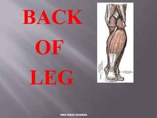
back of leg of human
- 2. The posterior compartment of the leg contains seven muscles, organised into two layers – superficial and deep. The two layers are separated by a band of fascia. The posterior leg is the largest of the three compartments. Collectively, the muscles in this area plantarflex and invert the foot. They are innervated by the tibial nerve, a terminal branch of the sciatic nerve. INTRODUCTION
- 3. SUPERFICIAL MUSCLES The superficial muscles form the characteristic ‘calf’ shape of the posterior leg. They all insert into the calcaneus of the foot (the heel bone), via the calcaneal tendon. The calcaneal reflex tests spinal roots S1-S2. To minimise friction during movement, there are two bursae (fluid filled sacs) associated with the calcaneal tendon: Subcutaneous calcaneal bursa – lies between the skin and the calcaneal tendon. Deep bursa of the calcaneal tendon – lies between the tendon and the calcaneus.
- 4. Tibia Medial mallelous Fibula Lateral mallelous Talus Calcaneous
- 5. Ligaments Lateral aspect Anterior tibiofibular Posterior tibiofibular Anterior talofibular Posterior talofibular Calcaneofibular
- 6. Medial aspect deltoid Tendons Achilles Peroneus longus Peroneus brevis Anterior tibialis Tibialis posterior Flexor digitorium Flexor hallicus longus
- 7. Muscles Gastrocnemius Soleus Tibialis anterior Tibialis posterior Flexor digitorium longus Flexor hallicus longus Peroneus longus Peroneus brevis
- 9. These muscles connect the tibia or fibula to the femur or pelvic girdle. The muscles either flex or extend the knee. The muscles include the hamstring group and the quadriceps femoris group.
- 10. superficial/ calf: gastrocnemius, soleus, plantaris
- 11. Gastrocnemius -is a very powerful superficial muscle .It runs from its two heads just above the knee to the heel, and is involved in standing, walking, running and jumping. Along with the soleus muscle it forms the calf muscle. Its function is plantar flexing the foot at the ankle joint and flexing the leg at the knee
- 12. . The Lateral Head originates from the Lateral Condyle of the femur, while the Medial Head originates from the Medial Condyle of the femur. Its other end forms a common tendon with the soleus muscle; this tendon is known as the calcaneal tendon or Achilles Tendon and inserts onto the posterior surface of the calcaneus, or mountain bone. NERVE SUPPLY : The nerve supply of the gastrocnemius muscle is the tibial nerve (S1, S2). ACTION : The gastrocnemius and soleus muscle act together as plantar flexors of foot at the ankle joint. The gastrocnemius also act as the flexor of the knee.
- 13. PLANTARIS The plantaris is a small muscle with a long tendon, which can be mistaken for a nerve as it descends down the leg. It is absent in 10% of people. •Attachments: Originates from the lateral supracondylar line of the femur. The muscle descends medially, condensing into a tendon that runs down the leg, between the gastrocnemius and soleus. The tendon blends with the calcaneal tendon. •Actions: It plantarflexes at the ankle joint, and because it crosses the knee, it is a flexor there. It is not a vital muscle for these movements. •Innervation: Tibial nerve.
- 14. the soleus is a powerful muscle in the back part of the lower leg (the calf). It runs from just below the knee to the heel, and is involved in standing and walking. It is closely connected to the gastrocnemius muscle.
- 15. ORIGIN : The soleus muscle has a dome-shaped origin from: back of head and upper one-fourth of the posterior surface of the shaft of the fibula soleal line and middle one- third of the medial border of the shaft of the tibia the tendinous soleal arch that stretches between the tibia and the fibula.
- 16. INSERTION : The tendon of this muscle fuses with the tendon of gastrocnemius to form tendoachilles, which is inserted into the middle one-third of the posterior surface of the calcaneum. NERVE SUPPLY : The nerve supply of the soleus muscle is the tibial nerve (S1, S2). ACTION : The soleus and gastrocnemius act as plantar flexors of the foot, at the ankle joint. The soleus is more powerful than gatrocnemius but is slow acting than the gastrocnemius. Soleus is chiefly a postural muscle, to steady the leg on the foot.
- 18. DEEP MUSCLES There are four muscles in the deep compartment of the posterior leg. One muscle, the popliteus, acts only on the knee joint. The remaining three muscles (tibialis posterior, flexor hallucis longus and flexor digitorum longus) act on the ankle and foot.
- 19. The popliteus is located superiorly in the leg. It lies behind the knee joint, forming the base of the popliteal fossa. There is a bursa (fluid filled sac) that lies between the popliteal tendon and the posterior surface of the knee joint. It is called the popliteus bursa POPLITEUS
- 20. Attachments: Originates from the lateral condyle of the femur and the posterior horn of the lateral meniscus. From there, it runs inferomedially towards the tibia and inserts above the origin of the soleus muscle. Actions: Laterally rotates the femur on the tibia – ‘unlocking’ the knee joint so that flexion can occur. Innervation: Tibial nerve.
- 21. Tibialis Posterior The tibialis posterior is the deepest out of the four muscles. It lies between the flexor digitorum longus and the flexor hallucis longus. Attachments: Originates from the interosseous membrane between the tibia and fibula, and posterior surfaces of the two bones. The tendon enters the foot posterior to the medial malleolus, and attaches to the plantar surfaces of the medial tarsal bones. Actions: Inverts and plantarflexes the foot, maintains the medial arch of the foot. Innervation: Tibial nerve.
- 22. Flexor Digitorum Longus The FDL is (surprisingly) a smaller muscle than the flexor hallucis longus. It is located medially in the posterior leg. Attachments: Originates from the medial surface of the tibia, attaches to the plantar surfaces of the lateral four digits. Actions: Flexes the lateral four toes. Innervation: Tibial nerve.
- 23. Flexor Hallucis Longus The flexor hallucis longus muscle is found on the lateral side of leg. This is slightly counter-intuitive, as it is opposite the great toe, which it acts on. Attachments: Originates from the posterior surface of the fibula, attaches to the plantar surface of the phalanx of the great toe. Actions: Flexes the great toe. Innervation: Tibial nerve.
- 25. The calf muscles do the work for movements like running, walking and cycling as it provides the initial propulsion for these movements. Most of the people train calf for aesthetic purposes. But calf muscle plays more important role than just to flaunt. Calf muscle is often called as the pseudo or periphery heart because of its very important function which is to return the blood from leg and foot to the heart. Blood flow from the lower body to heart has to work against the gravity, so the contraction of calf muscle builds up an external pressure that propels the blood through the veins to the heart. The gastrocnemius muscle is used more during dynamic, higher force activities and soleus muscle is more active during postural and static contractions. As the gastrocnemius crosses knee and ankle, the position of the knee during the plantar flexion resistance exercise affects activity of the muscle. At 90 degree of knee flexion, the gastrocnemius experiences passive insufficiency and hence less active. During calf raise exercise keeping knee 90 degree flexed to focus on soleus and zero degree flexed to focus on gastrocnemius.
- 26. The calves also act as a deceleration tool for the body. While doing a sprint when one need to stop or change directions quickly the calves absorb up to 10 to 12 times the body-weight. Trained calves are therefore required to bear this load and ensure the deceleration takes place safely to avoid injury caused by the eccentric phase of any exercise. The calves also stabilise the knees which is important for jumping exercises where unstable knees can result in poor form and injury. Here, a strong set of calves protect the joints. Well trained calves result in increased vertical jumping power. With the gastrocnemius mainly composed of fast-twitch muscle fibres, the calves can execute quick and explosive movements, such as those required during high jumps, squat jumps and sprints. Although genetics determine the amount of fast-twitch muscle fibres every person has, strengthening the calves helps everyone perform these power movements
- 27. soleus
- 30. The tibial nerve is a branch of the sciatic nerve. The tibial nerve passes through the popliteal fossa to pass below the arch of soleus. In the popliteal fossa the nerve gives off branches to gastrocnemius, popliteus, soleus and plantaris muscles, an articular branch to the knee joint, and a cutaneous branch that will become the sural nerve. The sural nerve is joined by fibres from the common fibular nerve and runs down the calf to supply the lateral side of the foot.
- 31. Lateral plantar nerve The lateral plantar nerve supplies quadratus plantae, flexor digiti minimi, adductor hallucis, the interossei, three lumbricals. and abductor digiti minimi. Cutaneous innervation is to the lateral sole and lateral one and one half toes
- 33. The posterior tibial artery –it is the large terminal branch of popliteal artery begins at the lower border of the Popliteus, opposite the interval between the tibia and fibula; it extends obliquely downward, and, as it descends, it approaches the tibial side of the leg, lying behind the tibia, .
- 34. and in the lower part of its course is situated midway between the medial malleolus and the medial process of the calcaneal tuberosity. Here it divides beneath the origin of the Adductor hallucis into the medial and lateral plantar arteries.
- 35. In anatomy, the fibular artery (also known as the peroneal artery) supplies blood to the lateral compartment of the leg and is typically a branch of posterior tibial artery
- 37. when a strain occurs, muscle fibers are torn to some degree. a pulled calf muscle happens when your internal muscles are overstretched from exercise. this is a common injury, especially among athletes and walkers. symptoms of a pulled calf muscle can depend on the severity of the injury. a mild strain can leave you with pain and feelings of pulling within the lower half of your leg. you can still walk with a mild strain, but it may be uncomfortable. other signs of a pulled calf muscle include: 1.mild swelling 2.redness 3.bruising 4.inability to stand up on the ball of your foot. a severe pull in your calf muscles can leave you with feelings of sharp pain. it can also affect your mobility, making you unable to walk. pulled calf muscles may be chronic from long-term injury or acute from brief over pulling .
- 38. Achilles Tendon Stretching A tight heel cord may limit dorsiflexion and may predispose athlete to ankle injury Should routinely stretch before and after practice Stretching should be performed with knee extended and flexed 15-30 degrees Strength Training Static and dynamic joint stability is important in preventing injury A balance in strength throughout the range, incorporating all muscles of the lower leg is critical
- 40. Ankle Fractures/Dislocations Cause of Injury Number of mechanisms – often similar to those seen in ankle sprains Signs of Injury Swelling and pain may be extreme with possible deformity Care Splint and refer to physician for X-ray and examination RICE to control hemorrhaging and swelling Once swelling is reduced, a walking cast or brace may be applied, w/ immobilization lasting 6-8 weeks Rehabilitation is similar to that of ankle sprains once range of motion is normal
- 44. Post reduction
- 47. Acute Leg Fractures Cause of Injury Result of direct blow or indirect trauma Fibular fractures seen with tibial fractures or as the result of direct trauma Signs of Injury Pain, swelling, soft tissue insult Leg will appear hard and swollen (Volkman’s contracture) Deformity – may be open or closed Care X-ray, reduction, casting up to 6 weeks depending on the extent of injury
- 49. Stress Fracture of Tibia or Fibula Cause of Injury Common overuse condition, particularly in those with structural and biomechanical insufficiencies Result of repetitive loading during training and conditioning Signs of Injury Pain with activity Pain more intense after exercise than before Point tenderness; difficult to discern bone and soft tissue pain Bone scan results (stress fracture vs. periostitis)
- 50. Care Eliminate offending activity Discontinue stress inducing activity 14 days Use crutch for walking Weight bearing may return when pain subsides After pain free for 2 weeks athlete can gradually return to activity Biomechanics must be addressed
- 51. Medial Tibial Stress Syndrome (Shin Splints) Cause of Injury Pain in anterior portion of shin Stress fractures, muscle strains, chronic anterior compartment syndrome, periosteum irritation Caused by repetitive microtrauma Weak muscles, improper footwear, training errors, varus foot, tight heel cord, hypermobile or pronated feet and even forefoot supination can contribute to MTSS May also involve stress fractures or exertional compartment syndrome
- 53. Achilles Tendonitis Cause of Injury Inflammatory condition involving tendon, sheath or paratenon Tendon is overloaded due to extensive stress Presents with gradual onset and worsens with continued use Decreased flexibility exacerbates condition Signs of Injury Generalized pain and stiffness, localized proximal to calcaneal insertion, warmth and painful with palpation, as well as thickened May progress to morning stiffness
- 55. Achilles Tendon Rupture Cause Occurs w/ sudden stop and go; forceful plantar flexion w/ knee moving into full extension Commonly seen in athletes > 30 years old Generally has history of chronic inflammation Signs of Injury Sudden snap (kick in the leg) w/ immediate pain which rapidly subsides Point tenderness, swelling, discoloration; decreased ROM Obvious indentation and positive Thompson test
- 58. Achilles Tendon Rupture Etiology Occurs w/ sudden stop and go; forceful plantar flexion w/ knee moving into full extension Commonly seen in athletes > 30 years old Generally has history of chronic inflammation Signs and Symptoms Sudden snap (kick in the leg) w/ immediate pain which rapidly subsides Point tenderness, swelling, discoloration; decreased ROM Obvious indentation and positive Thompson test Occurs 2-6 cm proximal the calcaneal insertion
- 60. Clinical Relevance: Ruptured Calcaneal Tendon Rupture of the calcaneal tendon refers to a partial or complete tear of the tendon. It is more likely to occur in people with a history of calcaneal tendinitis (chronic inflammation of the tendon). The injury is usually sustained during forceful plantarflexion of the foot. The patient will be unable to plantarflex the foot against resistance, and the affected foot will be permanently dorsiflexed. The soleus and gastrocnemius can contract to form a lump in the calf region. Treatment of a ruptured calcaneal tendon is usually non- surgical, except in those with active lifestyles.
- 61. Achilles Tendon Rupture (continued) Management Usual management involves surgical repair for serious injuries (return of 75-80% of function) Non-operative treatment consists of RICE, NSAID’s, analgesics, and a non-weight bearing cast for 6 weeks, followed up by a walking cast for 2 weeks (75-90% return to normal function) Rehabilitation lasts about 6 months and consists of ROM, PRE and wearing a 2cm heel lift in both shoes
- 62. Medial Tibial Stress Syndrome (Shin Splints) Etiology Pain in anterior portion of shin Catch all for stress fractures, muscle strains, chronic anterior compartment syndrome Accounts for 10-15% of all running injuries, 60% of leg pain in athletes Caused by repetitive microtrauma Weak muscles, improper footwear, training errors, varus foot, tight heel cord, hypermobile or pronated feet and even forefoot supination can contribute to MTSS May also involve, stress fractures or exertional compartment syndrome