muscles of the leg are categorized into anterior, lateral, and posterior groups, all these muscles are covered by the deep facial as shown in the image(1), these muscle groups are divided by the tibia and fibula, interosseous membrane, anterior and posterior intermuscular septa, which pass inwards from the deep fascia of the leg. in this article you gonna learn about muscles of the leg and their clinical correlates
Plant propagation: Sexual and Asexual propapagation.pptx
muscles of leg anatomy
1. 1/12
February 9, 2021
muscles of leg
kyrmed.com/muscles-of-leg/
muscles of the leg are categorized
into anterior, lateral, and posterior
groups, all these muscles are
covered by the deep facial as shown
in the image(1), these muscle
groups are divided by the tibia and
fibula, interosseous membrane,
anterior and posterior
intermuscular septa, which pass
inwards from the deep fascia of the
leg.
Anterior compartment muscles of the leg
we have four muscles in the anterior compartment of our leg, they are as follows:
1. tibialis anterior.
2. extensor digitorum longus.
3. extensor hallucis longus.
4. fibularis tertius.
All these muscles are supplied by branches of the fibular nerve( also called common
peroneal) and blood is supplied by the anterior tibial artery. when the foot is not
bearing weight they help to dorsiflex and invert the foot and toes.
During normal walking, these muscles help to pull the leg forwards when the foot is in
contact with the ground
2. 2/12
muscles of leg-Tibialis anterior
Origin- tibialis anterior is originated from the Lateral condyle and upper
lateral surface of the tibia
Insertion- tibialis anterior is inserted onto the Medial cuneiform and base of
1st metacarpal
nerve innervation- tibialis anterior is innervated by the Peroneal or fibulae
nerve which is the branch of the common fibular nerve
function- tibialis anterior is the strongest dorsiflexor of the foot and it also
inverts the foot
extensor digitorum longus
Origin- extensor digitorum longus is originated from the Lateral condyle of the
tibia and upper 3/4 of anterior interosseous membrane and fibula
insertion- extensor digitorum longus is inserted onto the Middle and distal
phalanges of lateral four digits
nerve innervation- extensor digitorum longus is innervated by the same
peroneal nerve with the root value of L5 and S1
function- this muscle helps in the extension of the toes and dorsiflexions of
the ankle
extensor hallucis longus
Origin- extensor hallucis longus is originated from the Middle of the anterior
surface of the fibula and interosseous membrane
insertion- extensor hallucis longus is inserted onto the Base of the distal phalanx
of hallux
nerve innervation- extensor hallucis longus is innervated by the same peroneal
nerve with the root value of L5 and S1
function- this muscle helps in the extension of the hallux and dorsiflexions
of the ankle
peroneus tertius
Origin- peroneus Tertius is originated from the Lower anterior fibula and
interosseous membrane
insertion- it is inserted onto the base of the fifth metacarpal
nerve innervation- peroneus Tertius is innervated by the same peroneal nerve
with the root value of L5 and S1
function- this muscle is the weak evertor of foot and dorsiflexions of the
ankle
3. 3/12
ANTERIOR COMPARTMENT MUSCLES- WORK
BY OpenStax College, CC BY 3.0, via Wikimedia
Commons
Lateral compartment muscles of leg
these muscles play a very important role in maintaining balance while we are standing, all
muscles in this group are innervated by the superficial peroneal nerve
peroneus longus
this muscle is also called as fibularis lobgus muscle and is most superficial muscle in
this compartment
Origin- peroneus longus is originated from the head and upper 2/3rd of the
lateral fibula
insertion- its tendon travelsbehind the lateral malleolus and ultimately inserted
laterally to the medial cuneiform and base of 1st metatarsal
4. 4/12
nerve innervation- peroneus longus is innervated by the same superficial
peroneal nerve with the root value of L5 and S1, S2
function- this muscle helps in the eversion of the foot and plantar flexions of
the ankle
peroneus brevis
this muscle is also called fibularis brevis muscle and is a deep muscle in this
compartment and it is shorter than PL
Origin- peroneus brevis is originated from the lower 2/3rd of the lateral fibula
insertion- it travelsbehind the lateral malleolus and ultimately inserted to the
base of the 5th metatarsal
nerve innervation- peroneus brevis is innervated by the same superficial
peroneal(fibular) nerve with the root value of L5 and S1, S2
function- this muscle helps in the eversion of the foot and weak plantar
flexions of the ankle
LATERAL AND ANTERIOR COMPARTMENT MUSCLES – Polygon data were generated
by Database Center for Life Science (DBCLS)[2], CC BY-SA 2.1 JP, via Wikimedia Commons
clinical correlate– twisted ankles
these fractures often result in pulling off the base of the 5th metatarsal, attached to the
tendon of peroneus (fibularis) brevis muscle, this may lead to the fracture of the base of
the 5th metatarsal bone
5. 5/12
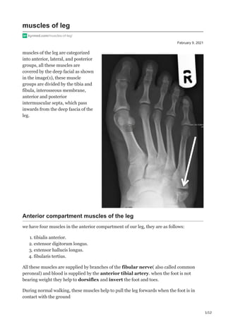
fracture of the base of the 5th metatarsal bone(
see the arrow)
Posterior compartment muscles of leg
all the muscles in this compartment are divided into superficial and deep muscles,
there are a total of seven muscles in this compartment that helps in pushing our body
forward during plantar flexion of the foot while walking
all these muscles commonly helps in plantar flexion and invertion of the foot
Superficial layer
gastrocnemius muscle
this muscle is the most superficial muscle in this compartment which lies over the
popliteus and soleus muscles, this muscle consists of two head- medial and lateral.
Origin- lateral head originates from the lateral condyle of the femur and the
medial head originated from the proximal to the medial condyle of the femur.
6. 6/12
insertion- the two heads unite at the inferior margin of the popliteal fossa to form
a single belly, and then its aponeurosis which narrows and joins with the
aponeurosis of soleus to form the Achilles tendon that inserts onto the posterior
surface of the calcaneus
nerve innervation- peroneus longus is innervated by the tibial nerve with the
root value of S1, S2
function- as this muscle crosses the knee joint and ankle joint it helps in the
flexion of the knee and plantar flexions of the ankle respectively
GASTROCNE,IOUS MUSCLE – Polygon data were generated by Database Center for Life Science
(DBCLS)[2], CC BY-SA 2.1 JP, via Wikimedia Commons
soleus muscle
this muscle is present deep to the gastrocnemius muscle, it is a broad triangular muscle
that resembles the sole fish( so its name)
7. 7/12
Origin- soleus is originated from the soleal line present of the posterior surface of
the tibia
insertion- this muscle forms the aponeurosis roughly at the level of mid-calf and
joins with the tendon of gastrocnemius to form the calcaneal tendon also called as
ACHILLES TENDON which inserts onto the posterior surface of the calcaneus
nerve innervation- soleus is innervated by the same tibial nerve with the root
value of L5 and S1, S2
function- this muscle helps in the eversion of the foot and weak plantar
flexions of the ankle
SOLEUS MUSCLE – Polygon data is generated by Database Center for Life
Science (DBCLS), CC BY-SA 2.1 JP, via Wikimedia Commons
clinical correlate– achilles tendon rupture
Rupture of the calcaneal tendon (Achilles tendon) usually occurs due to the acute
contraction of a muscle that is not exercised, such as may occur during a middle-aged
person’s first football game and also due to the presence of the history of calcaneal
tendonitis.
Rupture may be partial or complete; supportive care is enough for partial tears but
complete tears may require surgical treatment. The ankle reflex is demonstrated by
tapping the tendon with a tendon hammer.
8. 8/12
plantaris muscle
Origin- this small muscle is originated from the lateral supracondylar line of
the femur
insertion- the long tendon of this muscle runs medial to the gastrocnemius and
finally inserts onto the posterior surface of the calcaneus medial to the
Achilles tendon.
nerve innervation- soleus is innervated by the same tibial nerve with the root
value of L5 and S1, S2
function- this muscle helps in the eversion of the foot and weak plantar
flexions of the ankle
PLANTARIS MUSCLE- Polygon data is generated by Database Center for Life Science
(DBCLS), CC BY-SA 2.1 JP, via Wikimedia Commons
Deep layer
these muscles play a very important role in maintaining balance while we are standing, all
muscles in this group are innervated by the superficial peroneal nerve
poplitEus muscle
9. 9/12
this muscle is present behind the knee joint forming the base of the popliteal fossa
Origin- popliteus muscle is originated from the Lateral condyle of the femur and
lateral meniscus
insertion- this muscle inserts onto the upper posterior surface of the tibia.
nerve innervation- popliteus muscle is innervated by the tibial nerve with the
root value of L4, L5.
function- this muscle helps in the weak flexion of knee and unlocking the
knee joint- lateral rotation of femur on the tibia
POPLITEUS MUSCLE
Popliteal bursa
popliteal bursa also called is the fluid filled sac present behind the knee joint, there are
many bursae in the knee joint, so it will be discussed in knee joint article
flexor hallucis longus
10. 10/12
this muscle is the unipennate muscle present a bit laterally along the fibular side
Origin- flexor hallucis longus is originated from the lower 2/3rd of the
posterior surface of the fibula.
insertion- as you can see in the image, this muscle inserts onto the plantar surface
of the distal phalanx of hallux
nerve innervation- peroneus brevis is innervated by the tibial nerve with the
root value of S2, S3.
function- this muscle helps in the Flexion of the hallux, plantar flexion of the
ankle, and also an inversion of the foot, this muscle also play an important role
in supporting the medial longitudinal arch
Flexor digitorum longus
Origin- Flexor digitorum longus is originated from the lower posterior surface
of the tibia and fibula
insertion- as you can see in the image, this muscle inserts onto the Distal
phalanges of lateral four toes
11. 11/12
nerve innervation- Flexor digitorum longus is innervated by the tibial nerve
with the root value of S2, S3.
function- this muscle helps in the flexion of lateral four toes, plantar flexion
of the ankle, and also an inversion of the foot.
tibialis posterior
Origin- tibialis posterior is originated from the Posterior tibia below soleal
line, posterior surface of fibula
insertion- as you can see in the image, this muscle inserts onto the Tuberosity of
navicular, cuneiform, cuboid, and also inset onto the bases of 2nd, 3rd and
4th metatarsals
nerve innervation- tibialis posterior is innervated by the tibial nerve with the
root value of L4, L5
function- this muscle helps in the plantar flexion of the ankle, and inversion
of the foot.