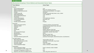
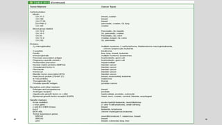
Tumor markers are substances produced by tumor cells or the body's response to tumors that can be detected in blood, urine, or tissue. They are used to screen for cancers, help diagnose cancer, determine prognosis, detect recurrence, and monitor treatment response. An ideal tumor marker would be highly sensitive and specific to a particular cancer, correlate with tumor size, respond rapidly to treatment, and predict recurrence before clinical detection. Examples of commonly used tumor markers include AFP, CEA, CA125, PSA, and CA15-3. Their levels can be elevated in both cancer and some benign conditions.
Tumour Markers are substances present in the tumour, produced by the tumour or by the host as a response to the presence of the tumour, providing information about biological characteristics of the tumour. these tumour markers may specific for the tissue but often get elevated in neoplastic as well non-neoplastic lesions, further Various analytical platforms available for serum tumour markers lack standardisation. These factors add to interpretative challenges in serum tumour markers
Tumor Markers include a wide range of biomacromolecules orchestrated in abundance fixation by a wide assortment of neoplastic cells. The markers could be endogenous results of exceptionally dynamic metabolic threatening cells or the results of recently turned on qualities, which stayed unexpressed in early life or recently obtained antigens at cell and sub-cell levels. The presence of tumor marker and their focus are identified with the beginning and development of dangerous tumors in patients. A perfect tumor marker ought to be profoundly delicate, explicit, dependable with high prognostic worth, organ particularity and it should relate with tumor stages. Be that as it may, none of the tumor markers answered to date has every one of these attributes. Inspite of these impediments, numerous tumor markers have indicated incredible clinical significance in checking adequacy of various methods of treatments during whole course of sickness in malignant growth patients. Moreover, assurance of markers additionally helps in early discovery of malignant growth repeat and in anticipation.
Tumor markers
Many cancers are associated with the abnormal production of some molecules which can be measured in plasma. These molecules are known as tumor markers.
A good tumor maker should have those properties:
1. A tumor marker should be present in or produced by tumor itself.
2. A tumor marker should not be present in healthy tissues.
3. Plasma level of a tumor marker should be at a minimum level in healthy subjects and in benign conditions.
4. A tumor marker should be specific for a tissue, it should have different immunological properties when it is synthesized in other tissues.
5. Plasma level of the tumor marker should be in proportion to the both size of the tumor and the activity of the tumor.
6. Half-life of a tumor should not be very long
7. A tumor marker should be present in plasma at a detectable level, even though tumor size is very small
Tumor markers are substances, such as proteins, biochemicals, hormones or enzymes, produced by tumor cells or by the body in response to tumor cells. As tumor cells multiply, cancer spreads, and tissue is damaged, these substances increase and leak into the bloodstream. Tumor marker levels in blood help physicians evaluate people for certain types of cancer
The presentation aims to be of introductory level. Classification of tumor markers along with brief description of selected tumor markers are provided.Applications and methods are listed but not discussed at length.
Tumor Biomarkers For Screening, Progression and Prognosis Vivek Misra
Tumor markers are substances that can be found in the body (usually in the blood or urine) when cancer is present. Along with other tests, tumor markers can be used to help show if cancer is present, to determine the type of cancer, and in some cases to help show if treatment is working. Some of the more common tumor markers are discussed here.
PREDICTIVE AND DIAGNOSTIC BIOMARKERS FOR OVARIAN CANCERDr. Girija Wagh
OVARIAN CANCER HAS ELUDED SCREENING AND EARLY DETECTION . SEVERAL BIOMARKERS ARE PROPOSED AND HERE IS A AN UPDATED REVIEW OF WHAT EXISTS IN THE CURRENT CLIMATE FOR THE SAME Ovarian carcinomas relate to highest death rate in gynecologic malignancies as absence of symptoms shield the disease in the early stage. Current evidences have been devoted to discovering early effective screening mechanism prior to the onset of clinical symptoms.Serum biomarkers may aid in the diagnosis of Early ovarian cancer
Distinguish malignant from benign disease
Prevent unnecessary surgery
Improve rates of early detection. Prof Girija Wagh is the Head of the Department of OBG at Bharati University Medical College and Hospital and a well acknowledged teacher and a researcher.This overview will certainly help the learners to approach this condition with more promise
ARTIFICIAL INTELLIGENCE IN HEALTHCARE.pdfAnujkumaranit
Artificial intelligence (AI) refers to the simulation of human intelligence processes by machines, especially computer systems. It encompasses tasks such as learning, reasoning, problem-solving, perception, and language understanding. AI technologies are revolutionizing various fields, from healthcare to finance, by enabling machines to perform tasks that typically require human intelligence.
Explore natural remedies for syphilis treatment in Singapore. Discover alternative therapies, herbal remedies, and lifestyle changes that may complement conventional treatments. Learn about holistic approaches to managing syphilis symptoms and supporting overall health.
NVBDCP.pptx Nation vector borne disease control programSapna Thakur
NVBDCP was launched in 2003-2004 . Vector-Borne Disease: Disease that results from an infection transmitted to humans and other animals by blood-feeding arthropods, such as mosquitoes, ticks, and fleas. Examples of vector-borne diseases include Dengue fever, West Nile Virus, Lyme disease, and malaria.
These lecture slides, by Dr Sidra Arshad, offer a quick overview of physiological basis of a normal electrocardiogram.
Learning objectives:
1. Define an electrocardiogram (ECG) and electrocardiography
2. Describe how dipoles generated by the heart produce the waveforms of the ECG
3. Describe the components of a normal electrocardiogram of a typical bipolar leads (limb II)
4. Differentiate between intervals and segments
5. Enlist some common indications for obtaining an ECG
Study Resources:
1. Chapter 11, Guyton and Hall Textbook of Medical Physiology, 14th edition
2. Chapter 9, Human Physiology - From Cells to Systems, Lauralee Sherwood, 9th edition
3. Chapter 29, Ganong’s Review of Medical Physiology, 26th edition
4. Electrocardiogram, StatPearls - https://www.ncbi.nlm.nih.gov/books/NBK549803/
5. ECG in Medical Practice by ABM Abdullah, 4th edition
6. ECG Basics, http://www.nataliescasebook.com/tag/e-c-g-basics
Report Back from SGO 2024: What’s the Latest in Cervical Cancer?bkling
Are you curious about what’s new in cervical cancer research or unsure what the findings mean? Join Dr. Emily Ko, a gynecologic oncologist at Penn Medicine, to learn about the latest updates from the Society of Gynecologic Oncology (SGO) 2024 Annual Meeting on Women’s Cancer. Dr. Ko will discuss what the research presented at the conference means for you and answer your questions about the new developments.
263778731218 Abortion Clinic /Pills In Harare ,sisternakatoto
263778731218 Abortion Clinic /Pills In Harare ,ABORTION WOMEN’S CLINIC +27730423979 IN women clinic we believe that every woman should be able to make choices in her pregnancy. Our job is to provide compassionate care, safety,affordable and confidential services. That’s why we have won the trust from all generations of women all over the world. we use non surgical method(Abortion pills) to terminate…Dr.LISA +27730423979women Clinic is committed to providing the highest quality of obstetrical and gynecological care to women of all ages. Our dedicated staff aim to treat each patient and her health concerns with compassion and respect.Our dedicated group ABORTION WOMEN’S CLINIC +27730423979 IN women clinic we believe that every woman should be able to make choices in her pregnancy. Our job is to provide compassionate care, safety,affordable and confidential services. That’s why we have won the trust from all generations of women all over the world. we use non surgical method(Abortion pills) to terminate…Dr.LISA +27730423979women Clinic is committed to providing the highest quality of obstetrical and gynecological care to women of all ages. Our dedicated staff aim to treat each patient and her health concerns with compassion and respect.Our dedicated group of receptionists, nurses, and physicians have worked together as a teamof receptionists, nurses, and physicians have worked together as a team wwww.lisywomensclinic.co.za/
Tom Selleck Health: A Comprehensive Look at the Iconic Actor’s Wellness Journeygreendigital
Tom Selleck, an enduring figure in Hollywood. has captivated audiences for decades with his rugged charm, iconic moustache. and memorable roles in television and film. From his breakout role as Thomas Magnum in Magnum P.I. to his current portrayal of Frank Reagan in Blue Bloods. Selleck's career has spanned over 50 years. But beyond his professional achievements. fans have often been curious about Tom Selleck Health. especially as he has aged in the public eye.
Follow us on: Pinterest
Introduction
Many have been interested in Tom Selleck health. not only because of his enduring presence on screen but also because of the challenges. and lifestyle choices he has faced and made over the years. This article delves into the various aspects of Tom Selleck health. exploring his fitness regimen, diet, mental health. and the challenges he has encountered as he ages. We'll look at how he maintains his well-being. the health issues he has faced, and his approach to ageing .
Early Life and Career
Childhood and Athletic Beginnings
Tom Selleck was born on January 29, 1945, in Detroit, Michigan, and grew up in Sherman Oaks, California. From an early age, he was involved in sports, particularly basketball. which played a significant role in his physical development. His athletic pursuits continued into college. where he attended the University of Southern California (USC) on a basketball scholarship. This early involvement in sports laid a strong foundation for his physical health and disciplined lifestyle.
Transition to Acting
Selleck's transition from an athlete to an actor came with its physical demands. His first significant role in "Magnum P.I." required him to perform various stunts and maintain a fit appearance. This role, which he played from 1980 to 1988. necessitated a rigorous fitness routine to meet the show's demands. setting the stage for his long-term commitment to health and wellness.
Fitness Regimen
Workout Routine
Tom Selleck health and fitness regimen has evolved. adapting to his changing roles and age. During his "Magnum, P.I." days. Selleck's workouts were intense and focused on building and maintaining muscle mass. His routine included weightlifting, cardiovascular exercises. and specific training for the stunts he performed on the show.
Selleck adjusted his fitness routine as he aged to suit his body's needs. Today, his workouts focus on maintaining flexibility, strength, and cardiovascular health. He incorporates low-impact exercises such as swimming, walking, and light weightlifting. This balanced approach helps him stay fit without putting undue strain on his joints and muscles.
Importance of Flexibility and Mobility
In recent years, Selleck has emphasized the importance of flexibility and mobility in his fitness regimen. Understanding the natural decline in muscle mass and joint flexibility with age. he includes stretching and yoga in his routine. These practices help prevent injuries, improve posture, and maintain mobilit
Lung Cancer: Artificial Intelligence, Synergetics, Complex System Analysis, S...Oleg Kshivets
RESULTS: Overall life span (LS) was 2252.1±1742.5 days and cumulative 5-year survival (5YS) reached 73.2%, 10 years – 64.8%, 20 years – 42.5%. 513 LCP lived more than 5 years (LS=3124.6±1525.6 days), 148 LCP – more than 10 years (LS=5054.4±1504.1 days).199 LCP died because of LC (LS=562.7±374.5 days). 5YS of LCP after bi/lobectomies was significantly superior in comparison with LCP after pneumonectomies (78.1% vs.63.7%, P=0.00001 by log-rank test). AT significantly improved 5YS (66.3% vs. 34.8%) (P=0.00000 by log-rank test) only for LCP with N1-2. Cox modeling displayed that 5YS of LCP significantly depended on: phase transition (PT) early-invasive LC in terms of synergetics, PT N0—N12, cell ratio factors (ratio between cancer cells- CC and blood cells subpopulations), G1-3, histology, glucose, AT, blood cell circuit, prothrombin index, heparin tolerance, recalcification time (P=0.000-0.038). Neural networks, genetic algorithm selection and bootstrap simulation revealed relationships between 5YS and PT early-invasive LC (rank=1), PT N0—N12 (rank=2), thrombocytes/CC (3), erythrocytes/CC (4), eosinophils/CC (5), healthy cells/CC (6), lymphocytes/CC (7), segmented neutrophils/CC (8), stick neutrophils/CC (9), monocytes/CC (10); leucocytes/CC (11). Correct prediction of 5YS was 100% by neural networks computing (area under ROC curve=1.0; error=0.0).
CONCLUSIONS: 5YS of LCP after radical procedures significantly depended on: 1) PT early-invasive cancer; 2) PT N0--N12; 3) cell ratio factors; 4) blood cell circuit; 5) biochemical factors; 6) hemostasis system; 7) AT; 8) LC characteristics; 9) LC cell dynamics; 10) surgery type: lobectomy/pneumonectomy; 11) anthropometric data. Optimal diagnosis and treatment strategies for LC are: 1) screening and early detection of LC; 2) availability of experienced thoracic surgeons because of complexity of radical procedures; 3) aggressive en block surgery and adequate lymph node dissection for completeness; 4) precise prediction; 5) adjuvant chemoimmunoradiotherapy for LCP with unfavorable prognosis.
Title: Sense of Taste
Presenter: Dr. Faiza, Assistant Professor of Physiology
Qualifications:
MBBS (Best Graduate, AIMC Lahore)
FCPS Physiology
ICMT, CHPE, DHPE (STMU)
MPH (GC University, Faisalabad)
MBA (Virtual University of Pakistan)
Learning Objectives:
Describe the structure and function of taste buds.
Describe the relationship between the taste threshold and taste index of common substances.
Explain the chemical basis and signal transduction of taste perception for each type of primary taste sensation.
Recognize different abnormalities of taste perception and their causes.
Key Topics:
Significance of Taste Sensation:
Differentiation between pleasant and harmful food
Influence on behavior
Selection of food based on metabolic needs
Receptors of Taste:
Taste buds on the tongue
Influence of sense of smell, texture of food, and pain stimulation (e.g., by pepper)
Primary and Secondary Taste Sensations:
Primary taste sensations: Sweet, Sour, Salty, Bitter, Umami
Chemical basis and signal transduction mechanisms for each taste
Taste Threshold and Index:
Taste threshold values for Sweet (sucrose), Salty (NaCl), Sour (HCl), and Bitter (Quinine)
Taste index relationship: Inversely proportional to taste threshold
Taste Blindness:
Inability to taste certain substances, particularly thiourea compounds
Example: Phenylthiocarbamide
Structure and Function of Taste Buds:
Composition: Epithelial cells, Sustentacular/Supporting cells, Taste cells, Basal cells
Features: Taste pores, Taste hairs/microvilli, and Taste nerve fibers
Location of Taste Buds:
Found in papillae of the tongue (Fungiform, Circumvallate, Foliate)
Also present on the palate, tonsillar pillars, epiglottis, and proximal esophagus
Mechanism of Taste Stimulation:
Interaction of taste substances with receptors on microvilli
Signal transduction pathways for Umami, Sweet, Bitter, Sour, and Salty tastes
Taste Sensitivity and Adaptation:
Decrease in sensitivity with age
Rapid adaptation of taste sensation
Role of Saliva in Taste:
Dissolution of tastants to reach receptors
Washing away the stimulus
Taste Preferences and Aversions:
Mechanisms behind taste preference and aversion
Influence of receptors and neural pathways
Impact of Sensory Nerve Damage:
Degeneration of taste buds if the sensory nerve fiber is cut
Abnormalities of Taste Detection:
Conditions: Ageusia, Hypogeusia, Dysgeusia (parageusia)
Causes: Nerve damage, neurological disorders, infections, poor oral hygiene, adverse drug effects, deficiencies, aging, tobacco use, altered neurotransmitter levels
Neurotransmitters and Taste Threshold:
Effects of serotonin (5-HT) and norepinephrine (NE) on taste sensitivity
Supertasters:
25% of the population with heightened sensitivity to taste, especially bitterness
Increased number of fungiform papillae
New Drug Discovery and Development .....NEHA GUPTA
The "New Drug Discovery and Development" process involves the identification, design, testing, and manufacturing of novel pharmaceutical compounds with the aim of introducing new and improved treatments for various medical conditions. This comprehensive endeavor encompasses various stages, including target identification, preclinical studies, clinical trials, regulatory approval, and post-market surveillance. It involves multidisciplinary collaboration among scientists, researchers, clinicians, regulatory experts, and pharmaceutical companies to bring innovative therapies to market and address unmet medical needs.
The prostate is an exocrine gland of the male mammalian reproductive system
It is a walnut-sized gland that forms part of the male reproductive system and is located in front of the rectum and just below the urinary bladder
Function is to store and secrete a clear, slightly alkaline fluid that constitutes 10-30% of the volume of the seminal fluid that along with the spermatozoa, constitutes semen
A healthy human prostate measures (4cm-vertical, by 3cm-horizontal, 2cm ant-post ).
It surrounds the urethra just below the urinary bladder. It has anterior, median, posterior and two lateral lobes
It’s work is regulated by androgens which are responsible for male sex characteristics
Generalised disease of the prostate due to hormonal derangement which leads to non malignant enlargement of the gland (increase in the number of epithelial cells and stromal tissue)to cause compression of the urethra leading to symptoms (LUTS
Knee anatomy and clinical tests 2024.pdfvimalpl1234
This includes all relevant anatomy and clinical tests compiled from standard textbooks, Campbell,netter etc..It is comprehensive and best suited for orthopaedicians and orthopaedic residents.
1. TUMOUR MARKERS
Dr. Ajit Kumar Singh
PGT-Laboratory medicine
CNCI , Kolkata-700160
Moderator
Dr. Garima Chauhan
MD (Biochemistry)
Assistant Professor
Department of laboratory medicine
Chittaranjan National Cancer Institute, Kolkata
2. DEFINITION
Substance that can be detected in higher than normal amount in serum ,
urine, or tissue of patient with certain types of cancer .
Tumor marker are produced either by the cancer cells themselves or by the
body in a response to the cancer.
No tumor marker is pathognomonic
Most used as a surveillance tool to monitor for recurrence of the neoplasm
3. USES OF TUMOUR MARKERS
o Screening populations at risk
Not all tumor markers are good screening tools
o Diagnosis
Use results from markers, imaging, risk factors, and symptoms
o Prognosis
Concentration of the marker determines prognosis
o Detection of recurrence
Once tumor is removed, elevations of marker can indicate regrowth
o Monitoring response to treatment
Decreased levels of tumor marker indicate therapy is working, increased levels of tumor
marker may indicate need for a change to therapy
o Clinical staging of cancer
o Estimating tumor volume (eg . CHOREOCARCINOMA )
4. 1. Detectable only if tumour is present
2. Identifies the type of tumour
3. Circulating concentration correlates with the mass of tumour present
4. Responds rapidly to treatment-induced remission
5. Responds rapidly to relapse (Predict recurrences before they are
clinically detectable)
6. Should predict outcome
7. In patients with stable disease
8. Test should be cost effective
What are the characteristics of an ideal tumor marker?
5. Classification according to type of the molecule:-
1 .Enzymes or isoenzymes (ALP,LDH, PAP)
2 .Hormones (calcitonin, β-Hcg)
3 .Oncofetal antigens (AFP, CEA)
4.Carbohydrate epitopes recognised by monoclonal antibodies
(CA 15-3,CA 19- 9,CA125)
5.Receptors (Estrogen, progesterone)
6.Protein Markers (Paraproteins)
9. Examples of some clinically important tumor
markers
1. AFP (alpha fetoprotein):
Alpha fetoprotein is protein that is produced in early fetal life.
AFP is the major serum protein of human fetus, and it falls to low levels by one
year of age.
The cut-off value ofAFP is 0-9 U/ml, but it reaches 500 U/ml in late pregnancy.
However, elevated levels (more than 500 U/ml) are found in many cases of 10HCC,
Hepatoblastoma (Fibrolamellar Variant – Neurotensin ) and GCT.
Prognosis -GCT
• Furthermore AFP may also be increased in serum of non-neoplastic conditions. In these
cases the rise may be transient, and associated with the tissue (usually the liver) response
to injury, and usually of lesser magnitude (100-300 U/ml )
10. 2. CEA ( Carcinoembryonic Antigen )
Itisaglycoprotein moleculenormallyfoundin thetissueofa developing fetus.
Levels of CEAin the blood decrease after birth.
CEAis normally found in small amounts in the blood of most healthy people.
Cut-off value of CEAis about 0- 3 ng/ml.
High CEA levels (above 5 ng/ml) have been found in colorectal adenocarcinoma and in
some tumors of the lung, pancreas, uterus, and breast.
Less marked elevations occurs in non-neoplastic diseases such as emphysema,
ulcerativecolitis,pancreatitisandalcoholism,andinserumofheavysmokers.
3. CA 15-3 (Cancer antigen 15-3)
CA15.3 is the most reliable and highly specific tumor marker for breast cancer.
The CA15.3 level can provide prognostic information in the follow-up management of
patients with breast cancer.
Cut-off value of CA15-3 is 0-35 units/ml.
11. 4. CA19-9 (Cancer antigen 19-9 )
Cancer antigen is a protein that is found in very small amounts on the surface of certain
cancer cells.
Cut-off value is 0-35 units/ml.
It may be found in the blood when it is shed by tumor cells.
A CA19-9 test may be done to check: a person’s response to treatment for pancreatic
cancer, especially advanced pancreatic cancer.
if pancreatic cancer is still growing or has come back (recurred) after treatment
It is also found in trace amounts in the pancreas, liver, gall bladder and lungs of
healthy adults.
5.CA 125 (cancer antigen 125)
Cancer antigen 125 is a protein produced by a variety of cells, particularly ovarian cancer
cells.
CA125 is most reliable and highly specific tumor marker for ovarian cancer.
Cut-off value is 0-35 units/ml.
12. 6.PSA (prostatic specific antigen)
It is widely accepted tumor marker in prostaticcancer.
PSA isglycoproteinproducedonlyby prostatic epithelialcells and it is organ specific.
Normal level: 0-4ng/ml.
Elevated Level (more than 4ng/ml) occurs in:
60% of prostatic cancer.
40% of benign prostatic hypertrophy.
7.Prostatic acid phosphatase(PAP)
One of the famous enzymes used as tumor marker.
The serum level of prostatic acid phosphatase (PAP) is elevated in prostatic carcinoma.
Certain precautions must be taken before taking serum samples for prostatic acid phosphatase
assay.
These include avoidance of rectal examination(DRE), and passage of catheter at least 7 days
before sampling and constipation must be avoided.
As these factors may result in false positive rise in serum acid phosphatsseactivity due to
squeezing of the prostatic tissues.
Cut-off value is 0.5-11 units/liter
13. 8.LDH (Lactate dehydrogenase)
An elevationin all five LDH isoenzymes (LDH1, LDH2, LDH3, LDH4, LDH5) canbe
seenin leukemia and lymphoma.
Selective increases in LDH isoenzymes LDH3 and LDH5 (liver enzyme) may
occur in liver carcinoma.
Cut-off value of total LDH ranges from 140 to 240units/liter.
9. Calcitonin
Patients with medullarythyroid carcinomacan be screenedby measuring
calcitonin levels.
10. HCG (Human chorionic gonadotropin)
Hormone produced by placenta, and used for detection of pregnancy.
Reaching maximum level at 8th week of gestation.
It increases in testicular carcinoma , GCT, GTN .
14. Human Chorionic Gonadotropin (βhCG)
Multiple forms in serum (intact heterodimer, free alpha, free
beta chains, various degradation products)
Function is to maintain progesterone production during first
2 weeks of pregnancy
Reference: <5 U/L
Elevated βhCG:
Physiological condition: pregnancy
Benign condition: pituitary adenoma
Malignancies:
Gestational trophoblastic disease
NSGCT
SGCT testis (20%)
15. 12. Cancer antigen CA 27-29
Ca 27-29 levels can be elevated in breast as well as in cancers of the colon, stomach, kidney, lung,
ovary, pancreas, uterus, and liver
First trimester pregnancy ,endometriosis, benign breast disease, kidney disease, and liver disease
also may be associated with elevated CA 27-29 levels.
13. P53
Tumor suppressor gene.
Because of its central role in preventing the propagation of cells with DNA damage, P53 has been
referred to as the “guardian of the genome”.
The P53 gene on chromosome 17p is the most commonly mutated gene in human cancer.
In about 50 to 70 percent of CRCs, P53 inactivation occurs by a mutation of one allele followed by
loss of the remaining wild type gene (Knudson’s 2 hit hypothesis).
In many but not all studies, patients whose tumors harbor P53 mutations have worse outcomes and
shorter survival than those without such mutations.
16. • It is unclear why patients with the Li Fraumeni syndrome (a condition caused by a
germline mutation in p53 in which patients frequently develop carcinomas,
sarcomas, and leukemias) are not at particularly increased risk of developing
CRCs.
• Li-Fraumeni syndrome (LFS) : is an inherited autosomal dominant disorder that is
manifested by a wide range of malignancies that appear at an unusually early age.
• Li-Fraumeni syndrome is also known as the Sarcoma, Breast, Leukemia, and
Adrenal Gland (SBLA) cancer syndrome.
• This cancer predisposition syndrome is inherited as an autosomal dominant
disorder and is associated with abnormalities in the tumor protein P53 gene (TP53).
17. PROTEIN MARKERS
1.Ferritin- Ferritin is a marker for Hodgkin lymphoma, leukemia, liver, lung and breast cancer.
2.Thyroglobulin- It is a useful marker for detection of differentiated thyroid cancer
(Follicular & papillary).
3.Paraproteins(M-protein Immunoglobulin- Bence-Jones protein) - is a free monoclonal
immunoglobulin light chain in the urine it is a reliable marker for multiple myeloma
4.β2 microglobulin
20. Biogenic amines
• Pheochromocytoma are diagnosed by testing 24-hour urine
samples for catecholamines and their metabolites as well as by
determinig plasma metanephrine levels.
• The 24-hour urinary vanillylmandelic acid (VMA) excretion has
poor diagnostic sensitivity and specificity compared with
fractionated 24-hour urinary metanephrines.
• False-positive VMA tests may result from ingestion of caffeine,
raw fruits, or medications (alpha-methyldopa).
• Urinary metanephrine are 98% sensitive and also about 98%
specific for pheochromocytomas, whereas VMA measurement
are slightly less sensitive and specific
21. Other common tumour markers
Neuron specific enolase – neuroendocrine tumours like small cell
carcinoma of lung, neuroblastoma, pheochromocytoma etc.
Chromogranin A
Inhibin
SCCA
S-100
HMB-45
Melan-A
Mitf-1
Vimentin
24. Some benign conditions associated with rise in
tumor markers
Associated non-malignant conditions
Viral Hepatitis, liver injury, pregnancy
Testicular failure, pregnancy
Smokers, hepatitis, cirrhosis, pancreatitis, gastritis
• Marker
• AFP
• β-hCG
• CEA
• PSA Prostatitis, benign prostatic hyper-plasia
25.
26.
27. • Specificity
• It is a measure of the incidence of negative results in persons known to be free of a disease, that is true negative (TN).
• Specificity = TN/ All without disease (FP +TN) X 100
• Efficiency
• The efficiency of a test is the number of correct results divided by the total number of tests.
• Efficiency = TP + TN/ Total number of tests (TP + TN + FP + FN) X 100
• Sensitivity
• It is a measure of the incidence of positive results in patients known to have a condition, that is true positive (TP)
• Sensitivity = TP/ All with disease (TP +FN) X 100
• Predictive values
• The predictive value of a test is a measure (%) of the times that the value (positive or negative) is the true value, i.e. the percent
of all positive tests that are true positives is the Positive Predictive Value (PV +ve).
• PV +ve = TP/ All positive (TP +FP) X 100
• PV -ve = TN/ All negative (TN +FN) X 100
28. Conclusions
Tumor markers have changed the diagnosis and management of patients with
malignancies
They play a vital role in staging testicular cancers, screening for prostate cancers,
prognosis in colorectal cancers.
They are being evaluated in screening for HCC, epithelial ovarian cancers.
Further research is needed to refine the role of these markers in management of
cancer patients.
Recent advances in technology provide additional malignant tumor markers in the
immediate future
29. 1. Have high disease sensitivity i.e.it should be positive in all patients with particular cancer.
Sensitivity of any tumor marker refers to the number of true positives, and it could be calculated as
follows:
• Number of true positives
X 100
• Number of true positives + False negatives
True positive isa malignant case which gives an elevated marker level above the
cut- off value.
False negative is a malignant case which gives a normal value for the marker
where as it is proved malignant histopathologically.
Cut-off value is the highest value obtained by the normal control.
2. Have high disease specificity i.e. it should be negative in all normal
population.
Specificity of any tumor marker refers to the number of true negatives (normal
individuals), and it could be calculated as follows:
• Number of true negatives
X 100
• Number of true negatives + False positives
True negative is a normal case which gives a normal value for the marker.
False positive is a normal case with elevated marker level abovethe cut-off value.
30. IDEAL TUMOR MARKERS
• Be specific to the tumor
• Level should change in response to tumor size
• An abnormal level should be obtained in the presence of
micrometastases
• The level should not have large fluctuations that are
independent of changes in tumor size
• Levels in healthy individuals are at much lower
concentrations than those found in cancer patients
• Predict recurrences before they are clinically detectable
• Test should be cost effective
31. Carcinoembryonic antigen (CEA)
• CEA is a highly glycosylated cell surface glycoprotein involved in intercellular
adhesion
• CEA is shed from the cell surface into the circulation and can be detected in
serum.
•It is thought that infiltrating tumour growth breaks down the normal
barriers that usually prevent CEA from entering the circulation.
Malignancies associated with
elevated CEA levels
Over-expressed primarily by
adenocarcinomas:
Colon
Rectum
Breast
Lung
Benign conditions associated with
elevated CEA levels
Pulmonary emphysema
Bronchitis
Acute ulcerative colitis
Rectal polyps
Hepatitis
Alcoholic cirrhosis
Obstructive jaundice
Smokers
Renal disease
32. CEA
Normal range: <5ng/ml
Smokers: <10ng/ml
Suspect malignancy at >20ng/ml
Suspect metastatic spread at >50ng/ml
T1/2: 3 days (1-5 days)
Dukes’ Grade
A
B
C
D
% patients
CEA >5ng/ml
4
26
44
65
33. Cancer Antigen 125 (CA125)
Glycoprotein, with unknown biological function
Marker developed through immunization of mice with cells from an
ovarian carcinoma line, to produce the OC 125 monoclonal antibody
Epithelial cells
• Fallopian tubes
• Endometrium
• Endocervix
• Normal ovary
Mesothelial cells
• Pleura
• Pericardium
• Peritoneum
34. CA125
‘Normal’ serum concentration: <35 kU/L
Elevated in menstruation
T1/2: 5 days (1-5 days)
Malignancies associated with elevated CA 125
80-85% ovarian serous adenocarcinomas
50% Stage I
>90% Stage II
Endometrium
Breast
Uterus
Liver
Stomach
Pancreas
Cervix
Fallopian tubes
Biliary tract
Colon
Lung
39. Prostatic Specific Antigen
(PSA)
Serine protease
Liquefy seminal coagulum
Production is androgen-dependent
Produced almost exclusively by epithelial component
of prostate gland
Raised in:
Benign prostatic hypertrophy
Acute and chronic prostatitis
Urinary retention
T½ = 2.5 days (radical prostatectomy)
40. Carcinoembryonic antigen (CEA)
• Glycoprotein
• Found in embryonic endodermal epithelium
• Serum levels disappear almost completely after birth, but
small amounts may be present in the colon.
• Elevated in primary colorectal cancer as well as with breast,
thyroid, lung, ovarian, prostate, liver, pancreatic cancer.
• Elevated in benign conditions including diverticulitis,
cholecystitis, Pancreatitis, Inflammatory bowel disease,
peptic ulcer disease, bronchitis, liver abscess, and alcoholic
cirrhosis, especially in smokers and in elderly persons
41. • Use of CEA level as a screening test for colorectal
cancer is not recommended
• Useful if obtained preoperatively and postoperatively in
patient with CR Ca
• Preoperative elevation indicator of poor prognosis
• The 2007 ASCO clinical practice guidelines state that the
data are insufficient to support the use of CEA to
determine whether to give a patient adjuvant therapy ,
that data stronger for the use of CEA for monitoring for
postoperative recurrence
• CEA measurement is the most cost-effective approach
for detecting metastasis.
42. • In cases in which the patient would be a candidate for
resection of recurrent colorectal cancer or systemic therapy,
the 2006 ASCO guidelines recommend the postoperative CEA
testing be performed every 3 moths in patient with stage two
or three disease for at least 3 years
• CEA is the marker of choice for monitoring metastatic CR Ca
during systemic therapy.
• CEA can be used in conjunction with diagnostic imaging and
history and physical exam for monitoring patient during active
therapy
• In the absence of measurable disease, an increase in CEA
level may be taken to indicate treatment failure.
43. • Individual laboratory assays vary slightly, but the typical
upper limit of normal for CEA in non-smokers is 3.8
micrograms per liter (mcg/L). For smokers, the upper
limit of normal is 5.5 mcg/L
44. In order to use a tumor marker for screening in the presence of cancer in
asymptomatic individuals in general population, the marker should be produced
by tumor cells and not be present in healthy people.
However, most tumor markers are present in normal, benign and cancer tissues
and are not specific enough to be used for screening cancer.
45. Applications in ovarian cancer detection
Early detection of ovarian cancer through the measurement of CA-125,
usually in combination with other modalities (eg, bimanual pelvic
examination, transvaginal ultrasonography), is the most promising
application of this tumor marker, permitting effective triage of patients for
primary surgery.
46. CA 125
1 Approx 90% of ovarian cancers are epithelial carcinomas and contain a
epithelium–related glycoprotein, cancer antigen 125.
2The major forms in serum have molecular weights of 200 kDa to 400 kDa.
3 Marked elevations (>1500 U/mL) are generally seen with ovarian cancer.
4 The ACOG and Society of Gynecologic Oncologists- recommend gyne-onco
referral for women with a pelvic mass suggestive of ovarian cancer and a serum
CA-125 value >35 U/mL in postmenopausal women or >200 U/mL in
premenopausal women.
47. Alpha-Fetoprotien
• glycoprotein that is normally produced during gestation by the fetal liver and
yolk sac.
• Levels decrease soon after birth in healthy adults.
• serum concentration of which is often elevated in patients with HCC. Serum
levels of AFP do not correlate well with other clinical features of HCC such as
size, stage, or prognosis.
• Elevated serum AFP occurs in pregnancy , with tumors of gonadal origin (both
germ cell(ovary or testicle) and non-germ cell) and in a variety of other
malignancies, of which gastric cancer is the most common.
• Elevated serum AFP may also be seen in patients with chronic liver disease
without HCC such as acute or chronic viral hepatitis . AFP may be slightly higher
in patients with cirrhosis due to hepatitis C .
• Benign conditions that can cause elevations of AFP : cirrhosis, hepatic necrosis,
acute hepatitis, chronic active hepatitis, ataxia telangiectasia, wiskott-aldrich
syndrome, neural tube defect, and pregnancy.
48. • The sensitivity of an elevated AFP level for detecting HCC is
60% .
• AFP is considered to be sensitive and specific enough To be
used for screening for HCC in high risk population .
• Current recommendations are to screen healthy hepatitis B
virus carriers with annual or semiannual measurement of AFP
and to screen carriers with cirrhosis of any etiology with twice-
yearly measurement of AFP level and liver ultrasonography.
• AFP efficacy in early diagnosis of HCC is limited
• Larger proportion of patients diagnosed with HCC are now
AFP seronegative due to imaging technology improvement
•
49. • It is generally accepted that serum levels greater than 500 mcg/L
(normal in most laboratories is between 10 and 20 mcg/L) in a high-risk
patient is diagnostic of HCC.However, HCC is often diagnosed at a
lower AFP level in patients undergoing screening.
• Not all tumors secrete AFP, and serum concentrations are normal in up
to 40 percent of small HCCs.
• Furthermore, an elevated AFP may be more likely in patients with HCC
due to viral hepatitis compared with alcoholic liver disease.
• Elevated levels were associated with the presence of stage III or IV
fibrosis, an elevated international normalized ratio, and an elevated
serum aspartate aminotransferase level.
• AFP levels are normal in the majority of patients with fibrolamellar
carcinoma, a variant of HCC.
• Patients with cirrhosis and persistently elevated AFP values have an
increased risk of developing HCC compared with those who have
fluctuating or normal levels.
• Despite the issues inherent in using AFP for the diagnosis of HCC, it
has emerged as an important prognostic marker, especially in patients
undergoing resection and those being considered for liver
transplantation. Patients with AFP levels >1000 mcg/L have an
extremely high risk of recurrent disease following transplantation,
irrespective of the tumor size
50. CA 19-9
• Cancer antigen 19-9 , sialylated Lewis (a) antigen
• Tumor related antigen
• Glycoprotein
• CA 19-9 levels are not a useful tumor marker.
• CA 19-9 requires the presence of the Lewis blood group antigen (a glycosyl
transferase) to be expressed. Among individuals with a Lewis-negative
phenotype (an estimated 5 to 10 percent of the population), CA 19-9 levels are
not a useful tumor marker
• One study found that serum concentrations above 37 U/mL represented the
most accurate cutoff value for discriminating pancreatic cancer from benign
pancreatic disease, but the sensitivity and specificity for pancreatic cancer at
this level were only 77 and 87 percent, respectively
• The data are insufficient to recommend use CA19-9 for screening, diagnosis,
surveillance, or monitoring of therapy for colon Ca
• 2006 ASCO guidelines there are insufficient data to recommend use of CA 19-9
for screening, diagnosis, or determination of operability of pancreatic cancer
51. • Patient with locally advanced or metastatic cancer
receiving active therapy, CA19-9 can be measured at the
start of therapy and every 1-3 months while therapy is
given, elevation in serial CA19-9 levels may indicate
progressive disease and should be confirmed by
additional studies.
• Although several marker candidates have emerged from
preclinical studies (and one, macrophage inhibitory
cytokine-1, appears particularly promising ), none has
replaced CA 19-9 to date
52.
53. C- KIT
• proto-oncogene c-Kit or tyrosine-protein kinase Kit or CD117, is a receptor
tyrosine kinase protein that in humans is encoded by the KIT gene.
• Antibodies to CD117 are widely used in immunohistochemistry to help
distinguish particular types of tumor in histological tissue sections. It is used
primarily in the diagnosis of GISTs, which are positive for CD117, but
negative for markers such as desmin and S-100, which are positive in
smooth muscle and neural tumors, which have a similar appearance. In
GISTs, CD117 staining is typically cytoplasmic, with stronger accentuation
along the cell membranes. CD117 antibodies can also be used in the
diagnosis of mast cell tumors and in distinguishing seminomas from
embryonal carcinomas
54. • around 95 percent of GISTs arising in adults
overexpress KIT.Approximately 80 percent of GISTs
have KIT gene mutations that lead to constitutive
activation of the KIT receptor.
55. PSA
• Androgen-regulated serine protease produced by the
prostate epithelium.
• PSA is normally present in low concentrations in the
blood of all adult males.
• PSA levels may be elevated in the blood of men with
benign prostatic hyperplasia, as well as in men with
prostate cancer
• PSA levels have been shown to be useful in evaluating
the effectiveness of prostate cancer treatment and
monitoring for recurrence after therapy.
56.
57. • In monitoring for recurrence, a trend of increasing levels is
considered more significant than a single absolute elevated
value.
• Although PSA has widely used for prostate Ca screening, the
utility of PSA screening remains controversial.
• In 2010 American cancer society updated its guidelines for the
early detection of prostate Ca to state that men who have at
least a 10 years life expectancy should have an opportunity to
make an informed decision with there health care provider
about whether to be screened for prostate Ca with DRE and
serum PSA, after receiving information about the benefits,
risks, and uncertainties associated with prostate Ca
screening, this recommendation was reinforced in their 2013
guideline.
58. • Studies have estimated that PSA elevations can precede
clinical disease by 5 to 10 years or even longer .
• PSA has a half-life of 2.2 days, and levels elevated by
different benign conditions will have variable recovery
times(DRE, Ejaculation,)
• The five-alpha reductase inhibitors finasteride and
dutasteride lower PSA levels.
• The traditional cutoff for an abnormal PSA level in the
major screening studies has been 4.0 ng/mL .
59. Screening for prostate cancer
• When a decision is made to screen, screening be performed with
prostate specific antigen (PSA) tests at intervals ranging from every
two to four years. We suggest not performing digital rectal examination
as part of screening.
• When a decision is made to screen, screening stop after age 69 or
earlier when comorbidities limit life expectancy to less than 10 years,
or the patient decides against further screening. Stopping screening at
age 65 may be appropriate if the PSA level is less than 1.0 ng/mL.
• Men with an abnormal DRE (if performed) or PSA level above 7 ng/mL
should be referred, without further testing, to an interventional
specialist who can evaluate them for a transrectal ultrasound-guided
prostate biopsy.
• men with a PSA level between 4 ng/mL and 7 ng/mL undergo repeat
testing several weeks later . Prior to repeat PSA testing, men should
abstain from ejaculation and bike riding for at least 48 hours. Men with
symptomatic prostatitis should be treated with antibiotics before
retesting. Men with a repeat PSA level above 4 ng/mL should be
referred for transrectal ultrasound-guided prostate biopsy.
60. • Numerous strategies have been proposed to improve the
diagnostic performance of PSA when levels are less than 10.0
ng/mL. These strategies include measuring PSA velocity
(change in PSA over time), PSA density (PSA per unit volume
of prostate), free PSA, complexed PSA, and using age- and
race-specific reference ranges.
• There is no consensus on using any of the PSA modifications,
and none of them has been shown in clinical trials to reduce
the number of unnecessary biopsies or improve clinical
outcomes. The total PSA cutoff of 4.0 ng/mL has been the
most accepted standard because it balances the tradeoff
between missing important cancers at a curable stage and
avoiding both detection of clinically insignificant disease and
subjecting men to unnecessary prostate biopsies.
61. CA 125
• A protein that is higher than normal in approximately 80 percent of women with ovarian
cancer. It can be measured with a blood test. CA 125 is commonly used to monitor
women with ovarian cancer.
• The normal values for CA 125 may vary slightly among individual
laboratories. In most laboratories, the normal value is less than 35 U
/ml.
• The most common use of the test is the monitoring of people with a known cancer that
elevates CA 125 level, such as cancer of the ovary. In the patient who is known to have a
malignancy, such as ovarian cancer, the CA 125 level can be monitored periodically. A
decreasing level generally indicates that therapy, including chemotherapy, has been
effective, while an increasing level indicates tumor recurrence. Because of normal test
variation, small changes are usually not considered significant. A doubling or halving of the
previous value would be important.
62. • In the patient who is being evaluated for a pelvic mass, a CA
125 level greater than 65 is associated with malignancy in
approximately 90% of cases. However, without a
demonstrable mass, the association is much weaker.
• A number of benign conditions can cause elevations of the CA
125 level, including pregnancy, endometriosis, uterine
fibroids(benign tumors), pancreatitis, normal menstruation,
pelvic inflammatory disease, and liver disease. Benign tumors
or cysts of the ovaries can also cause an abnormal test result.
• Increases can also be seen in cancers other than ovarian
cancer, including malignancies of the uterine tubes,
endometrium, lung, breast, pancreas, and gastrointestinal
tract.
63. • So if you have a higher than normal CA 125 level, you could have
ovarian cancer or a more common and less serious condition. Pelvic
ultrasound may be helpful to know if you have ovarian cancer, although
the only way to know for sure if you have ovarian cancer is to have
surgery. As a result, CA 125 is not recommended as a stand-alone
screening test for ovarian cancer.
• Combined CA 125 and pelvic ultrasound — Several studies have
looked at using CA 125 and pelvic ultrasound together to detect ovarian
cancer. However, the results of these studies have been somewhat
disappointing:
1) Many women had unnecessary surgery because of false positive
test results (the CA 125 or pelvic ultrasound was abnormal but no
cancer was found).
2) Some studies of CA 125 and pelvic ultrasound have found more
cancers at an early, more treatable stage, while others have not. No
published studies have shown that these tests reduce the risk of dying
of ovarian cancer.
64. Cancer antigen CA 27-29
• The MUC-1 gene product in the serum may be
quantitated by using radioimmunoassay with a
monoclonal antibody against the CA27-29
• Ca 27-29 levels can be elevated in breast as well as in
cancers of the colon, stomach, kidney, lung, ovary,
pancreas, uterus, and liver
• First trimester pregnancy ,endometriosis, benign breast
disease, kidney disease, and liver disease also may be
associated with elevated CA 27-29 levels.