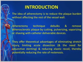
Atherectomy Devices
Presented by:- Dr. Himanshu Rana
INTRODUCTION
• The idea of atherectomy is to reduce the plaque burden
without affecting the rest of the vessel wall.
• Atherectomy technique debulks & remove
atherosclerotic plaque by cutting, pulverizing, vaporizing
or shaving with catheter deliverable devices.
• They offer theoretical advantages of eliminating stretch
injury, limiting acute dissection (& the need for
adjunctive stenting) & reducing elastic recoil, thereby
potentially reducing the rate of restenosis.
Differential Cutting DIFFERENTIAL CUTTING
The ability to cut one material
while sparing another based on
differences in composition.
ELASTIC TISSUE
Normal vascular tissue has elastic
properties that allow it to deflect
away from the advancing
diamonds on the rotating burr.
INELASTIC TISSUE
Selectively ablates inelastic tissue
(i.e. plaque) whether composed of
calcium, fibrotic tissue, neointima
or lipid-rich material.
Elastic Tissue
Inelastic Tissue
Results in preferential cutting of inelastic substrate
are functional at low and high speeds
PRINCIPLE
TYPES OF ATHERECTOMY
• Directional atherectomy
– Simpson catheter
– SilverHawk; TurboHawk
• Rotational atherectomy
– Rotablator
– CSI diamondback 360
– Pathway – Jetstream
• (Excimer) Laser
• Orbital atherectomy
“Historically, neither rotational nor
directional atherectomy has shown any
significant long term benefit over PTA alone
in the coronary or peripheral arteries”
- ARTIST Trial; Jueren et al.
Results of the Angioplasty versus rotational atherectomy for treatment of
diffuse in-stent re-stenosis trial (ARTIST Trial) Circulation. 2002; 105:583-588
Directional atherectomy systems
• SilverHawk & TurboHawk
• US Food and Drug Administration-approved.
• Approved for peripheral vasculature & not for
coronary, carotid, iliac, or renal arteries.
• Minimally invasive treatment that removes plaque
SilverHawk plaque excision system
• Forward cutting directional atherectomy device.
• Used with or without concurrent percutaneous
balloon angioplasty and stenting.
• Consists of a rotating blade inside a tubular housing
with a collection area (nosecone).
• This catheter is connected to a battery-driven motor
which spins the cutter.
SILVERHAWK PLAQUE EXCISION SYSTEM
TurboHawk system
• Similar except in the no. of inner blades (four
contoured blades)
• Favors use in highly calcified lesions and more plaque
removal per pass.
Directional atherectomy systems
• Both devices come in various sizes to enable
atherectomy in vessels with diameters of 1.5–7
mm.
• SilverHawk has advantage of directional control
for eccentric lesions.
• As the device is advanced, plaque is excised &
packed in the nosecone.
• Distal embolization remains a major problem.
Directional atherectomy systems
Advantages
• Lack of barotrauma, which decreases the risk of
neointimal hyperplasia &
• Lesser risk of dissection.
COMPLICATIONS
• Distal embolization
• Distal limb ischemia
• Dissection
• Perforation
• Pseudo-aneurysm formation
• No flow, and
• Ischemia
CLINICAL DATA
• TALON (Treating PeripherAls with SiLverHawk: Outcomes
CollectioN) registry
• 601 patients with over 1,258 infrainguinal lesions
• Patients with both claudication and critical limb ischemia.
• Both above- & below-knee lesions were treated.
• Achieved 50% or less diameter stenosis in 94.7% lesions.
• The procedural success rate was 97.6%.
• Stent placement was required in only 6.3% lesions
• 6- & 12-mo. survival free of TLR rates were 90% & 80%,
respectively.
CLINICAL DATA
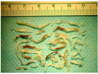
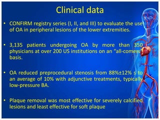
• DEFINITIVE LE (Determination of Effectiveness of
SilverHawk Peripheral Plaque Excision [SilverHawk
Device] for the Treatment of Infrainguinal
Vessels/Lower Extremities) registry – largest ever
conducted across 50 sites US & Europe
• Enrolled 799 patients with both claudication and CLI.
• Lesions up to 20 cm in length and multilevel lesions
• Success was reported at 89%, with a post-atherectomy
BA rate of 33% & bail-out stenting rate of 3%.
• Rates of distal embolization, dissection &
perforation were 3.8%, 2.3% & 5.3%
• All-complication rate needing treatment was 7.6%.
• At 12 months, SFA patency was 83% & IFA patency
was 78%.
• Limb salvage rate in CLI patients was 95%.
• Diabetics performed equally well as compared to
nondiabetics.
Rotational atherectomy
• Consists of an elliptical, nickel-plated, brass burr
• Coated with 2,000–3,000 microscopic diamond
crystals on the leading edge
• Available burr sizes are; 1.25, 1.5, 1.75, 2.0, 2.15,
2.25, 2.38, and 2.5 mm
• Burr rotates at 140,000–190,000 rpm
• Burr is advanced over the RotaWire
drive shaft
diamond coated burr
1.25 mm - 2.5 mm
(0.25 mm increments)
sheath
4.3 french O.D.
• Wire clip torquer should be placed to prevent the
guide wire from spinning
• A cocktail of Rotaglide® lubricant (nitroglycerine,
verapamil, and heparin) infuses through the Teflon
sheath which minimizes vasospasm, cools the
turbine, lubricates the driveshaft, and flushes the
particulate
• DIFFERENTIAL CUTTING
• Debris generated is < 10 μm, which traverses the
coronary microvasculature & is cleared by RES
Clinical data
• Single center Excimer Laser, Rotational Atherectomy, and
Balloon Angioplasty Comparison (ERBAC) study,
• 685 patients randomized to various atherectomy methods
• RA had the greatest initial success – 89% (RA) versus 77%
(excimer laser) versus 80% (BA)
• No difference in major in-hospital complications & at 6-mo.
follow-up.
• TLR was more frequent in the RA (42.4%) & the excimer
laser group (46.0%) than angioplasty group (31.9%,
P=0.013)
• Multicenter, prospective, comparison of balloon
angioplasty vs rotational atherectomy in complex
coronary lesions (COBRA) trial
• 502 patients were enrolled
• Similar results were found
Currently, due to lack of benefit in preventing
restenosis in native & restenotic lesions,
‘RA is used to prepare the calcified lesions for
stenting (drug eluting stents) when the stent is not
deliverable/expandable or the lesion is not dilatable
with conventional BA’
Absolute contraindications to RA are:
– Saphenous vein graft lesions;
– Presence of thrombus;
– Dissection; and
– Inability to cross the lesion with the guide wire.
Relative contraindications include:
– Lesion length > 25 mm;
– Lesion angulation > 45°;
– Severe LV dysfunction;
– Severe TVD or unprotected LM disease; and
– No candidacy to CABG, either because of patient
ineligibility or lack of onsite surgical backup
• Some of the reported complications of RA are:
– Q wave myocardial infarction (MI) (0.8%),
– Urgent CABG(2.0%),
– Non-Q MI (8.9%),
– Acute closures (1.1%),
– Slow flow (2%),
– Perforation (1.0%),
– Side-branch closure (5%),
– Dissection (4%), and
– Spasm (5%).
• Peripheral rotablator atherectomy has also recently
started being used for calcified below-knee arteries
Pathway Jetstream PV
Atherectomy System
• Pathway Jetstream PV Atherectomy System
(Pathway Medical Technologies)
• Consists of a single-use catheter with control pod
and a reusable, compact console
• The system is indicated for both thrombectomy and
RA by the same catheter
• The catheter is advanced maximum rate of 1
mm/second to avoid significant drops in rotational
speeds
• It has a front-cutting tip that makes it go through
tight lesions without predilation
• The electric motor spins for every 40 sec f/b a 10 sec.
pause.
• During treatment, saline solution is delivered to the
proximal end of the catheter using two lines:
– To flush the motor assembly to maintain airtight seal for
embolic protection;
– To infuse in the treatment area at the distal body of the
catheter to facilitate the catheter’s debulking and
aspiration capabilities
• Its differentially cutting catheter
• Above-knee catheters
– 2.1 mm/3.0 mm and 2.4 mm/3.4 mm have a catheter tip
that remains at a defined nominal diameter (2.1mm or–2.4
mm) when spinning clockwise, but
– expands to a defined maximum diameter (3.0 mm or 3.4
mm, respectively) when rotating counterclockwise
• For the below-knee use, there are fixed cutters
(single cutter) with sizes of 1.6 mm and 1.85 mm
• Pathway Jetstream is the only atherectomy device to
offer continuous active aspiration, and actively
removes atherosclerotic debris and thrombus from
the treatment site and delivers it to a collection bag
located on the console.
CLINICAL DATA
• Zeller et al21
• Treatment of 172 patients – 210 lesions
(femoropopliteal and infrapopliteal vessels) –
• 99% device success at 6-month and 12-month
• TLR rates of 15% and 26%, respectively.
• The 1-year restenosis rate was 38.2%
• In a review of 2,137 lesions treated in 1,029 patients,
• Jetstream devices had a combined embolization rate
of 22% (eight of 36), four of 18 (22%) in each group,
which was significantly higher than with BA alone
(five of 570, 0.9%),
• The use of embolization protection may be beneficial
when using this device.
COMPLICATIONS
• Abrupt vessel occlusion
• Dissection
• Distal emboli
• Hematoma at access site
• Infection
• Perforation
• Pseudoaneurysm
• Renal failure
• Restenosis
• Thrombus formation
EXCIMER LASER ATHERECTOMY
• Uses high energy, monochromatic light beam to alter or
dissolve (vaporize) the plaque without damaging the
surrounding tissue.
• Fiber-optic catheters are used to deliver this light beam.
• For endovascular applications, xenon chloride excimer
laser is used and its fiber-optic catheter has multiple
small fibers, in order to be flexible enough to navigate in
the arterial tree
• Laser can be used in both coronary and peripheral
applications
• It uses ultraviolet (UV) B region with a shorter
wavelength (300 nm), the absorption depth is less
• Another advantage is, it uses direct photochemical
lytic affect to break molecular bonds rather than
thermal affect
• UV B photons absorbed by the proteins and lipids in
cells actually break chemical bonds which facilitates
lyses of cellular structures
• When laser is used, two factors are controlled by the
operators:
– number of pulses per second (frequency) given and
– energy amount (fluence)
• the high energy is delivered with short interaction
time (pulsed).
• In this way, chemical bonds are broken only in the
tissue that the laser is touching without damaging
surrounding material or increasing the heat
• The excimer laser catheter removes tissue with a
thickness of 10 μm with each pulse of energy.
• Pulsed-wave xenon chloride laser is commonly used
in clinical practice;
• it operates within a wavelength of 308 nm, with
relatively long pulses (pulse duration of 135
nanoseconds),
• produces an output of 165 mJ per pulse.
• The long pulse length is required for successful delivery of
the UV light by silica fibers at the fluences necessary for
therapy – typically between 30 and 80 mJ/mm2.
• After this pulse of 135 nanoseconds, laser energy is not
emitted.
• Typically, pulse repetition rates of 25–40 pulses/second
are used.
• By doing this, the total power emitted from the catheter
tip is less than 3 W for the largest catheters and this
minimizes thermal effects during the tissue-ablation
process
• The size of the laser catheters used are chosen based
on reference coronary/peripheral vessel diameter,
and available sizes are 0.9 mm, 1.4 mm, 1.7 mm, 2.0
mm, 2.3 mm, and 2.5 mm.
• While using excimer laser, advancement of the
catheter should be slow (0.5 mm/second and no
faster than 1 mm/second) for effective plaque
removal due to shallow penetration energy depth
(35–50 μm).
• iodinated dye absorbs the excimer laser energy nearly
completely and will cause cavitation bubbles, vapor
bubbles, and percussive waves which will lead to
dissections/perforations,
• laser catheter should never be activated in contrast
media
• blood (hemoglobin) strongly absorbs excimer laser light
at 308 nm.
• saline flushes need to be given in order to remove blood
and contrast
• The excimer laser has been also used to facilitate
crossing of the wire through chronic total occlusions
by using the “step-by-step” technique
• There are newer designs for ablating larger lumen,
such as the TURBO-Booster catheter (Spectranetics)
which uses a custom guide catheter that allows the
laser to move in different directions and ablate more
tissue.
CLINICAL DATA
• CARMEL multicenter study, excimer laser angioplasty
was successfully used in more than 90% of the
enrolled 151 acute myocardial infarction (AMI)
patients with a large thrombus burden with a
relatively low rate (8.6%) of major cardiac adverse
events (MACE).
• For in-stent restenosis: in the Laser Angioplasty for
Restenotic Stents multicenter registry (LARS), laser
angioplasty reduced 30-day repeat-target-site
coronary intervention, but it did not decrease in 1
year
• CORAL study, excimer laser atherectomy was used in
diseased vein grafts with comparable 30-day MACE
(18.4%) to that of the control population (19.4%) from
the SAFER trial.
• It is important to notice that use of a distal embolic
protection device in the SAFER trial, where the filter wire
was used, showed a reduction of MACE (42% relative
reduction) compared to the control group, which further
supports the use of the protection device
• The device was studied in the multicenter clinical trial
ClirPath Excimer Laser to Enlarge Lumen Openings
(CELLO) with high procedural success rate,
• It can also be used to assist endovascular treatment
of peripheral arterial disease.
• In the Laser Angioplasty for Critical Limb Ischemia
(LACI) trial, 155 critically ischemic limbs with above-
or below-knee disease that were poor candidates for
surgical revascularization were treated.
• Despite the fact that mean treatment length was .16
cm and most of the patients had multiple
stenosis/occlusions, a limb-salvage rate of 93% was
achieved at 6 months.
ORBITAL ATHERECTOMY
• Diamondback 360° OAS is very similar to RA devices,
as it uses a crown that is equivalent to the RA burr
• The crown is eccentrically mounted, diamond coated,
and rotates at speeds varying from 60,000 to 200,000
rpm
• OAS uses its unique orbiting action to remove plaque,
and it has the ability to increase lumen diameter by
increasing the orbital speed
• A crown is eccentric in shape, in contrast to an RA
burr
• Theoretically, the less diseased, more elastic arterial
wall flexes away from the crown and minimizes the
risk of vessel trauma
• Perhaps the most-unique feature of OA is the ability
to create variable lumen size with the same catheter
by only changing the speed of rotation
• used in diseased peripheral, coronary arteries and in
diseased artificial arteriovenous dialysis fistulae
• Some other advantages of the orbital motion of the
crown is
– its being in contact with only one part of the vessel wall at
any given moment, and
– not obstructing flow in a diseased vessel (100% occluded
vessel which does not have flow anyway),
– which will minimize heat generation and also lead to the
– continuous clearance of the sanded microscopic particulate
matter rather than having it build up into a large load of
matter
• Crowns come in sizes of 1.25, 1.5, 2.0, and 2.25 mm.
The crowns need to be advanced over the wire. As a
guide wire, ViperWire is used
Clinical data
• CONFIRM registry series (I, II, and III) to evaluate the use
of OA in peripheral lesions of the lower extremities.
• 3,135 patients undergoing OA by more than 350
physicians at over 200 US institutions on an “all-comers”
basis.
• OA reduced preprocedural stenosis from 88%±12% s to
an average of 10% with adjunctive treatments, typically
low-pressure BA.
• Plaque removal was most effective for severely calcified
lesions and least effective for soft plaque
• dissection (11.3%),
• spasm (6.3%),
• slow flow (4.4%),
• embolism (2.2%),
• vessel closure (1.5%),
• thrombus (1.2%), and
• perforation (0.7%).52
• Other complications reported with OA use are
hemolysis and hemolysis-induced pancreatitis
“Atherrectomy devices are a good
alternative when the traditional measures
of angioplasty fails but should not not be
used as a first hand measure to treat a
lesion”
Circulation 2002 “Atherectomy devices and their future”
Thank you

Atherectomy devices

  • 1.
  • 2.
    INTRODUCTION • The ideaof atherectomy is to reduce the plaque burden without affecting the rest of the vessel wall. • Atherectomy technique debulks & remove atherosclerotic plaque by cutting, pulverizing, vaporizing or shaving with catheter deliverable devices. • They offer theoretical advantages of eliminating stretch injury, limiting acute dissection (& the need for adjunctive stenting) & reducing elastic recoil, thereby potentially reducing the rate of restenosis.
  • 3.
    Differential Cutting DIFFERENTIALCUTTING The ability to cut one material while sparing another based on differences in composition. ELASTIC TISSUE Normal vascular tissue has elastic properties that allow it to deflect away from the advancing diamonds on the rotating burr. INELASTIC TISSUE Selectively ablates inelastic tissue (i.e. plaque) whether composed of calcium, fibrotic tissue, neointima or lipid-rich material. Elastic Tissue Inelastic Tissue Results in preferential cutting of inelastic substrate are functional at low and high speeds PRINCIPLE
  • 4.
    TYPES OF ATHERECTOMY •Directional atherectomy – Simpson catheter – SilverHawk; TurboHawk • Rotational atherectomy – Rotablator – CSI diamondback 360 – Pathway – Jetstream • (Excimer) Laser • Orbital atherectomy
  • 5.
    “Historically, neither rotationalnor directional atherectomy has shown any significant long term benefit over PTA alone in the coronary or peripheral arteries” - ARTIST Trial; Jueren et al. Results of the Angioplasty versus rotational atherectomy for treatment of diffuse in-stent re-stenosis trial (ARTIST Trial) Circulation. 2002; 105:583-588
  • 6.
    Directional atherectomy systems •SilverHawk & TurboHawk • US Food and Drug Administration-approved. • Approved for peripheral vasculature & not for coronary, carotid, iliac, or renal arteries. • Minimally invasive treatment that removes plaque
  • 7.
    SilverHawk plaque excisionsystem • Forward cutting directional atherectomy device. • Used with or without concurrent percutaneous balloon angioplasty and stenting. • Consists of a rotating blade inside a tubular housing with a collection area (nosecone). • This catheter is connected to a battery-driven motor which spins the cutter.
  • 8.
  • 11.
    TurboHawk system • Similarexcept in the no. of inner blades (four contoured blades) • Favors use in highly calcified lesions and more plaque removal per pass.
  • 12.
    Directional atherectomy systems •Both devices come in various sizes to enable atherectomy in vessels with diameters of 1.5–7 mm. • SilverHawk has advantage of directional control for eccentric lesions. • As the device is advanced, plaque is excised & packed in the nosecone. • Distal embolization remains a major problem.
  • 13.
    Directional atherectomy systems Advantages •Lack of barotrauma, which decreases the risk of neointimal hyperplasia & • Lesser risk of dissection.
  • 14.
    COMPLICATIONS • Distal embolization •Distal limb ischemia • Dissection • Perforation • Pseudo-aneurysm formation • No flow, and • Ischemia
  • 15.
    CLINICAL DATA • TALON(Treating PeripherAls with SiLverHawk: Outcomes CollectioN) registry • 601 patients with over 1,258 infrainguinal lesions • Patients with both claudication and critical limb ischemia. • Both above- & below-knee lesions were treated. • Achieved 50% or less diameter stenosis in 94.7% lesions. • The procedural success rate was 97.6%. • Stent placement was required in only 6.3% lesions • 6- & 12-mo. survival free of TLR rates were 90% & 80%, respectively.
  • 16.
    CLINICAL DATA • DEFINITIVELE (Determination of Effectiveness of SilverHawk Peripheral Plaque Excision [SilverHawk Device] for the Treatment of Infrainguinal Vessels/Lower Extremities) registry – largest ever conducted across 50 sites US & Europe • Enrolled 799 patients with both claudication and CLI. • Lesions up to 20 cm in length and multilevel lesions • Success was reported at 89%, with a post-atherectomy BA rate of 33% & bail-out stenting rate of 3%.
  • 17.
    • Rates ofdistal embolization, dissection & perforation were 3.8%, 2.3% & 5.3% • All-complication rate needing treatment was 7.6%. • At 12 months, SFA patency was 83% & IFA patency was 78%. • Limb salvage rate in CLI patients was 95%. • Diabetics performed equally well as compared to nondiabetics.
  • 18.
    Rotational atherectomy • Consistsof an elliptical, nickel-plated, brass burr • Coated with 2,000–3,000 microscopic diamond crystals on the leading edge • Available burr sizes are; 1.25, 1.5, 1.75, 2.0, 2.15, 2.25, 2.38, and 2.5 mm • Burr rotates at 140,000–190,000 rpm • Burr is advanced over the RotaWire
  • 19.
    drive shaft diamond coatedburr 1.25 mm - 2.5 mm (0.25 mm increments) sheath 4.3 french O.D.
  • 20.
    • Wire cliptorquer should be placed to prevent the guide wire from spinning • A cocktail of Rotaglide® lubricant (nitroglycerine, verapamil, and heparin) infuses through the Teflon sheath which minimizes vasospasm, cools the turbine, lubricates the driveshaft, and flushes the particulate • DIFFERENTIAL CUTTING • Debris generated is < 10 μm, which traverses the coronary microvasculature & is cleared by RES
  • 23.
    Clinical data • Singlecenter Excimer Laser, Rotational Atherectomy, and Balloon Angioplasty Comparison (ERBAC) study, • 685 patients randomized to various atherectomy methods • RA had the greatest initial success – 89% (RA) versus 77% (excimer laser) versus 80% (BA) • No difference in major in-hospital complications & at 6-mo. follow-up. • TLR was more frequent in the RA (42.4%) & the excimer laser group (46.0%) than angioplasty group (31.9%, P=0.013)
  • 24.
    • Multicenter, prospective,comparison of balloon angioplasty vs rotational atherectomy in complex coronary lesions (COBRA) trial • 502 patients were enrolled • Similar results were found
  • 25.
    Currently, due tolack of benefit in preventing restenosis in native & restenotic lesions, ‘RA is used to prepare the calcified lesions for stenting (drug eluting stents) when the stent is not deliverable/expandable or the lesion is not dilatable with conventional BA’
  • 26.
    Absolute contraindications toRA are: – Saphenous vein graft lesions; – Presence of thrombus; – Dissection; and – Inability to cross the lesion with the guide wire. Relative contraindications include: – Lesion length > 25 mm; – Lesion angulation > 45°; – Severe LV dysfunction; – Severe TVD or unprotected LM disease; and – No candidacy to CABG, either because of patient ineligibility or lack of onsite surgical backup
  • 27.
    • Some ofthe reported complications of RA are: – Q wave myocardial infarction (MI) (0.8%), – Urgent CABG(2.0%), – Non-Q MI (8.9%), – Acute closures (1.1%), – Slow flow (2%), – Perforation (1.0%), – Side-branch closure (5%), – Dissection (4%), and – Spasm (5%). • Peripheral rotablator atherectomy has also recently started being used for calcified below-knee arteries
  • 28.
    Pathway Jetstream PV AtherectomySystem • Pathway Jetstream PV Atherectomy System (Pathway Medical Technologies) • Consists of a single-use catheter with control pod and a reusable, compact console • The system is indicated for both thrombectomy and RA by the same catheter
  • 29.
    • The catheteris advanced maximum rate of 1 mm/second to avoid significant drops in rotational speeds • It has a front-cutting tip that makes it go through tight lesions without predilation • The electric motor spins for every 40 sec f/b a 10 sec. pause.
  • 30.
    • During treatment,saline solution is delivered to the proximal end of the catheter using two lines: – To flush the motor assembly to maintain airtight seal for embolic protection; – To infuse in the treatment area at the distal body of the catheter to facilitate the catheter’s debulking and aspiration capabilities • Its differentially cutting catheter
  • 31.
    • Above-knee catheters –2.1 mm/3.0 mm and 2.4 mm/3.4 mm have a catheter tip that remains at a defined nominal diameter (2.1mm or–2.4 mm) when spinning clockwise, but – expands to a defined maximum diameter (3.0 mm or 3.4 mm, respectively) when rotating counterclockwise • For the below-knee use, there are fixed cutters (single cutter) with sizes of 1.6 mm and 1.85 mm
  • 35.
    • Pathway Jetstreamis the only atherectomy device to offer continuous active aspiration, and actively removes atherosclerotic debris and thrombus from the treatment site and delivers it to a collection bag located on the console.
  • 36.
    CLINICAL DATA • Zelleret al21 • Treatment of 172 patients – 210 lesions (femoropopliteal and infrapopliteal vessels) – • 99% device success at 6-month and 12-month • TLR rates of 15% and 26%, respectively. • The 1-year restenosis rate was 38.2%
  • 37.
    • In areview of 2,137 lesions treated in 1,029 patients, • Jetstream devices had a combined embolization rate of 22% (eight of 36), four of 18 (22%) in each group, which was significantly higher than with BA alone (five of 570, 0.9%), • The use of embolization protection may be beneficial when using this device.
  • 38.
    COMPLICATIONS • Abrupt vesselocclusion • Dissection • Distal emboli • Hematoma at access site • Infection • Perforation • Pseudoaneurysm • Renal failure • Restenosis • Thrombus formation
  • 39.
    EXCIMER LASER ATHERECTOMY •Uses high energy, monochromatic light beam to alter or dissolve (vaporize) the plaque without damaging the surrounding tissue. • Fiber-optic catheters are used to deliver this light beam. • For endovascular applications, xenon chloride excimer laser is used and its fiber-optic catheter has multiple small fibers, in order to be flexible enough to navigate in the arterial tree • Laser can be used in both coronary and peripheral applications
  • 40.
    • It usesultraviolet (UV) B region with a shorter wavelength (300 nm), the absorption depth is less • Another advantage is, it uses direct photochemical lytic affect to break molecular bonds rather than thermal affect • UV B photons absorbed by the proteins and lipids in cells actually break chemical bonds which facilitates lyses of cellular structures
  • 41.
    • When laseris used, two factors are controlled by the operators: – number of pulses per second (frequency) given and – energy amount (fluence) • the high energy is delivered with short interaction time (pulsed). • In this way, chemical bonds are broken only in the tissue that the laser is touching without damaging surrounding material or increasing the heat
  • 42.
    • The excimerlaser catheter removes tissue with a thickness of 10 μm with each pulse of energy. • Pulsed-wave xenon chloride laser is commonly used in clinical practice; • it operates within a wavelength of 308 nm, with relatively long pulses (pulse duration of 135 nanoseconds), • produces an output of 165 mJ per pulse.
  • 43.
    • The longpulse length is required for successful delivery of the UV light by silica fibers at the fluences necessary for therapy – typically between 30 and 80 mJ/mm2. • After this pulse of 135 nanoseconds, laser energy is not emitted. • Typically, pulse repetition rates of 25–40 pulses/second are used. • By doing this, the total power emitted from the catheter tip is less than 3 W for the largest catheters and this minimizes thermal effects during the tissue-ablation process
  • 44.
    • The sizeof the laser catheters used are chosen based on reference coronary/peripheral vessel diameter, and available sizes are 0.9 mm, 1.4 mm, 1.7 mm, 2.0 mm, 2.3 mm, and 2.5 mm. • While using excimer laser, advancement of the catheter should be slow (0.5 mm/second and no faster than 1 mm/second) for effective plaque removal due to shallow penetration energy depth (35–50 μm).
  • 45.
    • iodinated dyeabsorbs the excimer laser energy nearly completely and will cause cavitation bubbles, vapor bubbles, and percussive waves which will lead to dissections/perforations, • laser catheter should never be activated in contrast media • blood (hemoglobin) strongly absorbs excimer laser light at 308 nm. • saline flushes need to be given in order to remove blood and contrast
  • 46.
    • The excimerlaser has been also used to facilitate crossing of the wire through chronic total occlusions by using the “step-by-step” technique • There are newer designs for ablating larger lumen, such as the TURBO-Booster catheter (Spectranetics) which uses a custom guide catheter that allows the laser to move in different directions and ablate more tissue.
  • 48.
    CLINICAL DATA • CARMELmulticenter study, excimer laser angioplasty was successfully used in more than 90% of the enrolled 151 acute myocardial infarction (AMI) patients with a large thrombus burden with a relatively low rate (8.6%) of major cardiac adverse events (MACE). • For in-stent restenosis: in the Laser Angioplasty for Restenotic Stents multicenter registry (LARS), laser angioplasty reduced 30-day repeat-target-site coronary intervention, but it did not decrease in 1 year
  • 49.
    • CORAL study,excimer laser atherectomy was used in diseased vein grafts with comparable 30-day MACE (18.4%) to that of the control population (19.4%) from the SAFER trial. • It is important to notice that use of a distal embolic protection device in the SAFER trial, where the filter wire was used, showed a reduction of MACE (42% relative reduction) compared to the control group, which further supports the use of the protection device • The device was studied in the multicenter clinical trial ClirPath Excimer Laser to Enlarge Lumen Openings (CELLO) with high procedural success rate,
  • 50.
    • It canalso be used to assist endovascular treatment of peripheral arterial disease. • In the Laser Angioplasty for Critical Limb Ischemia (LACI) trial, 155 critically ischemic limbs with above- or below-knee disease that were poor candidates for surgical revascularization were treated. • Despite the fact that mean treatment length was .16 cm and most of the patients had multiple stenosis/occlusions, a limb-salvage rate of 93% was achieved at 6 months.
  • 51.
    ORBITAL ATHERECTOMY • Diamondback360° OAS is very similar to RA devices, as it uses a crown that is equivalent to the RA burr • The crown is eccentrically mounted, diamond coated, and rotates at speeds varying from 60,000 to 200,000 rpm • OAS uses its unique orbiting action to remove plaque, and it has the ability to increase lumen diameter by increasing the orbital speed
  • 52.
    • A crownis eccentric in shape, in contrast to an RA burr • Theoretically, the less diseased, more elastic arterial wall flexes away from the crown and minimizes the risk of vessel trauma • Perhaps the most-unique feature of OA is the ability to create variable lumen size with the same catheter by only changing the speed of rotation • used in diseased peripheral, coronary arteries and in diseased artificial arteriovenous dialysis fistulae
  • 53.
    • Some otheradvantages of the orbital motion of the crown is – its being in contact with only one part of the vessel wall at any given moment, and – not obstructing flow in a diseased vessel (100% occluded vessel which does not have flow anyway), – which will minimize heat generation and also lead to the – continuous clearance of the sanded microscopic particulate matter rather than having it build up into a large load of matter • Crowns come in sizes of 1.25, 1.5, 2.0, and 2.25 mm. The crowns need to be advanced over the wire. As a guide wire, ViperWire is used
  • 56.
    Clinical data • CONFIRMregistry series (I, II, and III) to evaluate the use of OA in peripheral lesions of the lower extremities. • 3,135 patients undergoing OA by more than 350 physicians at over 200 US institutions on an “all-comers” basis. • OA reduced preprocedural stenosis from 88%±12% s to an average of 10% with adjunctive treatments, typically low-pressure BA. • Plaque removal was most effective for severely calcified lesions and least effective for soft plaque
  • 57.
    • dissection (11.3%), •spasm (6.3%), • slow flow (4.4%), • embolism (2.2%), • vessel closure (1.5%), • thrombus (1.2%), and • perforation (0.7%).52 • Other complications reported with OA use are hemolysis and hemolysis-induced pancreatitis
  • 58.
    “Atherrectomy devices area good alternative when the traditional measures of angioplasty fails but should not not be used as a first hand measure to treat a lesion” Circulation 2002 “Atherectomy devices and their future”
  • 59.