The document provides a detailed overview of anesthesia management for patients with asthma and chronic obstructive pulmonary disease (COPD), including definitions, causes, pathophysiology, and preoperative assessment strategies. It emphasizes the importance of understanding the dynamic interactions between environmental factors and these respiratory conditions, and outlines the necessary preoperative optimization techniques like smoking cessation and the use of bronchodilators. Additionally, it discusses the anesthetic techniques and medications preferred for managing these patients, aiming to minimize complications while ensuring effective ventilation and oxygenation during surgery.

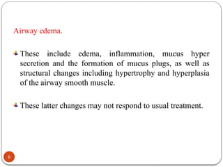

![Dynamic hyperinflation
In COPD there is 'air trapping' (increased residual volume and increased ratio of
residual volume to total lung capacity) and progressive hyperinflation (increased
total lung capacity).
During spontaneous breathing, the high expiratory airway resistance, combined
with expiratory flow limitation, low elastic recoil, high ventilatory demands and
short expiratory time due to the increased respiratory rate, may not permit the
respiratory system to reach the elastic equilibrium volume (i.e., passive
functional residual capacity [FRC]) at end-expiration.
This phenomenon is commonly referred to as dynamic hyperinflation.
Thus, an elastic threshold load (intrinsic positive end expiratory pressure
[PEEPi]) is imposed on the inspiratory muscles at the beginning of inspiration
and increases the amount of the inspiratory effort needed for gas flow
32](https://image.slidesharecdn.com/asthmaandcopd-250116091850-7c30dddd/85/ASTHMA-and-Chronic-obstructive-lung-disease-pptx-32-320.jpg)


![CTEV [ clubfoot] DR ARUN LAL ,DR MOHAMED ASHRAF travancore medical college k...](https://cdn.slidesharecdn.com/ss_thumbnails/ctevclubfootdrarunlaldrmohamedashraftravancoremedicalcollegekollamkeralaindia-260208063247-18fc466c-thumbnail.jpg?width=640&height=640&fit=bounds)


![ONFH[AVN HIP] -TRIPLE REGIME -A NOVAL SURGICAL CONCEPT .pptx](https://cdn.slidesharecdn.com/ss_thumbnails/onfhavnhip2026koaconcalicutdrgokuldevdrmashraf-260210064517-213ec005-thumbnail.jpg?width=640&height=640&fit=bounds)


