Downloaded 191 times
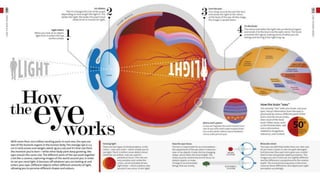
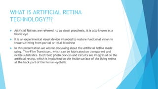
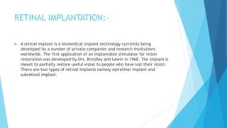
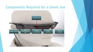
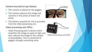


The document covers the concept of the artificial retina, explaining how the human eye works, the diseases affecting it, and the technology behind bionic eyes. It details the history of bionic eye development, components like cameras and video processing units, and the working mechanism of retinal implants. Additionally, it highlights the advantages of this technology in restoring vision, including the ability to identify objects and perform daily activities.