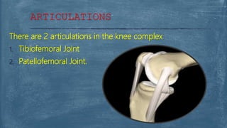
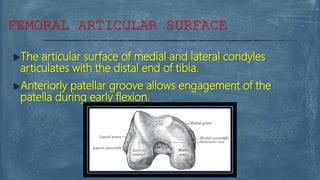
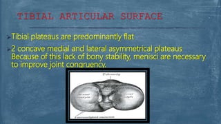
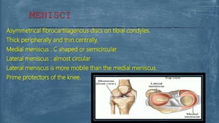
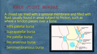
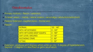
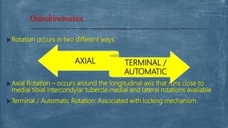
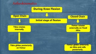
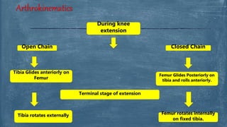
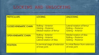
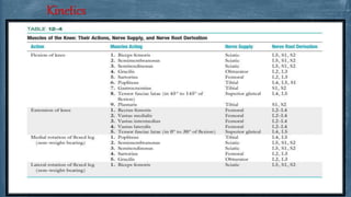
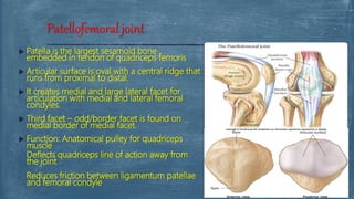
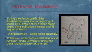
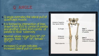
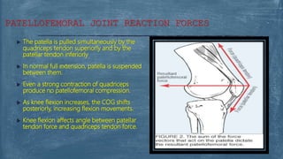
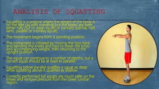
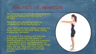
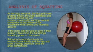
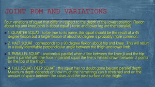
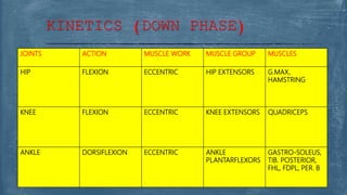
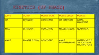
- NITHIN NAIR (MPT-1)
BIOMECHANICS
The Knee Complex
Able to:
 Know and describe the type of joint
 Know and describe the articulations of the knee joint
 The structures of the knee joint including ligaments, menisci &
bursae.
 Know and describe the different movements of the knee.
 Know and describe Kinetics and kinematics of the knee
complex.
 Recall the kinetics and Kinematics of squatting
OBJECTIVES
Provide mobility.
Support body during Static and Dynamic activities.
In Closed Kinematic Chain – works with hip and ankle
joints to support body weight in static erect posture.
Dynamically support during sitting and squatting
activities and transferring body weight during
locomotor activities.
In Open Kinematic Chain – provide mobility for foot
in space.
INTRODUCTION
The knee is classified as a;
 1. Synovial /diarthroses- a freely movable joint.
 2. Double condyloid – 3 degrees of movement.
 3. Modified Hinge joint- moves in one plane with slight
rotational movement, but the rotation is not enough to be
considered significant.
Type of Joint
There are 2 articulations in the knee complex
1. Tibiofemoral Joint
2. Patellofemoral Joint.
ARTICULATIONS
The articular surface of medial and lateral condyles
articulates with the distal end of tibia.
Anteriorly patellar groove allows engagement of the
patella during early flexion.
FEMORAL ARTICULAR SURFACE
Tibial plateaus are predominantly flat
2 concave medial and lateral asymmetrical plateaus
Because of this lack of bony stability, menisci are necessary
to improve joint congruency.
TIBIAL ARTICULAR SURFACE
Asymmetrical fibrocartilagenous discs on tibial condyles.
Thick peripherally and thin centrally.
Medial meniscus : C shaped or semicircular
Lateral meniscus : almost circular
Lateral meniscus is more mobile than the medial meniscus.
Prime protectors of the knee.
MENISCI
Functions :
Maintains the bony contact and potential joint space.
Escorts the articular surface.
Nourishes the articular surface.
Increases the concavity of tibial condyle for better
articulation with femoral condyle.
Serves as a cushion and overcomes the thrust.
Deepens joint Cavity.
Uniformly – spread synovial fluid.
Serves as Shock absorbers
MENISCI
Wide and lax; thin in front and
at the side; and contains
the patella ("knee
cap"), ligaments, menisci,
and bursae.
 The capsule consists of
a synovial and a fibrous
membrane separated by fatty
deposits anteriorly and
posteriorly.
KNEE JOINT CAPSULE
Anteromedial & anterolateral portions of capsule are
known as extensor retinaculum or medial & lateral
patellar retinacula
2 layers: Deeper and Superficial
Deeper longitudinal fibers connects capsule anteriorly to
menisci and tibia via coronary ligaments. This is known as
patellomeniscal / patellotibial bands.
Superficial transverse fibers blends with fibers of vastus
medialis and lateralis and to post. Tibial condyle.
EXTENSOR RETINACULUM
Large and more complex than the fibrous layer
Synovium adheres to the inner wall of the fibrous layer.
Posteriorly, it invaginates anteriorly following the contour of femoral
intercondylar notch.
Embryonically, synovial lining is divided into 3 compartments by septa –
sup. Patellofemoral , medial tibiofemoral, lateral tibiofemoral
compartments.
By 12 weeks of gestation, the synovial septa are resorbed resulting in a
single joint cavity.
Superior compartment remain as superior recess of capsule –
suprapatellar bursa
SYNOVIAL LAYER OF KNEE JOINT.
Synovial septa which are not completely resorbed in
adulthood, exist as folds or pleats of synovial tissue – PLICAE /
PATELLAR PLICAE
Commonly known plicae are – inferior, superior, medial plica
Occasionally plica may become irritated and inflamed which
leads to pain, effusion and changes in joint structure and
function.
Plica on the medial side of the joint may cause knee joint pain
– plica syndrome.
SYNOVIAL LAYER OF KNEE JOINT.
The knee ligaments are credited with restricting and
controlling –
Excessive knee motion
Varus and valgus stresses at knee
Anterior and posterior displacement of tibia beneath femur
Medial and lateral rotation of tibia beneath femur
Stabilizations in anteroposterior displacements and
rotations of tibia known as rotatory stabilization
KNEE JOINT LIGAMENTS
 Medial collateral ligament
 Lateral collateral ligament
 Anterior cruciate ligament
 Posterior cruciate ligament
 Oblique popliteal ligament
 Arcuate popliteal ligament
 Ligament of Humphrey or anterior meniscofemoral ligament
 Ligament of Wrisberg or posterior meniscofemoral ligament
KNEE JOINT LIGAMENTS
MEDIAL COLLATERAL LIGAMENT Resists valgus stresses on the knee
Checks lateral rotation (tibial motion)
LATERAL COLLATERAL LIGAMENT Resist varus stresses
Checks lateral rotation (tibial motion)
ANTERIOR CRUCIATE LIGAMENT Primary restraint to anterior translation of tibia on femur
Checks medial rotation (tibial motion)
POSTERIOR CRUCIATE LIGAMENT Primary restraint to posterior displacement of tibia on femur
Checks medial rotation (tibial motion)
OBLIQUE POPLITEAL LIGAMENT Checks hyperextension
ARCUATE POPLITEAL LIGAMENT Checks hyperextension
LIGAMENT OF HUMPHREY Provide secondary reinforcement to PCL
They may also assist and control motion of the lateral meniscus
Work in conjunction with politeus muscle & prevent posterior tibial
translation
LIGAMENT OF WRISBERG Provide secondary reinforcement to PCL
They may also assist and control motion of the lateral meniscus
Work in conjunction with politeus muscle & prevent posterior tibial
translation
KNEE JOINT LIGAMENTS
A closed sac lined with a synovial membrane and filled with
fluid, usually found in areas subject to friction, such as
where a tendon passes over a bone.
 Infra patellar bursa
 Suprapatellar bursa
 Pre patellar bursa
 Pes anserious bursa
 Semimembranosus bursa
KNEE JOINT BURSAE
 Primary motions – flexion / extension
 To lesser extent – medial / lateral rotation, varus/valgus (abduction/adduction)
 Flexion/ Extension: (Sagittal plane – frontal axis)
 Flexion -
 Extension- produces at 0 degrees whilst some go into -5 degree of hyperextension,
beyond - 5 degree it is described as genu recurvatum.
Osteokinematics
NORMAL 130 - 140°
WITH HIP EXTENDED 0 - 120°
WITH HIP FLEXED (DEEP SQUATS) 0 - 160°
GAIT (LEVEL GROUND) 0 – 60°
ASCENDING STAIRS 0 - 80°
SITTING 0 - 90°
 Rotation occurs in two different ways.
 Axial Rotation – occurs around the longitudinal axis that runs close to
medial tibial intercondylar tubercle.medial and lateral rotations available
 Terminal / Automatic Rotation: Associated with locking mechanism.
Osteokinematics
AXIAL TERMINAL /
AUTOMATIC
Arthrokinematics
During Knee Flexion
Open Chain Closed Chain
Tibia glides posteriorly
on Femur
Femur glides anteriorly
on tibia and rolls
posteriorly.
Initial stage of flexion
Tibia rotates
internally
Femur rotates
externally on fixed
tibia.
Arthrokinematics
During knee
extension
Open Chain Closed Chain
Tibia Glides anteriorly on
Femur
Femur Glides Posteriorly on
tibia and rolls anteriorly.
Terminal stage of extension
Tibia rotates externally Femur rotates internally
on fixed tibia.
PARTICULARS LOCKING UNLOCKING
CLOSED KINEMATIC CHAIN Rolling – Anterior
Gliding - Posterior
Medial rotation of femur
Lateral rotation of femur
Rolling – Posterior
Gliding - Anterior
OPEN KINEMATIC CHAIN Rolling - Anterior
Gliding - Anterior
Lateral rotation of tibia
Medial rotation of tibia
Rolling – Posterior
Gliding - Posterior
POSITION In terminal stage of extension
of knee joint.
In initial flexion from extension
of knee joint
LOCKING AND UNLOCKING
Kinetics
 Patella is the largest sesamoid bone ,
embedded in tendon of quadriceps femoris
 Articular surface is oval with a central ridge that
runs from proximal to distal.
 It creates medial and large lateral facet for
articulation with medial and lateral femoral
condyles.
 Third facet – odd/border facet is found on
medial border of medial facet.
 Function: Anatomical pulley for quadriceps
muscle
• Deflects quadriceps line of action away from
the joint
• Reduces friction between ligamentum patellae
and femoral condyle
Patellofemoral joint
PATELLAR MOVEMENTS
 During knee flexion,patella glides
distally.Medial translation in beginning of
flexion. By 30 degree of knee flexion patella
begins lateral translation ,increases until 45
degrees of flexion.
 During extension – patella recoils proximally.
 Rotations – medial and lateral tilt, flexion and
extension (medio-lateral axis), medial and
lateral rotation (antero-posterior axis)
 Q angle estimates the lateral pull of
quadriceps muscle.
 It is formed by intersection of lines
drawn from ASIS to centre of patella
and another drawn from centre of
patella to tibial tuberosity.
 Normal values range from 10°-20°
degrees. Male – 10° -14°, female –
15°-17°
 Increased Q angle indicates
increased lateral pull on patella.
Q ANGLE
FACTORS INCREASING Q ANGLE
 Genu valgum
 Increased femoral anteversion
 External tibial torsion
 Laterally positioned tibial tuberosity
 Tight lateral retinaculum
Q ANGLE
 The patella is pulled simultaneously by the
quadriceps tendon superiorly and by the
patellar tendon inferiorly
 In normal full extension, patella is suspended
between them.
 Even a strong contraction of quadriceps
produce no patellofemoral compression.
 As knee flexion increases, the COG shifts
posteriorly, increasing flexion movements.
 Knee flexion affects angle between patellar
tendon force and quadriceps tendon force.
PATELLOFEMORAL JOINT REACTION FORCES
 Squatting is a posture where the weight of the body is
on the feet (as with standing) but the knees are bent
either fully (full or deep squat) or partially (partial, half,
semi, parallel or monkey squat).
 The movement begins from a standing position.
 The movement is initiated by moving the hips back
and bending the knees and hips to lower the torso
and accompanying weight, then returning to the
upright position.
 The squat can continue to a number of depths, but a
correct squat should be at least to parallel.
 Squatting below parallel qualifies a squat as deep
while squatting above it qualifies as shallow
 Correctly performed full squats are much safer on the
knees and remove pressure from the lower lumbar
region.
ANALYSIS OF SQUATTING
 Two common errors include descending too
rapidly and flexing the torso too far
forward.
 Rapid descent risks being unable to
complete the lift or causing injury.
 Over-flexing the torso greatly increases the
forces exerted on the lower back, risking
a spinal disc herniation.
 If the knee is not tracking over the toes
during the movement this results in
twisting/shearing of the joint and unwanted
torque affecting the ligaments which can
soon result in injury. The knee should always
follow the toe. Have your toes slightly
pointed out in order to track the knee
properly
ANALYSIS OF SQUATTING
 As the body descends, the hips and knees
undergo flexion, the ankle dorsiflexes and
muscles around the joint
contract eccentrically, reaching maximal
contraction at the bottom of the
movement while slowing and reversing
descent.
 If the knees slide forward or cave in then
tension is taken from the hamstrings,
hindering power on the ascent.
 Returning to vertical contracts the
muscles concentrically, and the hips and
knees undergo extension, while the
ankle plantarflexes.
ANALYSIS OF SQUATTING
Four variations of squat that differ in respect to the depth of the lowest position. Flexion
about hip and knee joints is about equal ( torso and lower leg are then parallel)
 1. QUARTER SQUAT : to be true to its name, this squat should be the result of a 45
degree flexion but a larger flexion of about 60 degree is probably more common.
 2. HALF SQUAT : corresponds to a 90 degree flexion about hip and knee . This will result
in a easily identifiable perpendicular angle between the thigh and lower limb.
 3. PARALLEL SQUAT : anatomical parallel when a line between the knee jt and the hip
joint is parallel with the floor. In parallel squat the line is instead drawn between 2 points
on the top of the thigh.
 4. FULL SQUAT/ DEEP SQUAT : this squat has no doubt gone beyond parallel depth.
Maximum depth depends on how much the hamstrings can b stretched and on the
amount of space between the calves and the post surface of the thighs.
JOINT ROM AND VARIATIONS
JOINTS ACTION MUSCLE WORK MUSCLE GROUP MUSCLES
HIP FLEXION ECCENTRIC HIP EXTENSORS G.MAX,
HAMSTRING
KNEE FLEXION ECCENTRIC KNEE EXTENSORS QUADRICEPS
ANKLE DORSIFLEXION ECCENTRIC ANKLE
PLANTARFLEXORS
GASTRO-SOLEUS,
TIB. POSTERIOR,
FHL, FDPL, PER. B
KINETICS (DOWN PHASE)
JOINTS ACTION MUSCLE WORK MUSCLE GROUP MUSCLES
HIP EXTENSION CONCENTRIC HIP EXTENSORS G.MAX,
HAMSTRING
KNEE EXTENSION CONCENTRIC KNEE EXTENSORS QUADRICEPS
ANKLE PLANTAR FLEXION CONCENTRIC ANKLE
PLANTARFLEXORS
GASTRO-SOLEUS,
TIB. POSTERIOR,
FHL, FDPL, PER. B
KINETICS (UP PHASE)
Biomechanics

Biomechanics

  • 1.
    - NITHIN NAIR(MPT-1) BIOMECHANICS The Knee Complex
  • 2.
    Able to:  Knowand describe the type of joint  Know and describe the articulations of the knee joint  The structures of the knee joint including ligaments, menisci & bursae.  Know and describe the different movements of the knee.  Know and describe Kinetics and kinematics of the knee complex.  Recall the kinetics and Kinematics of squatting OBJECTIVES
  • 3.
    Provide mobility. Support bodyduring Static and Dynamic activities. In Closed Kinematic Chain – works with hip and ankle joints to support body weight in static erect posture. Dynamically support during sitting and squatting activities and transferring body weight during locomotor activities. In Open Kinematic Chain – provide mobility for foot in space. INTRODUCTION
  • 4.
    The knee isclassified as a;  1. Synovial /diarthroses- a freely movable joint.  2. Double condyloid – 3 degrees of movement.  3. Modified Hinge joint- moves in one plane with slight rotational movement, but the rotation is not enough to be considered significant. Type of Joint
  • 5.
    There are 2articulations in the knee complex 1. Tibiofemoral Joint 2. Patellofemoral Joint. ARTICULATIONS
  • 6.
    The articular surfaceof medial and lateral condyles articulates with the distal end of tibia. Anteriorly patellar groove allows engagement of the patella during early flexion. FEMORAL ARTICULAR SURFACE
  • 7.
    Tibial plateaus arepredominantly flat 2 concave medial and lateral asymmetrical plateaus Because of this lack of bony stability, menisci are necessary to improve joint congruency. TIBIAL ARTICULAR SURFACE
  • 8.
    Asymmetrical fibrocartilagenous discson tibial condyles. Thick peripherally and thin centrally. Medial meniscus : C shaped or semicircular Lateral meniscus : almost circular Lateral meniscus is more mobile than the medial meniscus. Prime protectors of the knee. MENISCI
  • 9.
    Functions : Maintains thebony contact and potential joint space. Escorts the articular surface. Nourishes the articular surface. Increases the concavity of tibial condyle for better articulation with femoral condyle. Serves as a cushion and overcomes the thrust. Deepens joint Cavity. Uniformly – spread synovial fluid. Serves as Shock absorbers MENISCI
  • 10.
    Wide and lax;thin in front and at the side; and contains the patella ("knee cap"), ligaments, menisci, and bursae.  The capsule consists of a synovial and a fibrous membrane separated by fatty deposits anteriorly and posteriorly. KNEE JOINT CAPSULE
  • 11.
    Anteromedial & anterolateralportions of capsule are known as extensor retinaculum or medial & lateral patellar retinacula 2 layers: Deeper and Superficial Deeper longitudinal fibers connects capsule anteriorly to menisci and tibia via coronary ligaments. This is known as patellomeniscal / patellotibial bands. Superficial transverse fibers blends with fibers of vastus medialis and lateralis and to post. Tibial condyle. EXTENSOR RETINACULUM
  • 12.
    Large and morecomplex than the fibrous layer Synovium adheres to the inner wall of the fibrous layer. Posteriorly, it invaginates anteriorly following the contour of femoral intercondylar notch. Embryonically, synovial lining is divided into 3 compartments by septa – sup. Patellofemoral , medial tibiofemoral, lateral tibiofemoral compartments. By 12 weeks of gestation, the synovial septa are resorbed resulting in a single joint cavity. Superior compartment remain as superior recess of capsule – suprapatellar bursa SYNOVIAL LAYER OF KNEE JOINT.
  • 13.
    Synovial septa whichare not completely resorbed in adulthood, exist as folds or pleats of synovial tissue – PLICAE / PATELLAR PLICAE Commonly known plicae are – inferior, superior, medial plica Occasionally plica may become irritated and inflamed which leads to pain, effusion and changes in joint structure and function. Plica on the medial side of the joint may cause knee joint pain – plica syndrome. SYNOVIAL LAYER OF KNEE JOINT.
  • 14.
    The knee ligamentsare credited with restricting and controlling – Excessive knee motion Varus and valgus stresses at knee Anterior and posterior displacement of tibia beneath femur Medial and lateral rotation of tibia beneath femur Stabilizations in anteroposterior displacements and rotations of tibia known as rotatory stabilization KNEE JOINT LIGAMENTS
  • 15.
     Medial collateralligament  Lateral collateral ligament  Anterior cruciate ligament  Posterior cruciate ligament  Oblique popliteal ligament  Arcuate popliteal ligament  Ligament of Humphrey or anterior meniscofemoral ligament  Ligament of Wrisberg or posterior meniscofemoral ligament KNEE JOINT LIGAMENTS
  • 16.
    MEDIAL COLLATERAL LIGAMENTResists valgus stresses on the knee Checks lateral rotation (tibial motion) LATERAL COLLATERAL LIGAMENT Resist varus stresses Checks lateral rotation (tibial motion) ANTERIOR CRUCIATE LIGAMENT Primary restraint to anterior translation of tibia on femur Checks medial rotation (tibial motion) POSTERIOR CRUCIATE LIGAMENT Primary restraint to posterior displacement of tibia on femur Checks medial rotation (tibial motion) OBLIQUE POPLITEAL LIGAMENT Checks hyperextension ARCUATE POPLITEAL LIGAMENT Checks hyperextension LIGAMENT OF HUMPHREY Provide secondary reinforcement to PCL They may also assist and control motion of the lateral meniscus Work in conjunction with politeus muscle & prevent posterior tibial translation LIGAMENT OF WRISBERG Provide secondary reinforcement to PCL They may also assist and control motion of the lateral meniscus Work in conjunction with politeus muscle & prevent posterior tibial translation KNEE JOINT LIGAMENTS
  • 17.
    A closed saclined with a synovial membrane and filled with fluid, usually found in areas subject to friction, such as where a tendon passes over a bone.  Infra patellar bursa  Suprapatellar bursa  Pre patellar bursa  Pes anserious bursa  Semimembranosus bursa KNEE JOINT BURSAE
  • 18.
     Primary motions– flexion / extension  To lesser extent – medial / lateral rotation, varus/valgus (abduction/adduction)  Flexion/ Extension: (Sagittal plane – frontal axis)  Flexion -  Extension- produces at 0 degrees whilst some go into -5 degree of hyperextension, beyond - 5 degree it is described as genu recurvatum. Osteokinematics NORMAL 130 - 140° WITH HIP EXTENDED 0 - 120° WITH HIP FLEXED (DEEP SQUATS) 0 - 160° GAIT (LEVEL GROUND) 0 – 60° ASCENDING STAIRS 0 - 80° SITTING 0 - 90°
  • 19.
     Rotation occursin two different ways.  Axial Rotation – occurs around the longitudinal axis that runs close to medial tibial intercondylar tubercle.medial and lateral rotations available  Terminal / Automatic Rotation: Associated with locking mechanism. Osteokinematics AXIAL TERMINAL / AUTOMATIC
  • 20.
    Arthrokinematics During Knee Flexion OpenChain Closed Chain Tibia glides posteriorly on Femur Femur glides anteriorly on tibia and rolls posteriorly. Initial stage of flexion Tibia rotates internally Femur rotates externally on fixed tibia.
  • 21.
    Arthrokinematics During knee extension Open ChainClosed Chain Tibia Glides anteriorly on Femur Femur Glides Posteriorly on tibia and rolls anteriorly. Terminal stage of extension Tibia rotates externally Femur rotates internally on fixed tibia.
  • 22.
    PARTICULARS LOCKING UNLOCKING CLOSEDKINEMATIC CHAIN Rolling – Anterior Gliding - Posterior Medial rotation of femur Lateral rotation of femur Rolling – Posterior Gliding - Anterior OPEN KINEMATIC CHAIN Rolling - Anterior Gliding - Anterior Lateral rotation of tibia Medial rotation of tibia Rolling – Posterior Gliding - Posterior POSITION In terminal stage of extension of knee joint. In initial flexion from extension of knee joint LOCKING AND UNLOCKING
  • 24.
  • 25.
     Patella isthe largest sesamoid bone , embedded in tendon of quadriceps femoris  Articular surface is oval with a central ridge that runs from proximal to distal.  It creates medial and large lateral facet for articulation with medial and lateral femoral condyles.  Third facet – odd/border facet is found on medial border of medial facet.  Function: Anatomical pulley for quadriceps muscle • Deflects quadriceps line of action away from the joint • Reduces friction between ligamentum patellae and femoral condyle Patellofemoral joint
  • 26.
    PATELLAR MOVEMENTS  Duringknee flexion,patella glides distally.Medial translation in beginning of flexion. By 30 degree of knee flexion patella begins lateral translation ,increases until 45 degrees of flexion.  During extension – patella recoils proximally.  Rotations – medial and lateral tilt, flexion and extension (medio-lateral axis), medial and lateral rotation (antero-posterior axis)
  • 27.
     Q angleestimates the lateral pull of quadriceps muscle.  It is formed by intersection of lines drawn from ASIS to centre of patella and another drawn from centre of patella to tibial tuberosity.  Normal values range from 10°-20° degrees. Male – 10° -14°, female – 15°-17°  Increased Q angle indicates increased lateral pull on patella. Q ANGLE
  • 28.
    FACTORS INCREASING QANGLE  Genu valgum  Increased femoral anteversion  External tibial torsion  Laterally positioned tibial tuberosity  Tight lateral retinaculum Q ANGLE
  • 29.
     The patellais pulled simultaneously by the quadriceps tendon superiorly and by the patellar tendon inferiorly  In normal full extension, patella is suspended between them.  Even a strong contraction of quadriceps produce no patellofemoral compression.  As knee flexion increases, the COG shifts posteriorly, increasing flexion movements.  Knee flexion affects angle between patellar tendon force and quadriceps tendon force. PATELLOFEMORAL JOINT REACTION FORCES
  • 30.
     Squatting isa posture where the weight of the body is on the feet (as with standing) but the knees are bent either fully (full or deep squat) or partially (partial, half, semi, parallel or monkey squat).  The movement begins from a standing position.  The movement is initiated by moving the hips back and bending the knees and hips to lower the torso and accompanying weight, then returning to the upright position.  The squat can continue to a number of depths, but a correct squat should be at least to parallel.  Squatting below parallel qualifies a squat as deep while squatting above it qualifies as shallow  Correctly performed full squats are much safer on the knees and remove pressure from the lower lumbar region. ANALYSIS OF SQUATTING
  • 31.
     Two commonerrors include descending too rapidly and flexing the torso too far forward.  Rapid descent risks being unable to complete the lift or causing injury.  Over-flexing the torso greatly increases the forces exerted on the lower back, risking a spinal disc herniation.  If the knee is not tracking over the toes during the movement this results in twisting/shearing of the joint and unwanted torque affecting the ligaments which can soon result in injury. The knee should always follow the toe. Have your toes slightly pointed out in order to track the knee properly ANALYSIS OF SQUATTING
  • 32.
     As thebody descends, the hips and knees undergo flexion, the ankle dorsiflexes and muscles around the joint contract eccentrically, reaching maximal contraction at the bottom of the movement while slowing and reversing descent.  If the knees slide forward or cave in then tension is taken from the hamstrings, hindering power on the ascent.  Returning to vertical contracts the muscles concentrically, and the hips and knees undergo extension, while the ankle plantarflexes. ANALYSIS OF SQUATTING
  • 33.
    Four variations ofsquat that differ in respect to the depth of the lowest position. Flexion about hip and knee joints is about equal ( torso and lower leg are then parallel)  1. QUARTER SQUAT : to be true to its name, this squat should be the result of a 45 degree flexion but a larger flexion of about 60 degree is probably more common.  2. HALF SQUAT : corresponds to a 90 degree flexion about hip and knee . This will result in a easily identifiable perpendicular angle between the thigh and lower limb.  3. PARALLEL SQUAT : anatomical parallel when a line between the knee jt and the hip joint is parallel with the floor. In parallel squat the line is instead drawn between 2 points on the top of the thigh.  4. FULL SQUAT/ DEEP SQUAT : this squat has no doubt gone beyond parallel depth. Maximum depth depends on how much the hamstrings can b stretched and on the amount of space between the calves and the post surface of the thighs. JOINT ROM AND VARIATIONS
  • 34.
    JOINTS ACTION MUSCLEWORK MUSCLE GROUP MUSCLES HIP FLEXION ECCENTRIC HIP EXTENSORS G.MAX, HAMSTRING KNEE FLEXION ECCENTRIC KNEE EXTENSORS QUADRICEPS ANKLE DORSIFLEXION ECCENTRIC ANKLE PLANTARFLEXORS GASTRO-SOLEUS, TIB. POSTERIOR, FHL, FDPL, PER. B KINETICS (DOWN PHASE)
  • 35.
    JOINTS ACTION MUSCLEWORK MUSCLE GROUP MUSCLES HIP EXTENSION CONCENTRIC HIP EXTENSORS G.MAX, HAMSTRING KNEE EXTENSION CONCENTRIC KNEE EXTENSORS QUADRICEPS ANKLE PLANTAR FLEXION CONCENTRIC ANKLE PLANTARFLEXORS GASTRO-SOLEUS, TIB. POSTERIOR, FHL, FDPL, PER. B KINETICS (UP PHASE)