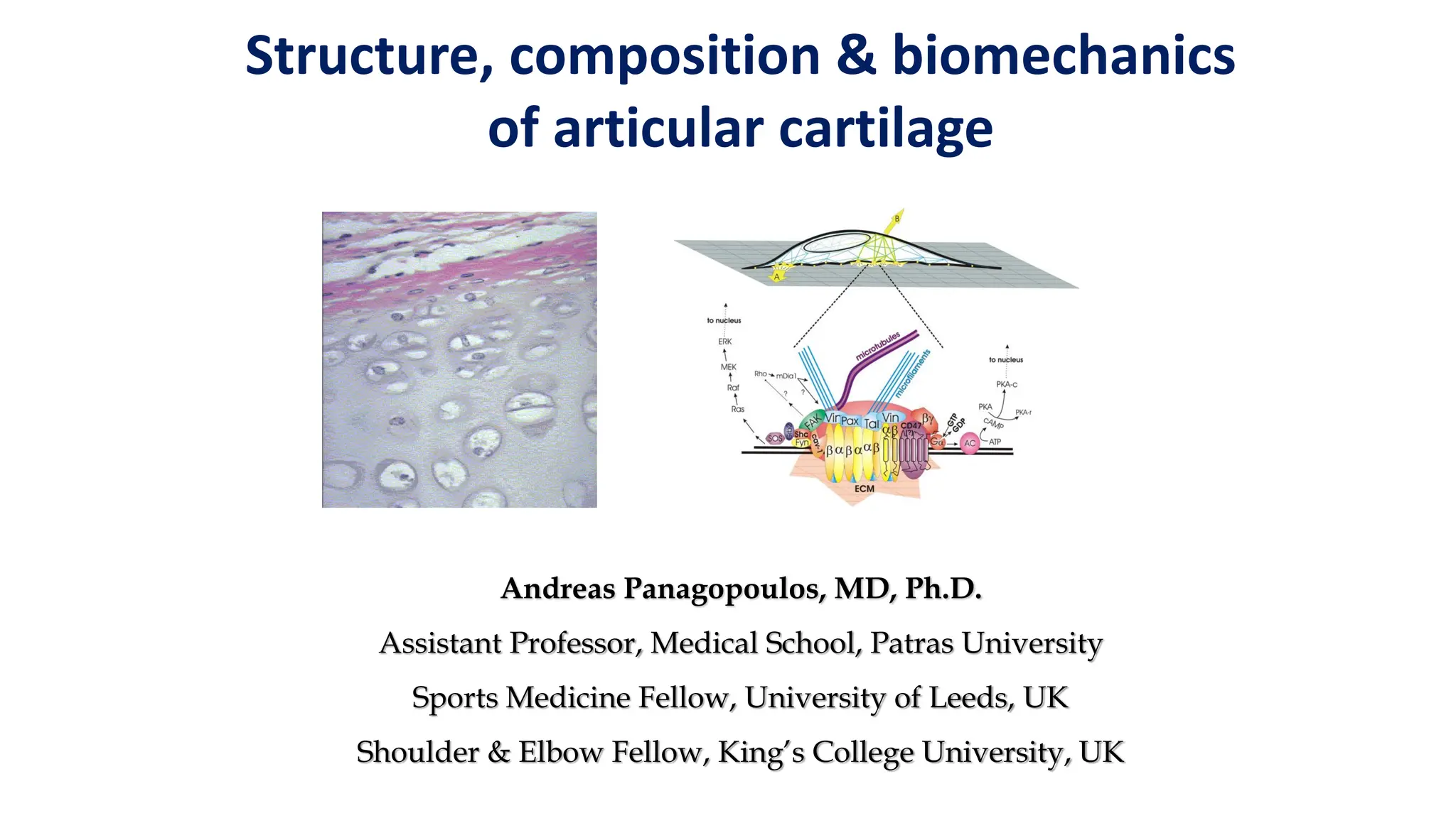
Structure, composition &biomechanics
of articular cartilage
Andreas Panagopoulos, MD, Ph.D.
Assistant Professor, Medical School, Patras University
Sports Medicine Fellow, University of Leeds, UK
Shoulder & Elbow Fellow, King’s College University, UK
2.
Cartilage functions
Tissue withspecial biomechanical and biochemical characteristics
1. Distributes joint loads over a wide area, decreasing
the stresses sustained by the contacting joint surfaces
2. Allows relative movement of the opposing joint surfaces with
minimal friction and wear
3. Minimizes peak stresses on subchondral bone
4. Provides a friction-reducing, weight-bearing surface with a
friction coefficient of 0.0025
5. Functions within a contact pressure range of 2-11 MPa
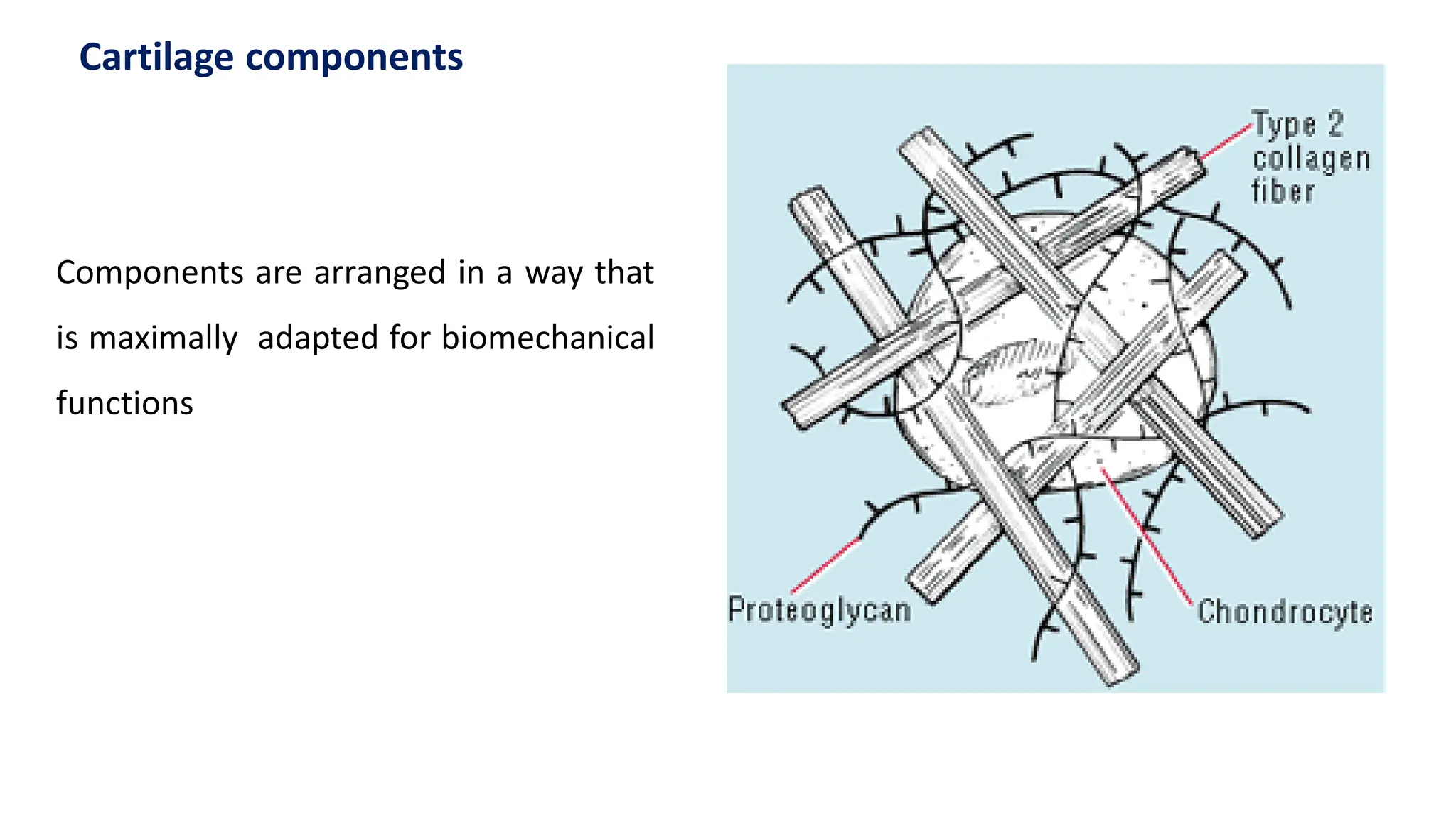
Components are arrangedin a way that
is maximally adapted for biomechanical
functions
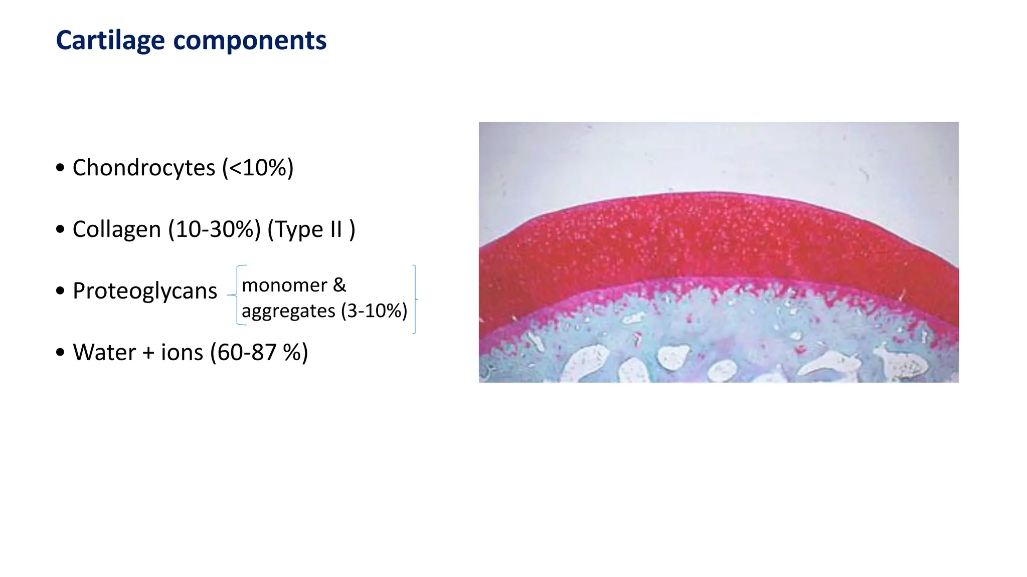
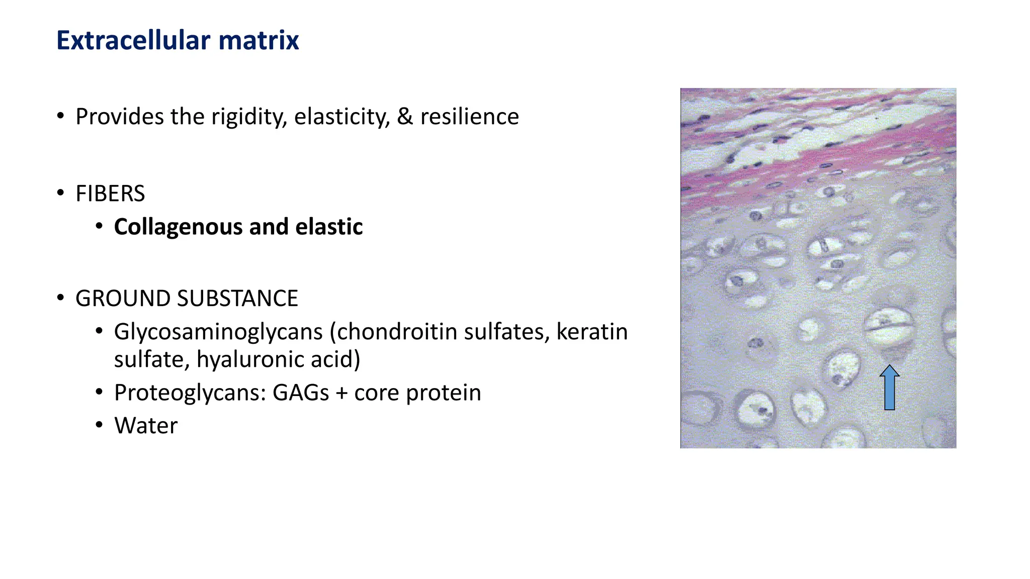
Cartilage components
5.
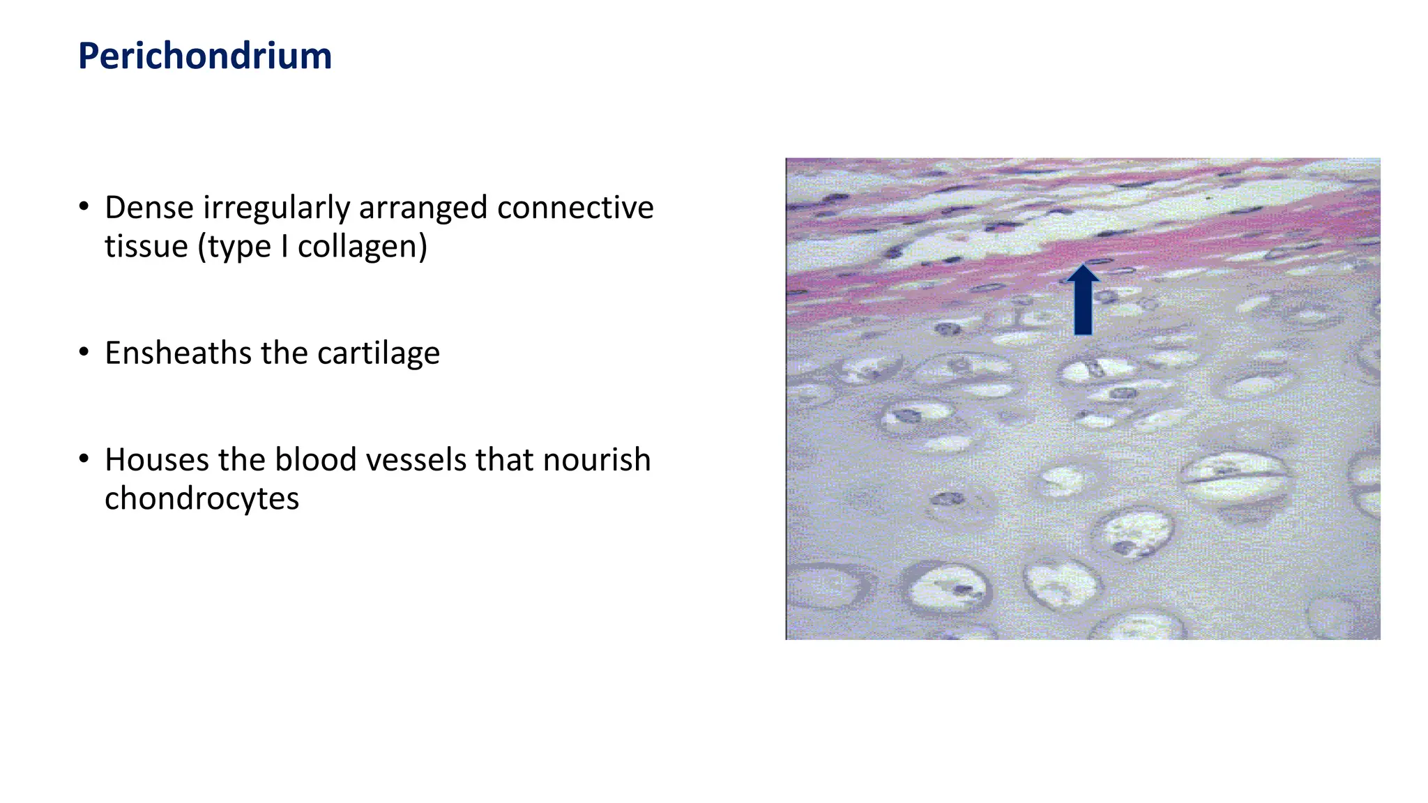
• Dense irregularlyarranged connective
tissue (type I collagen)
• Ensheaths the cartilage
• Houses the blood vessels that nourish
chondrocytes
Perichondrium
6.
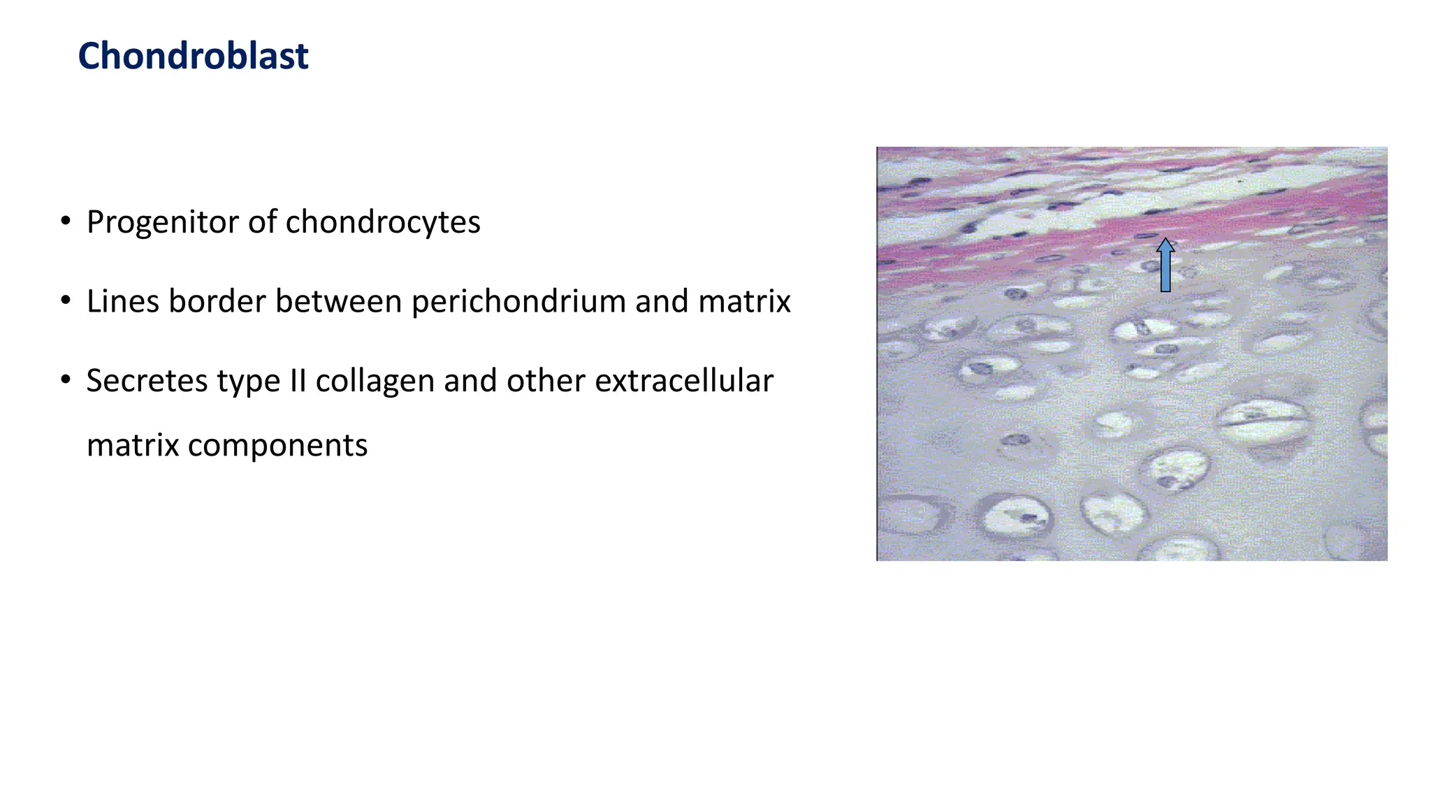
• Progenitor ofchondrocytes
• Lines border between perichondrium and matrix
• Secretes type II collagen and other extracellular
matrix components
Chondroblast
7.
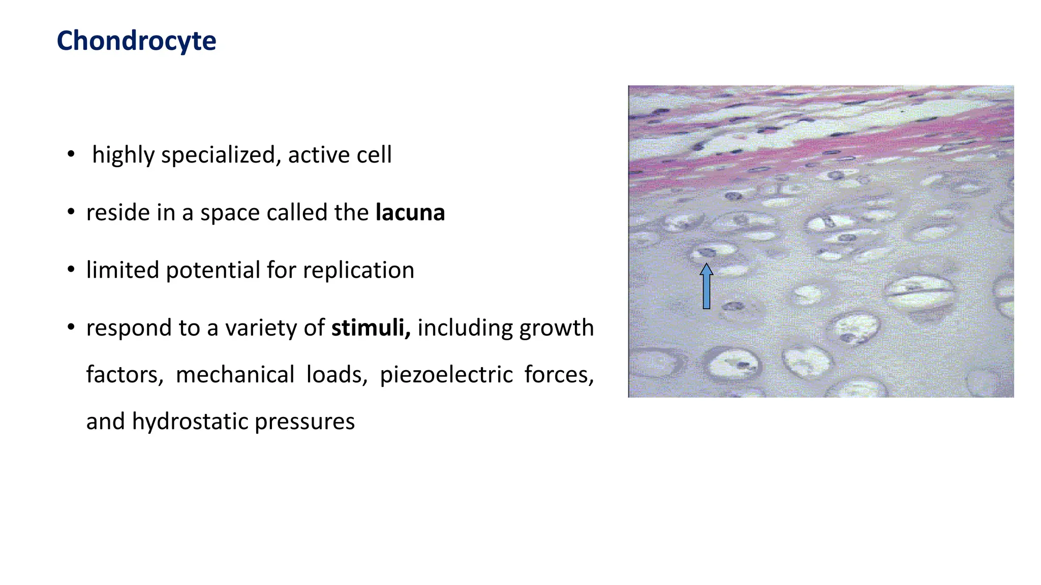
• highly specialized,active cell
• reside in a space called the lacuna
• limited potential for replication
• respond to a variety of stimuli, including growth
factors, mechanical loads, piezoelectric forces,
and hydrostatic pressures
Chondrocyte
8.
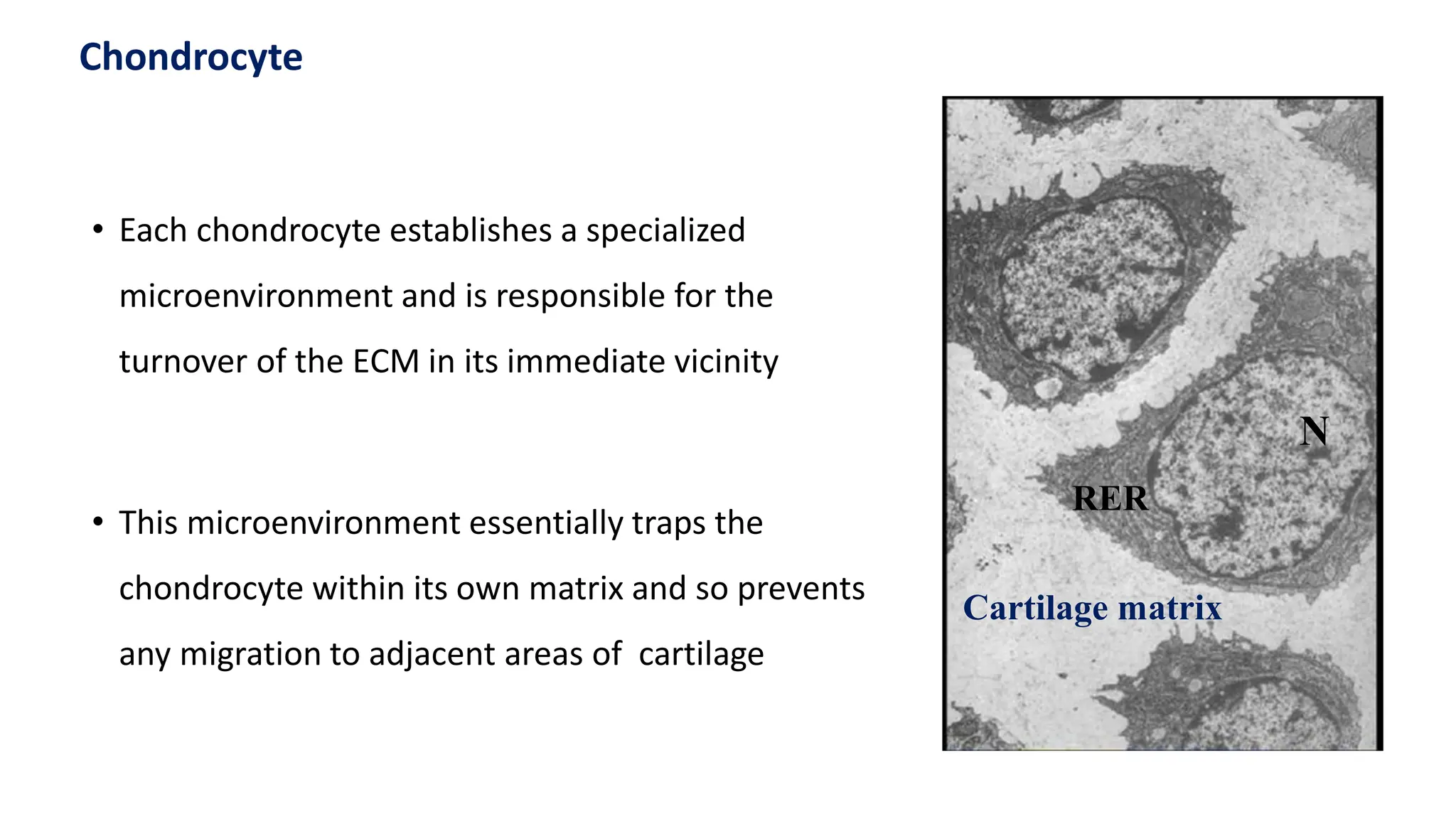
• Each chondrocyteestablishes a specialized
microenvironment and is responsible for the
turnover of the ECM in its immediate vicinity
• This microenvironment essentially traps the
chondrocyte within its own matrix and so prevents
any migration to adjacent areas of cartilage
Cartilage matrix
RER
N
Chondrocyte
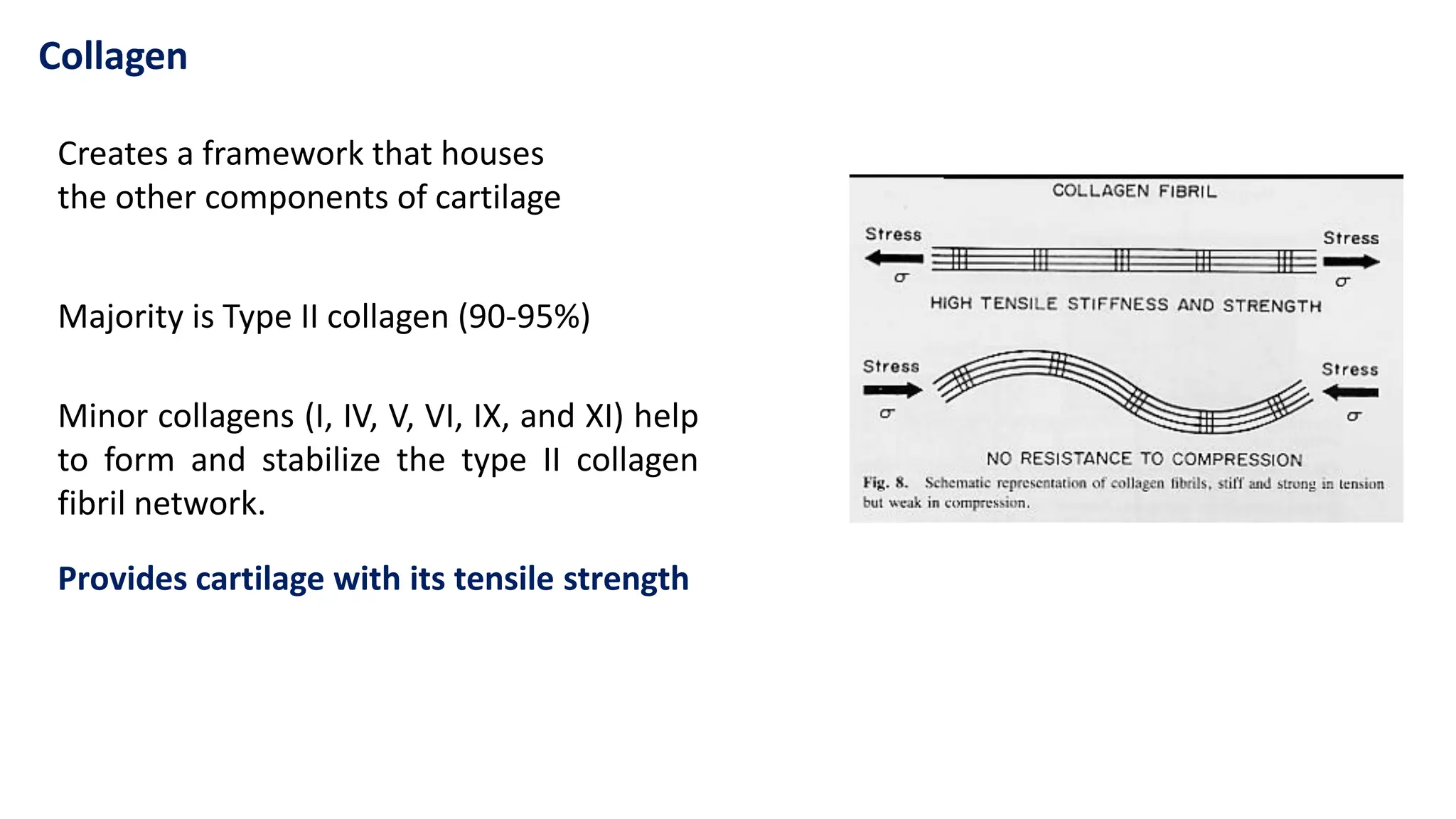
Collagen
Majority is TypeII collagen (90-95%)
Minor collagens (I, IV, V, VI, IX, and XI) help
to form and stabilize the type II collagen
fibril network.
Provides cartilage with its tensile strength
Creates a framework that houses
the other components of cartilage
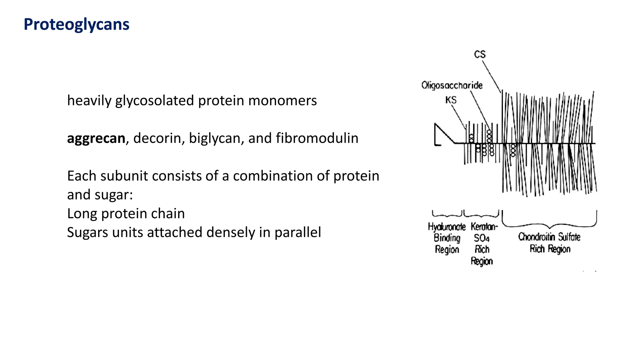
Proteoglycans
heavily glycosolated proteinmonomers
aggrecan, decorin, biglycan, and fibromodulin
Each subunit consists of a combination of protein
and sugar:
Long protein chain
Sugars units attached densely in parallel
13.
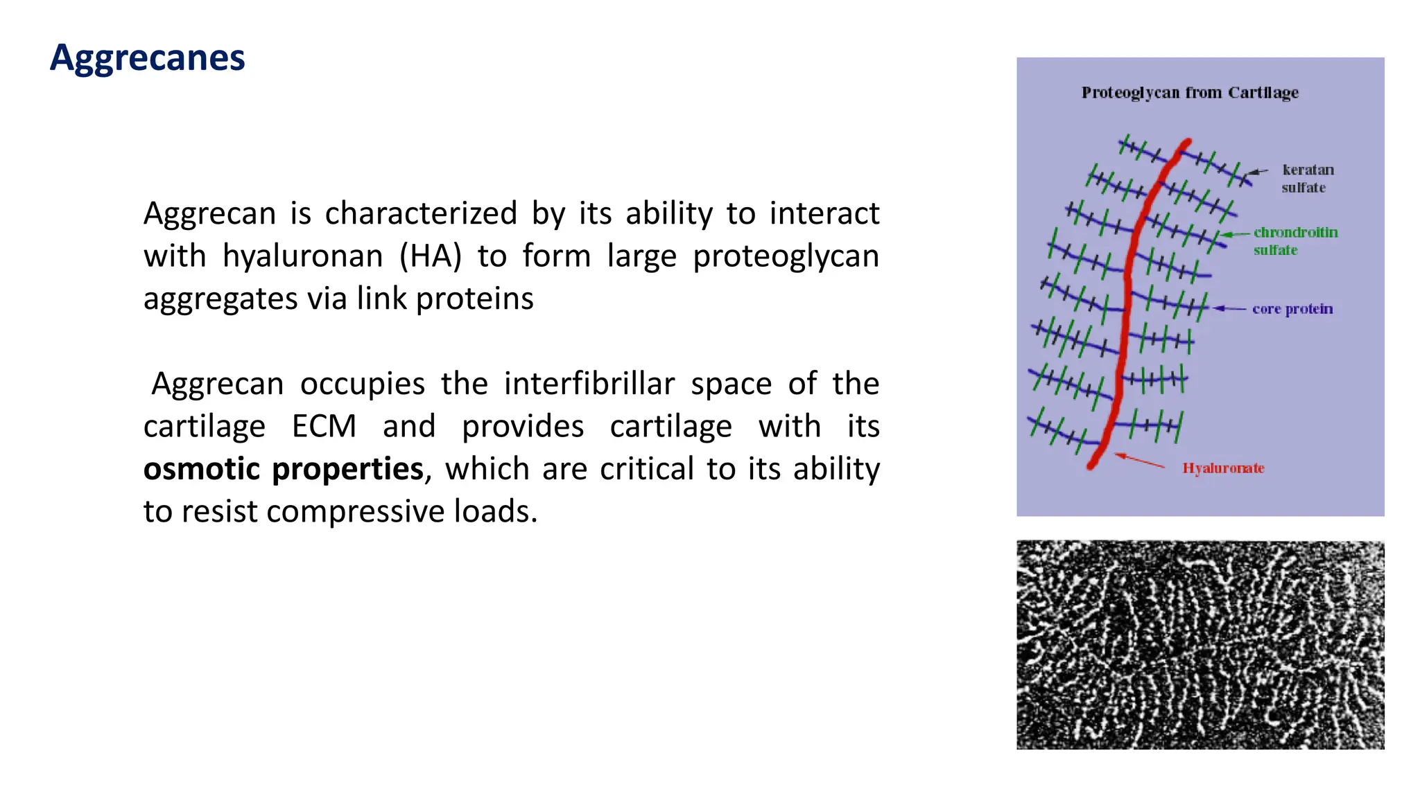
Aggrecan is characterizedby its ability to interact
with hyaluronan (HA) to form large proteoglycan
aggregates via link proteins
Aggrecan occupies the interfibrillar space of the
cartilage ECM and provides cartilage with its
osmotic properties, which are critical to its ability
to resist compressive loads.
Aggrecanes
14.
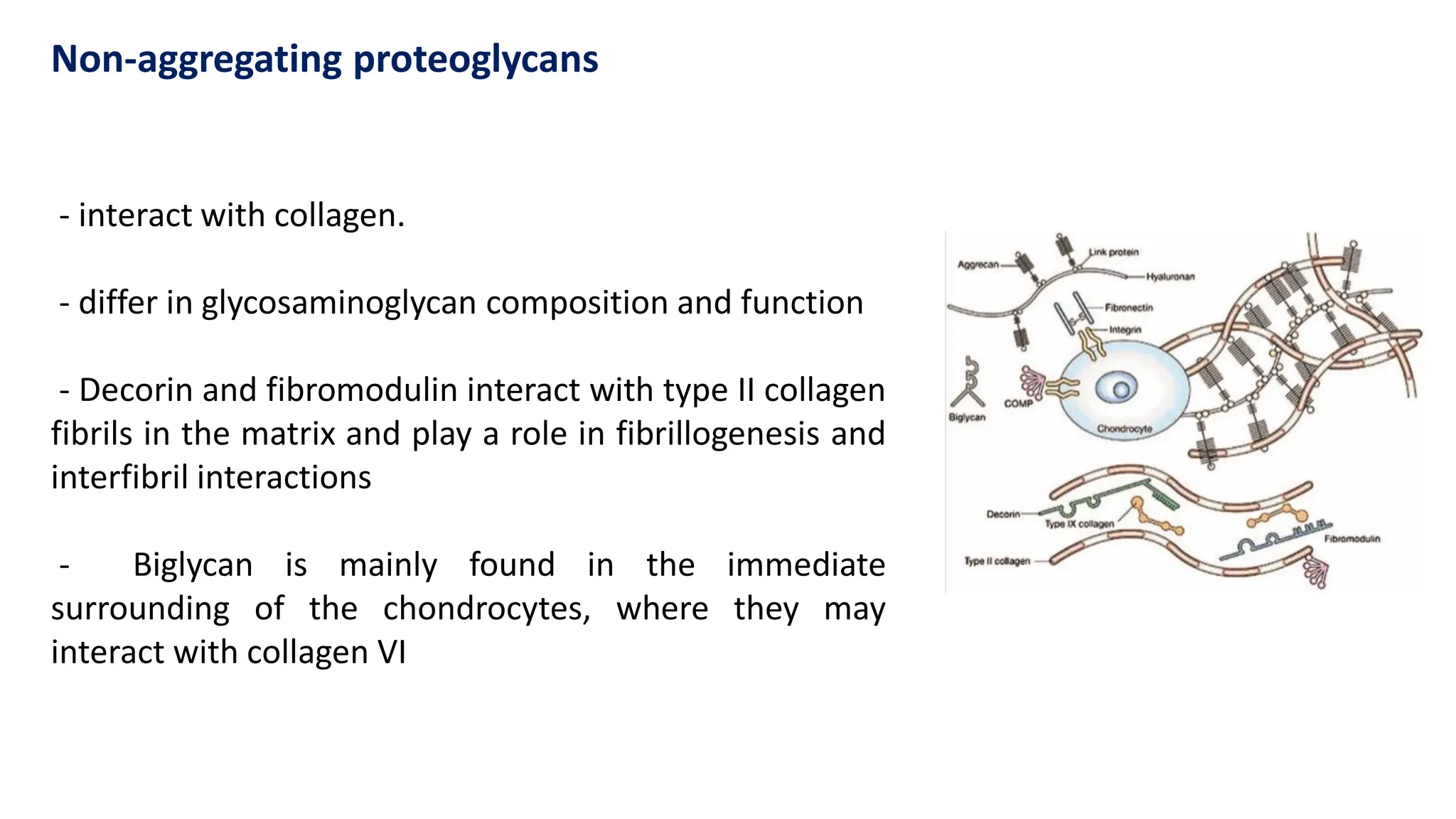
- interact withcollagen.
- differ in glycosaminoglycan composition and function
- Decorin and fibromodulin interact with type II collagen
fibrils in the matrix and play a role in fibrillogenesis and
interfibril interactions
- Biglycan is mainly found in the immediate
surrounding of the chondrocytes, where they may
interact with collagen VI
Non-aggregating proteoglycans
15.
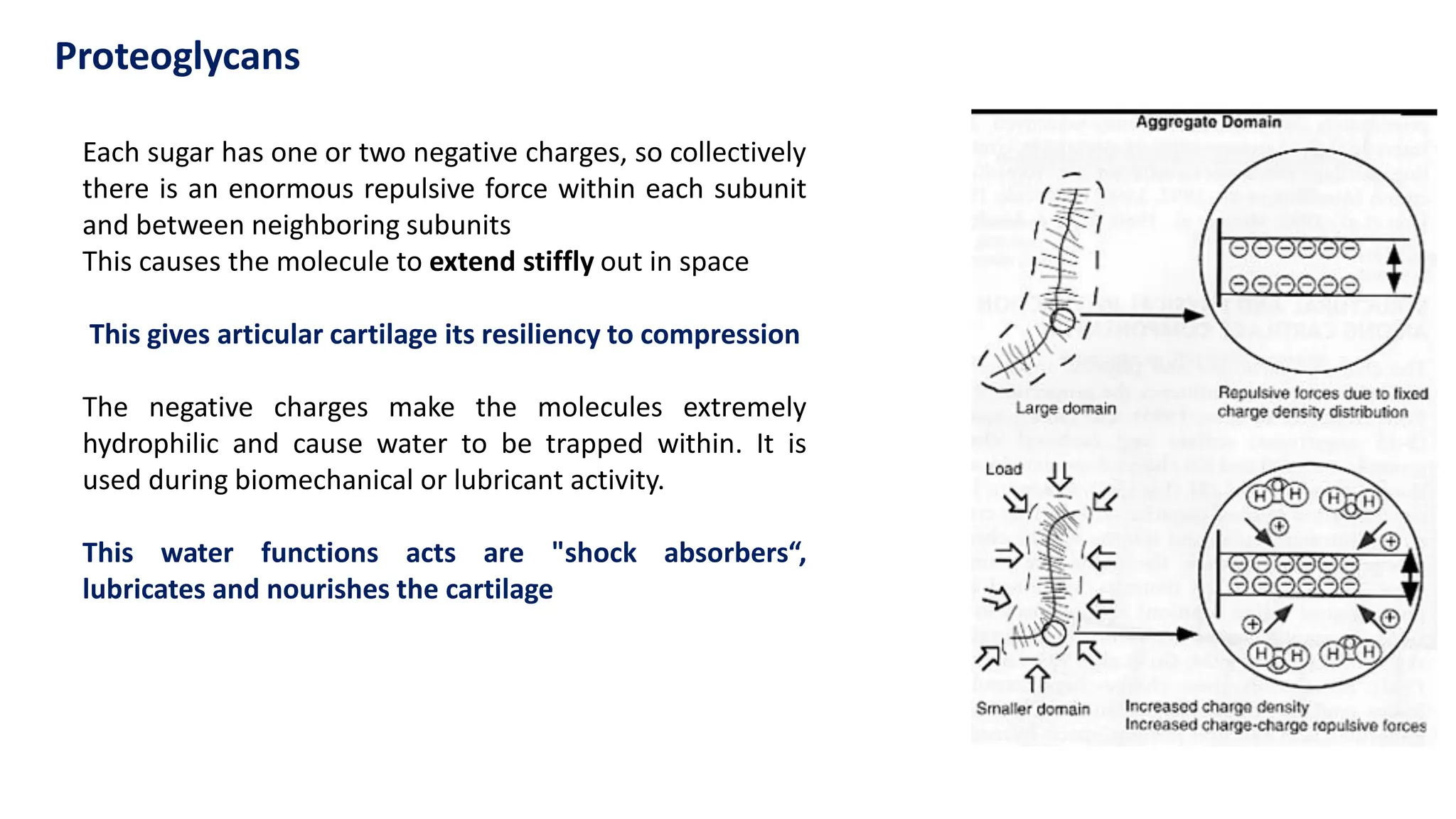
Each sugar hasone or two negative charges, so collectively
there is an enormous repulsive force within each subunit
and between neighboring subunits
This causes the molecule to extend stiffly out in space
This gives articular cartilage its resiliency to compression
The negative charges make the molecules extremely
hydrophilic and cause water to be trapped within. It is
used during biomechanical or lubricant activity.
This water functions acts are "shock absorbers“,
lubricates and nourishes the cartilage
Proteoglycans
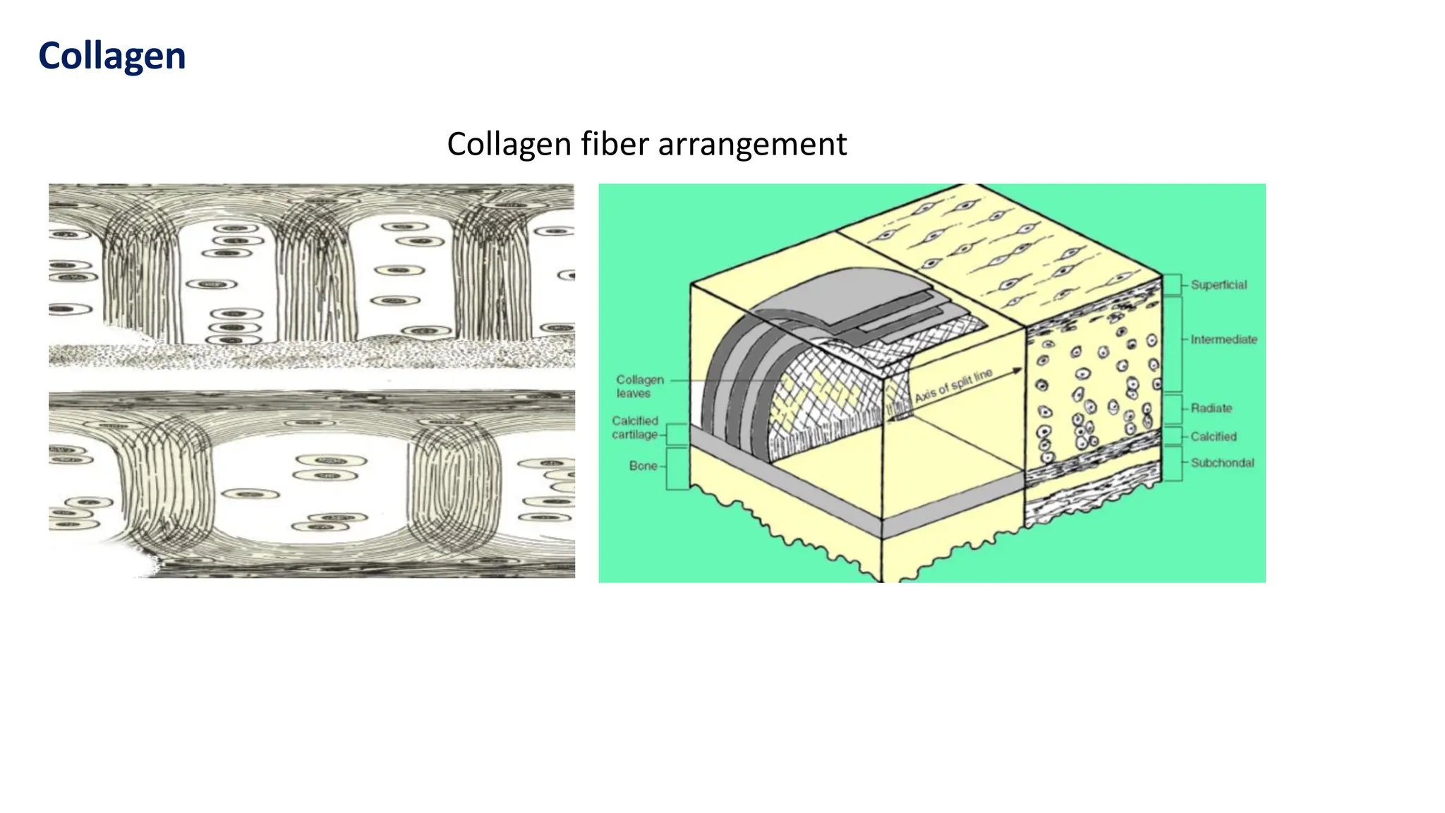
16.
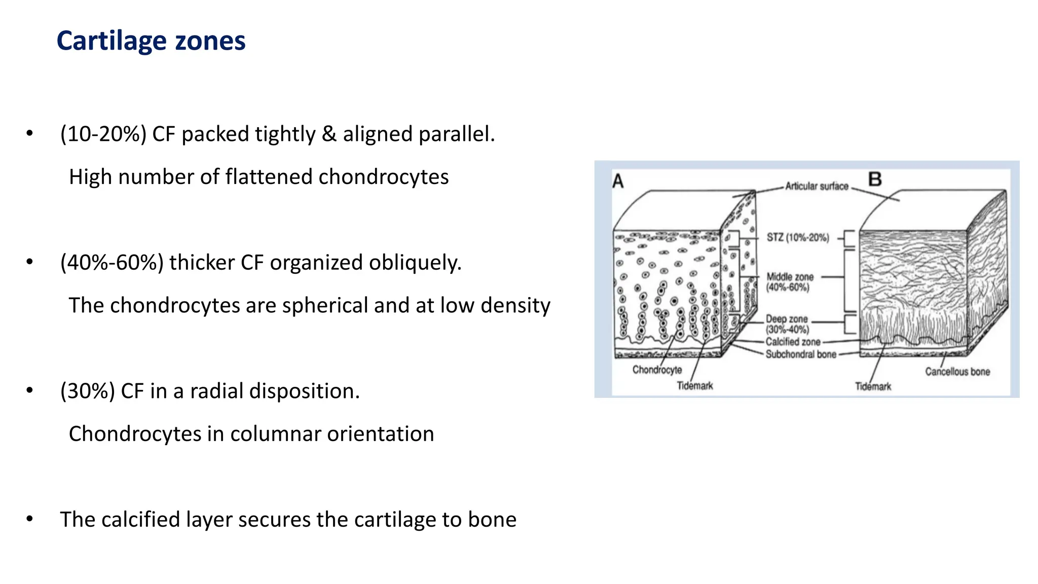
• (10-20%) CFpacked tightly & aligned parallel.
High number of flattened chondrocytes
• (40%-60%) thicker CF organized obliquely.
The chondrocytes are spherical and at low density
• (30%) CF in a radial disposition.
Chondrocytes in columnar orientation
• The calcified layer secures the cartilage to bone
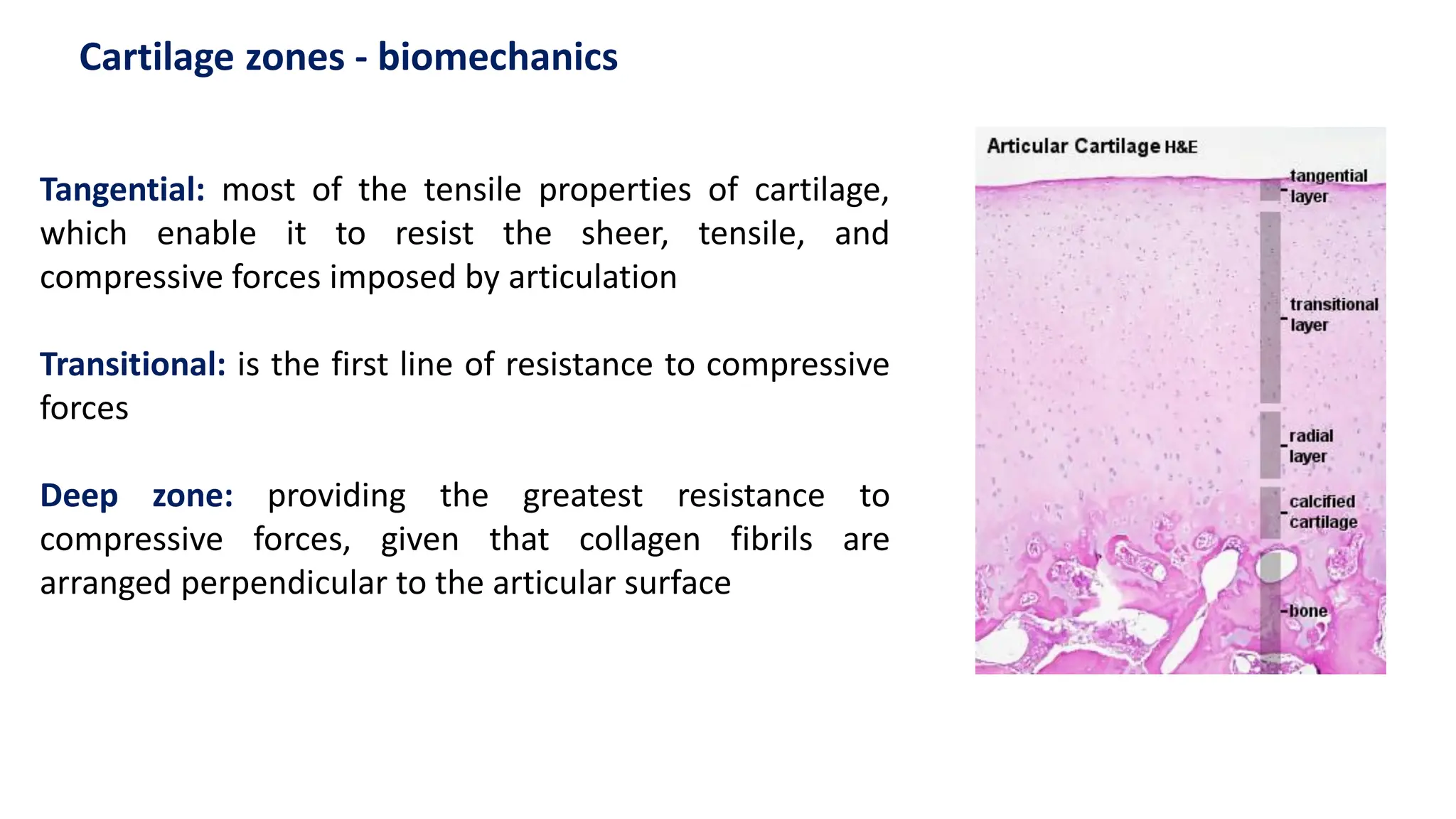
Cartilage zones
17.
Tangential: most ofthe tensile properties of cartilage,
which enable it to resist the sheer, tensile, and
compressive forces imposed by articulation
Transitional: is the first line of resistance to compressive
forces
Deep zone: providing the greatest resistance to
compressive forces, given that collagen fibrils are
arranged perpendicular to the articular surface
Cartilage zones - biomechanics
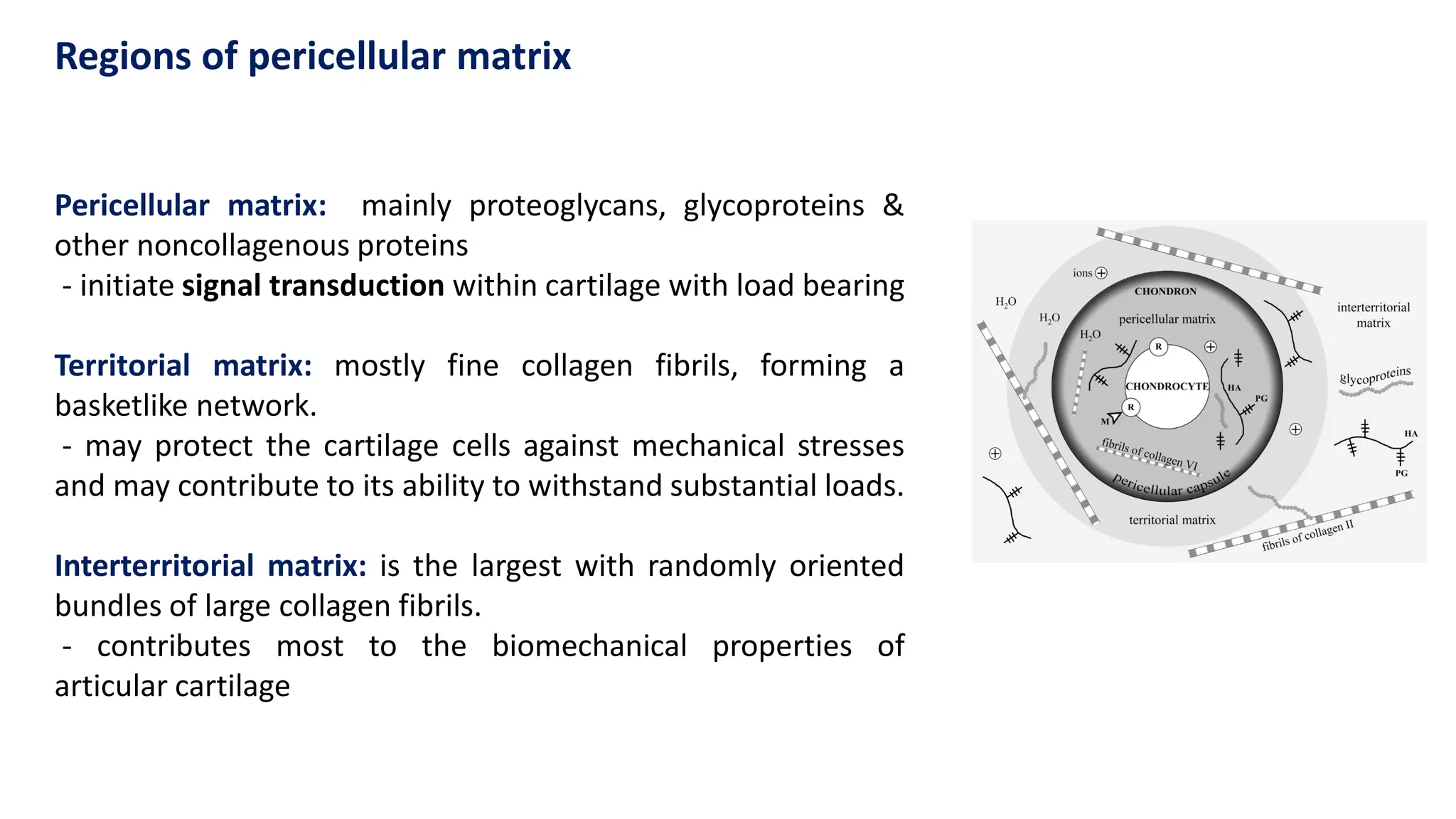
18.
Pericellular matrix: mainlyproteoglycans, glycoproteins &
other noncollagenous proteins
- initiate signal transduction within cartilage with load bearing
Territorial matrix: mostly fine collagen fibrils, forming a
basketlike network.
- may protect the cartilage cells against mechanical stresses
and may contribute to its ability to withstand substantial loads.
Interterritorial matrix: is the largest with randomly oriented
bundles of large collagen fibrils.
- contributes most to the biomechanical properties of
articular cartilage
Regions of pericellular matrix
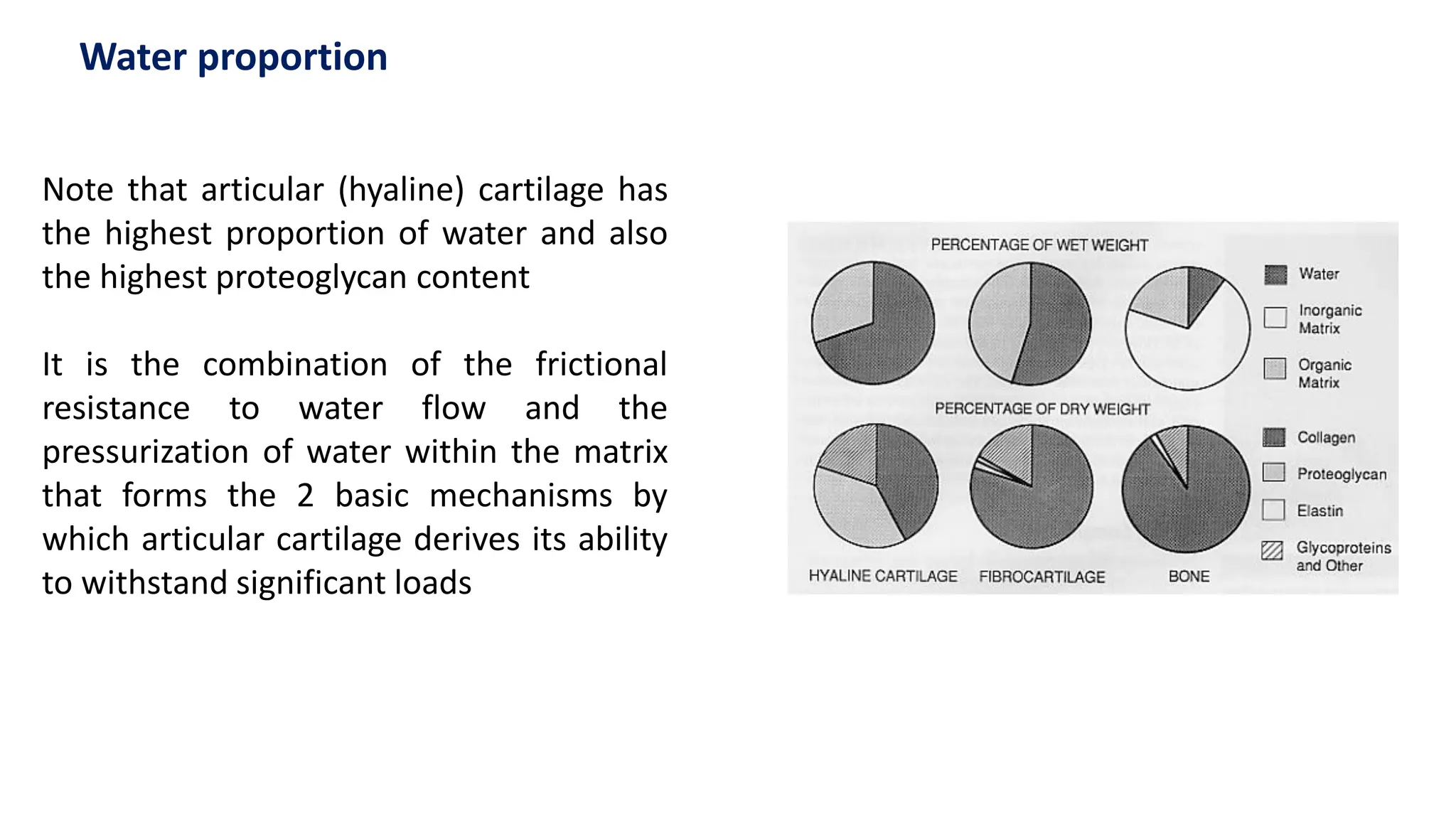
19.
Note that articular(hyaline) cartilage has
the highest proportion of water and also
the highest proteoglycan content
It is the combination of the frictional
resistance to water flow and the
pressurization of water within the matrix
that forms the 2 basic mechanisms by
which articular cartilage derives its ability
to withstand significant loads
Water proportion
20.
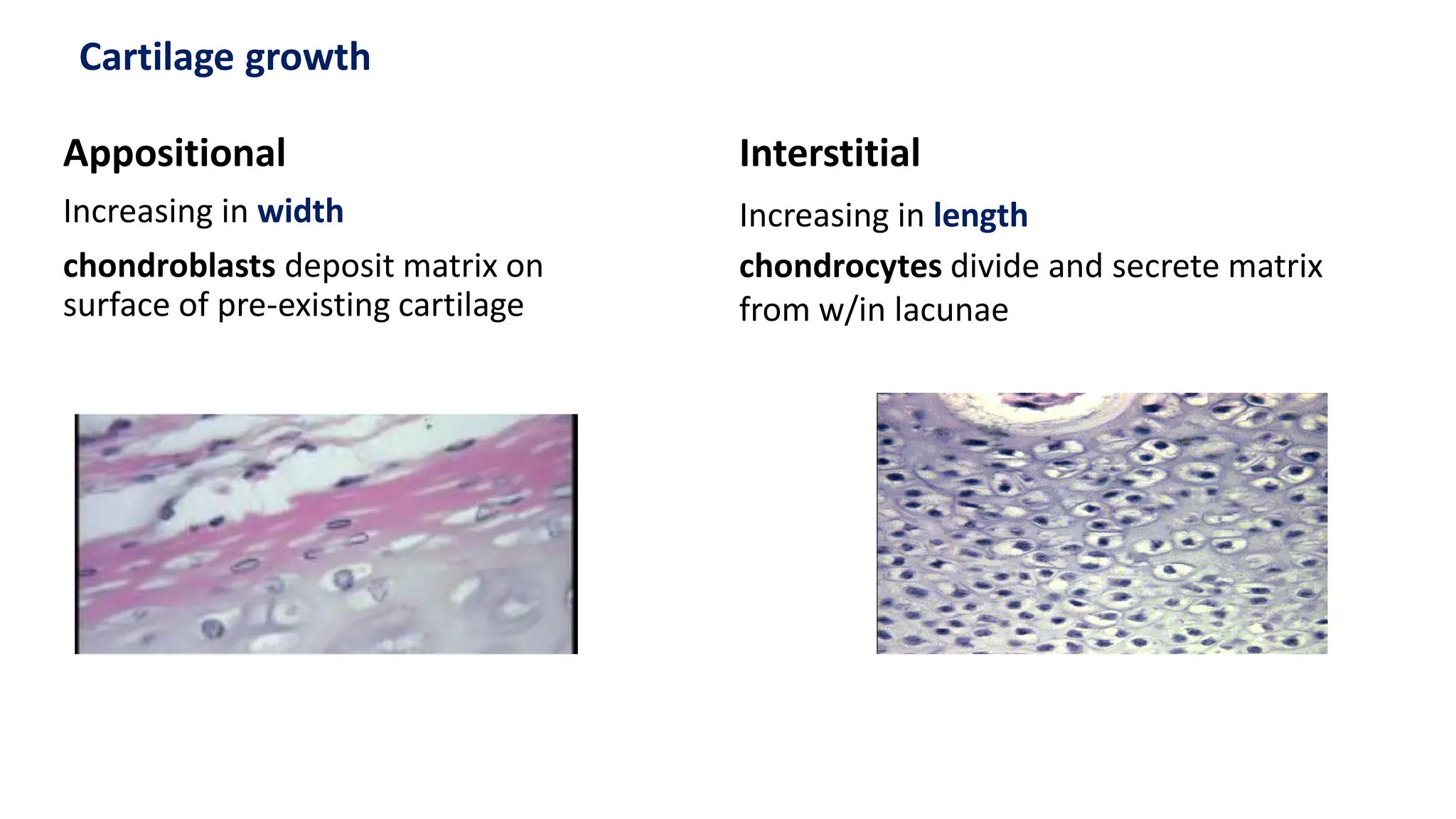
Appositional
Increasing in width
chondroblastsdeposit matrix on
surface of pre-existing cartilage
Cartilage growth
Interstitial
Increasing in length
chondrocytes divide and secrete matrix
from w/in lacunae
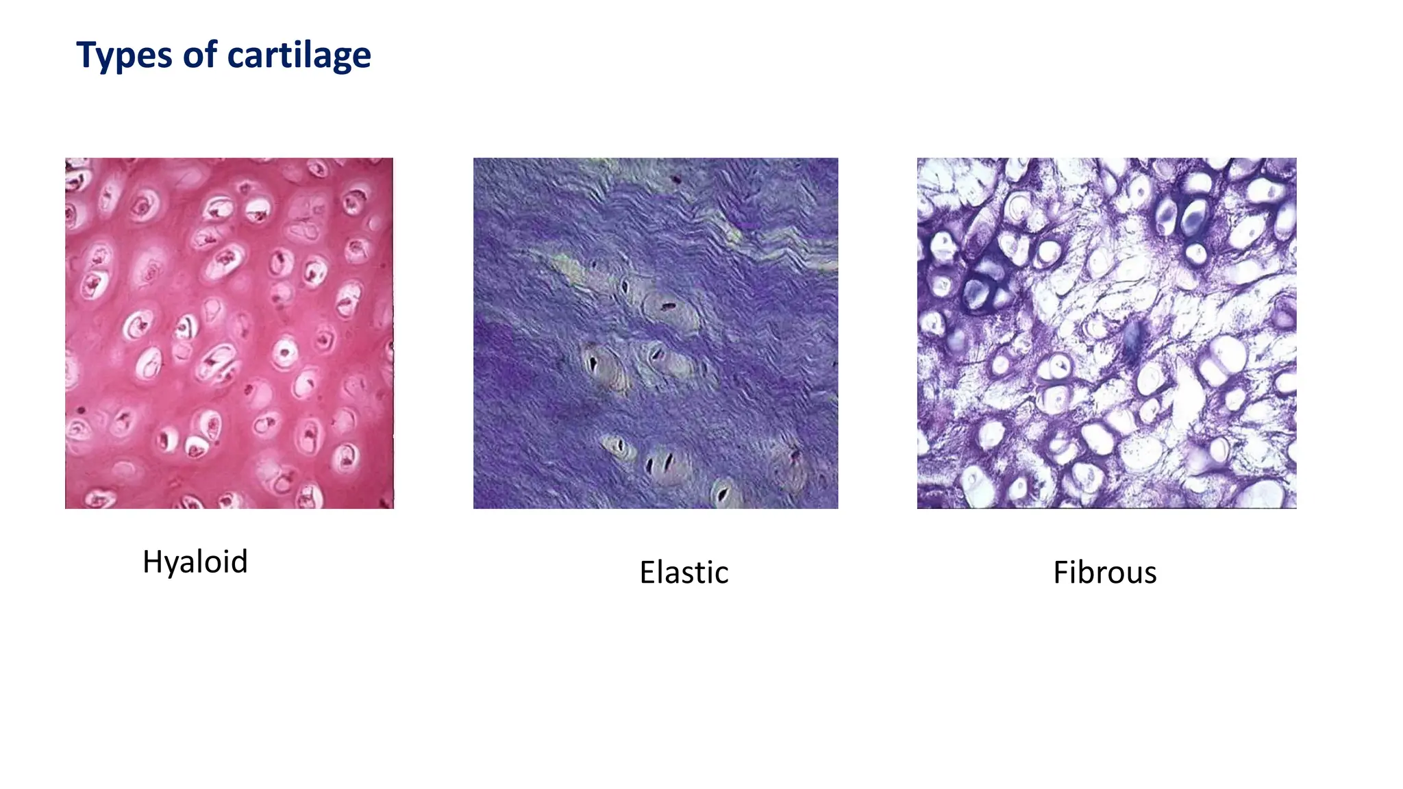
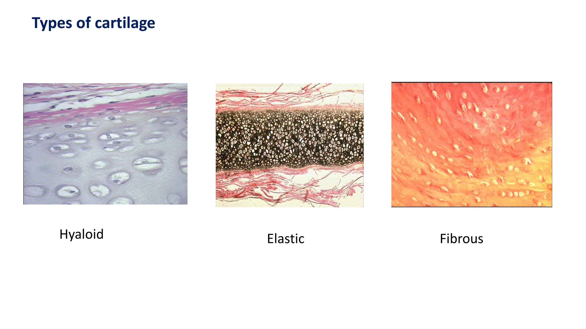
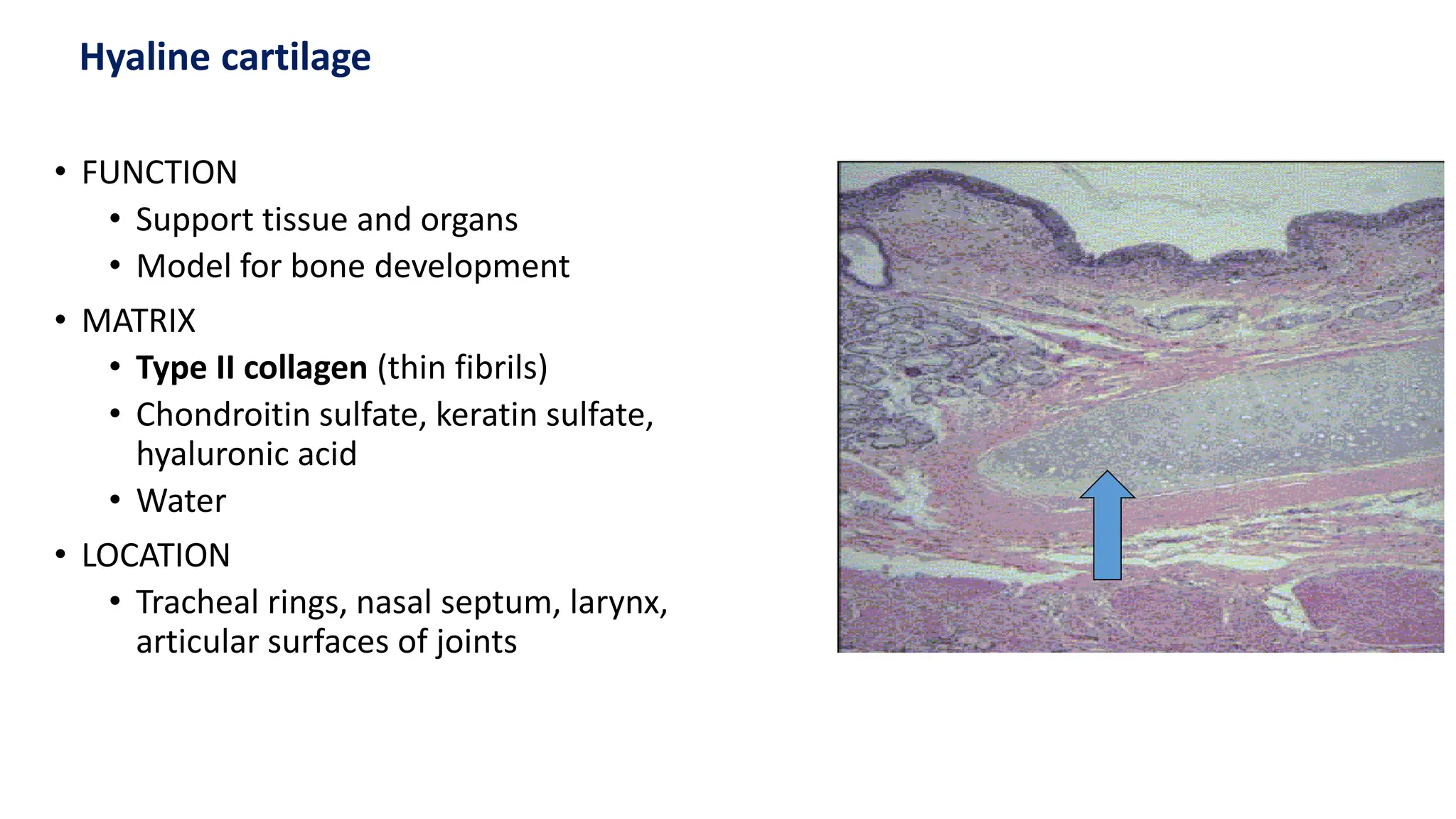
• FUNCTION
• Supporttissue and organs
• Model for bone development
• MATRIX
• Type II collagen (thin fibrils)
• Chondroitin sulfate, keratin sulfate,
hyaluronic acid
• Water
• LOCATION
• Tracheal rings, nasal septum, larynx,
articular surfaces of joints
Hyaline cartilage
24.
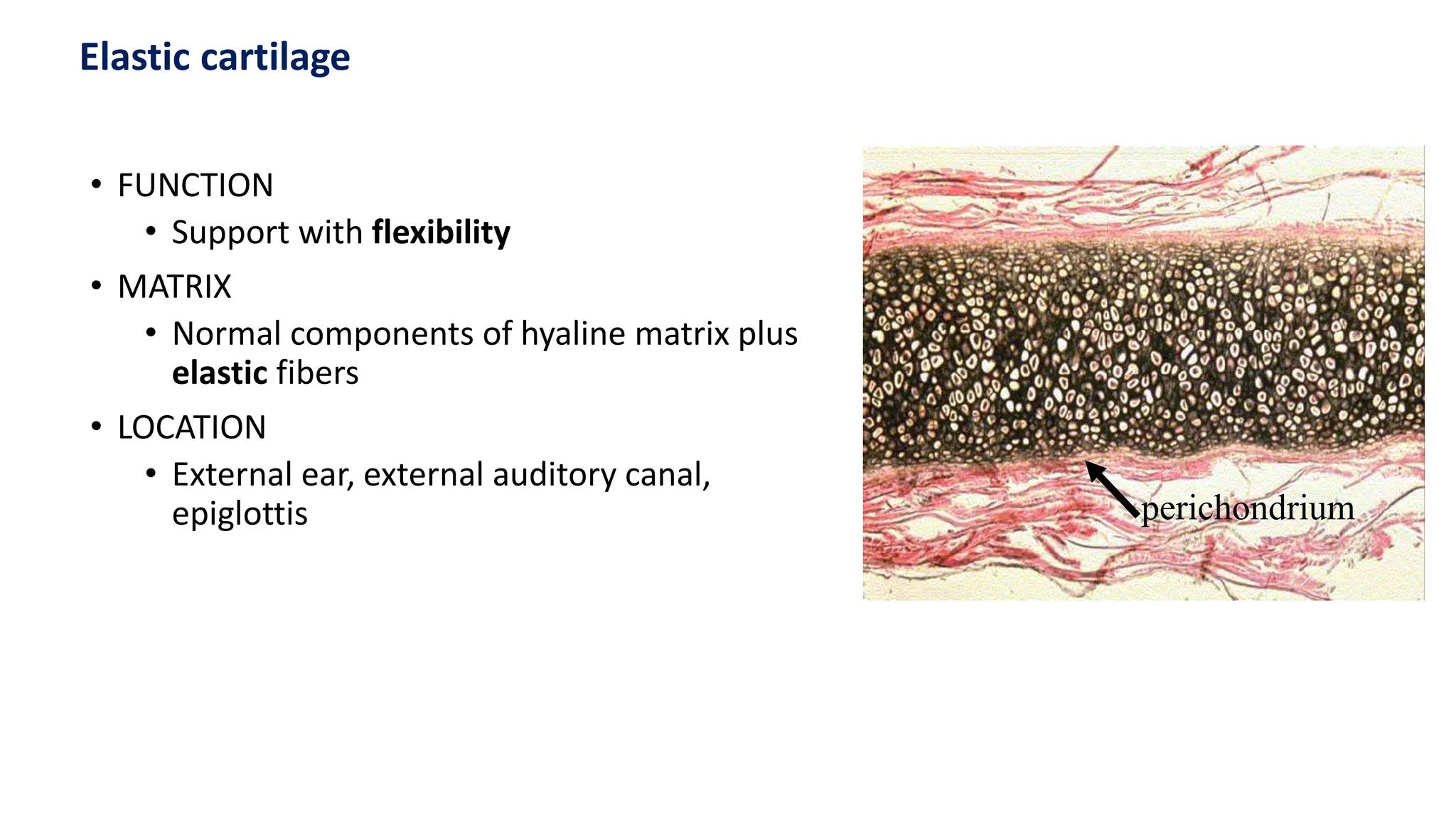
• FUNCTION
• Supportwith flexibility
• MATRIX
• Normal components of hyaline matrix plus
elastic fibers
• LOCATION
• External ear, external auditory canal,
epiglottis perichondrium
Elastic cartilage
25.
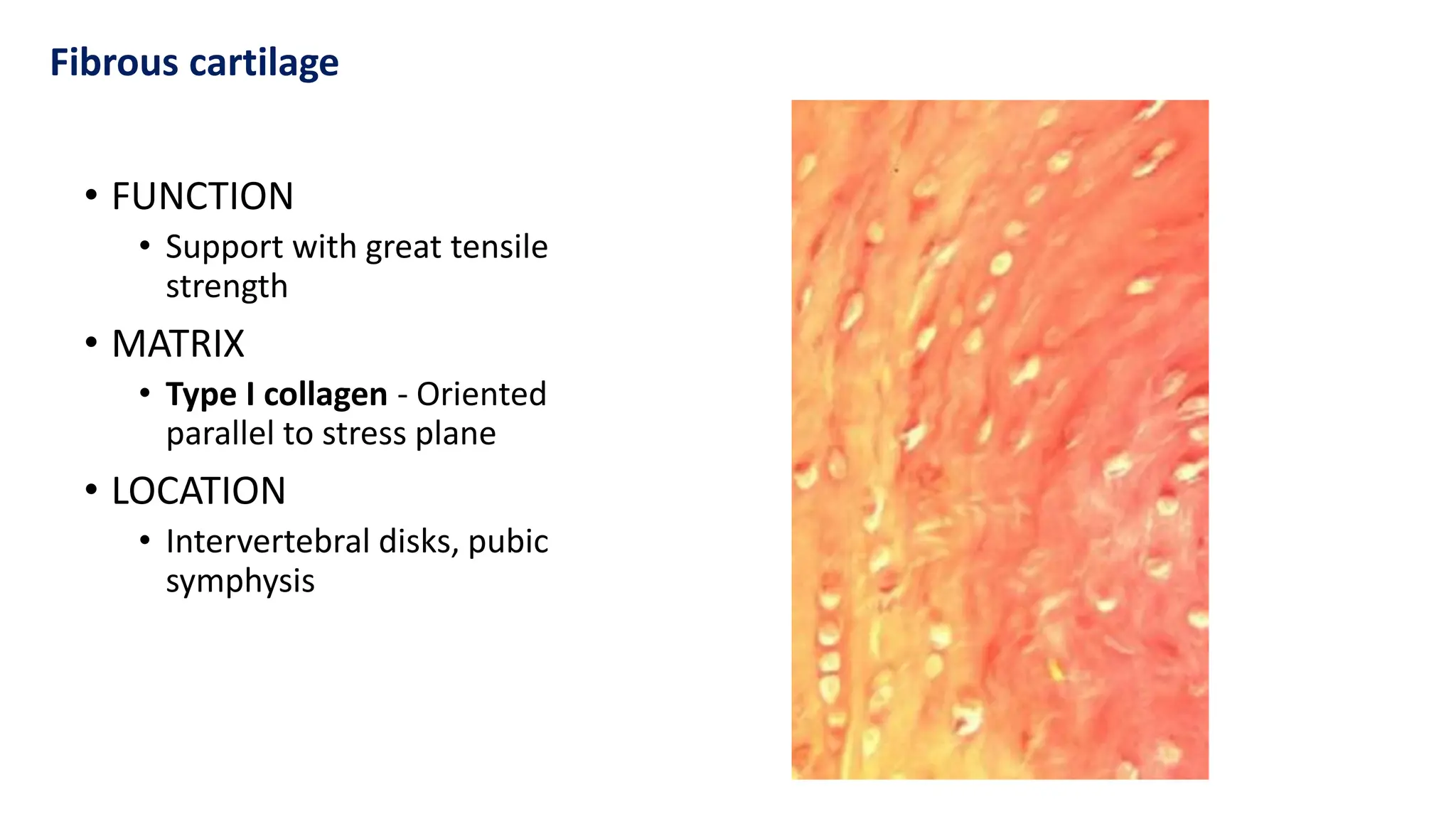
• FUNCTION
• Supportwith great tensile
strength
• MATRIX
• Type I collagen - Oriented
parallel to stress plane
• LOCATION
• Intervertebral disks, pubic
symphysis
Fibrous cartilage
26.
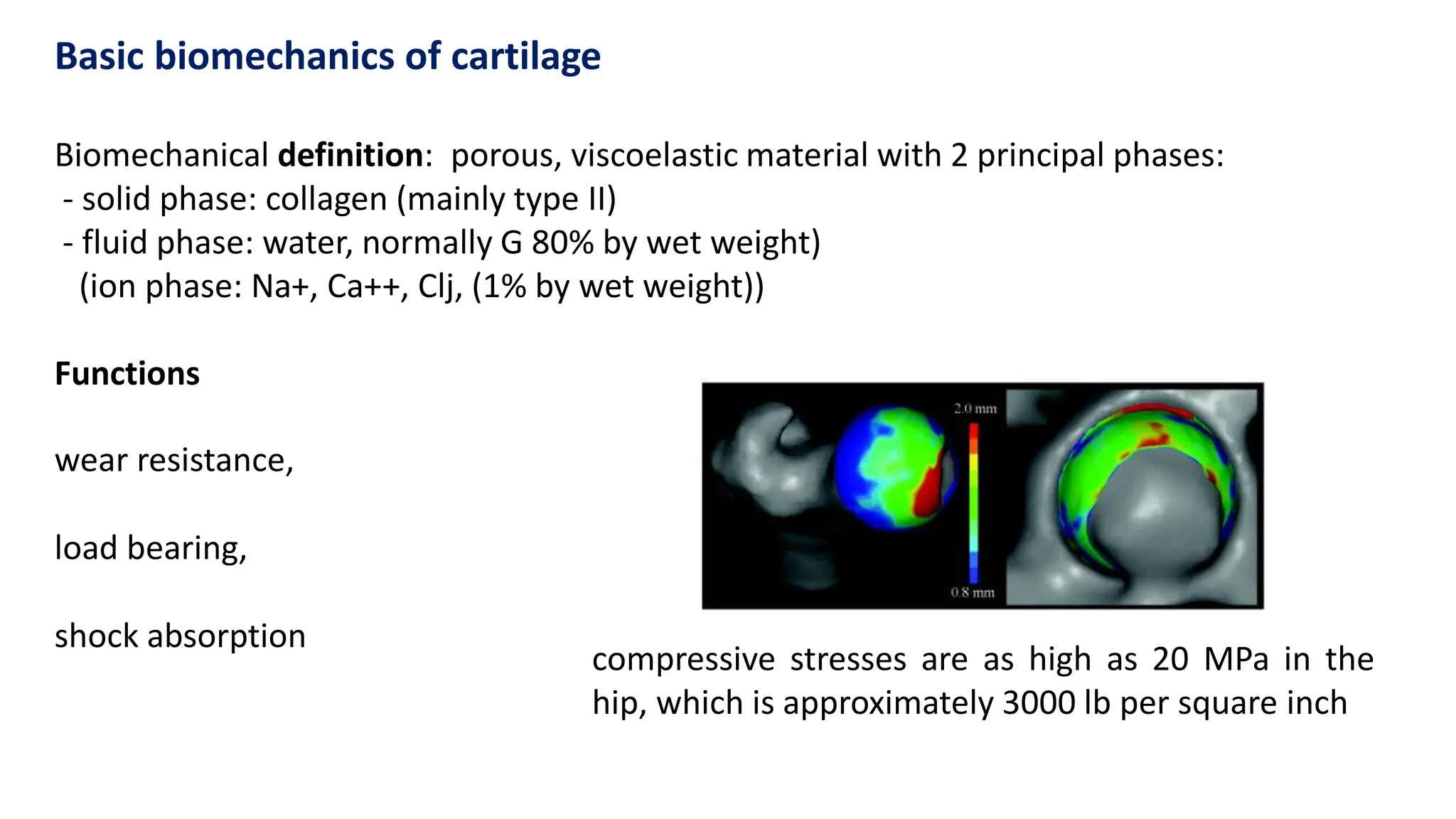
Biomechanical definition: porous,viscoelastic material with 2 principal phases:
- solid phase: collagen (mainly type II)
- fluid phase: water, normally G 80% by wet weight)
(ion phase: Na+, Ca++, Clj, (1% by wet weight))
Functions
wear resistance,
load bearing,
shock absorption
Basic biomechanics of cartilage
compressive stresses are as high as 20 MPa in the
hip, which is approximately 3000 lb per square inch
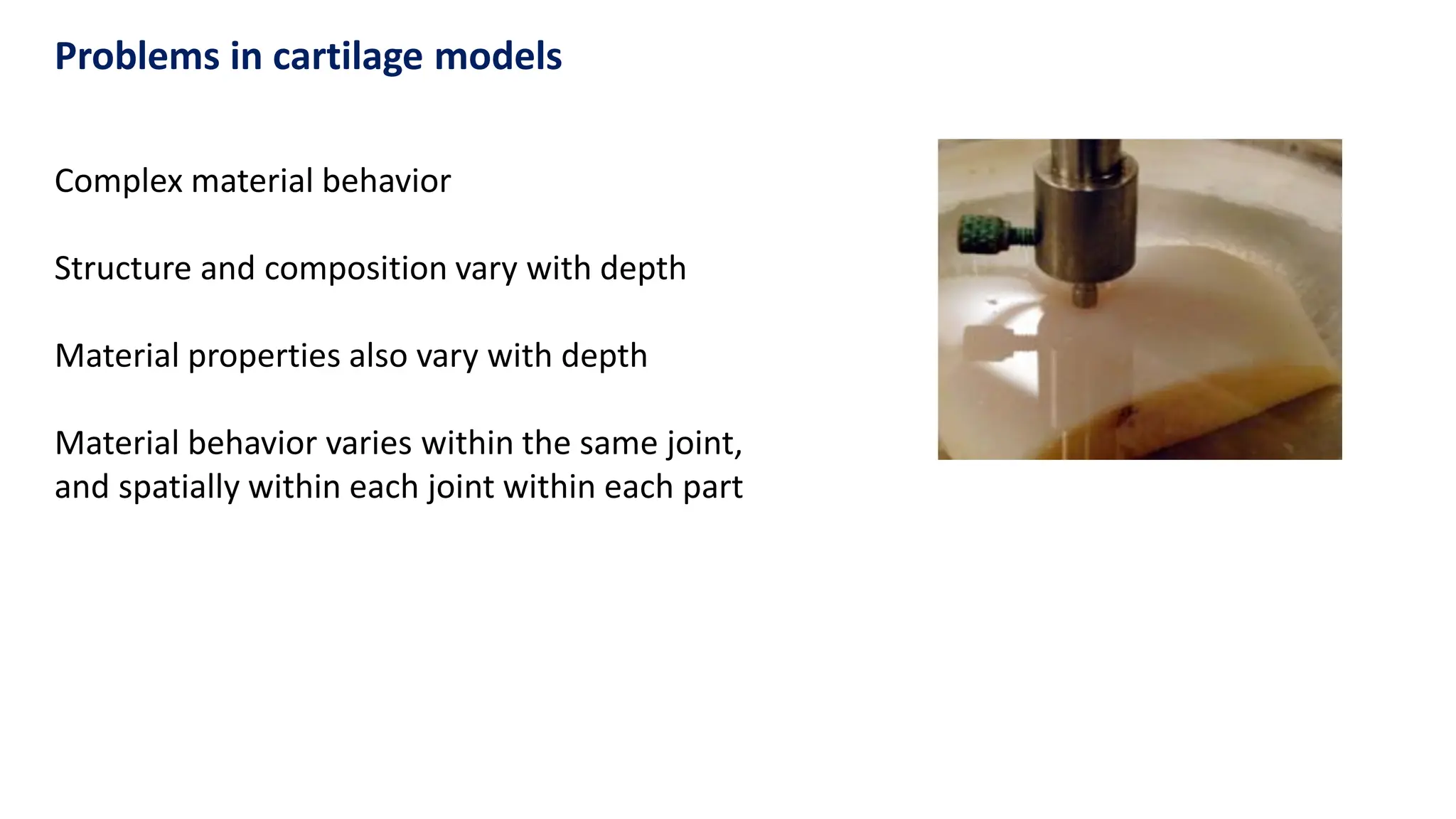
Complex material behavior
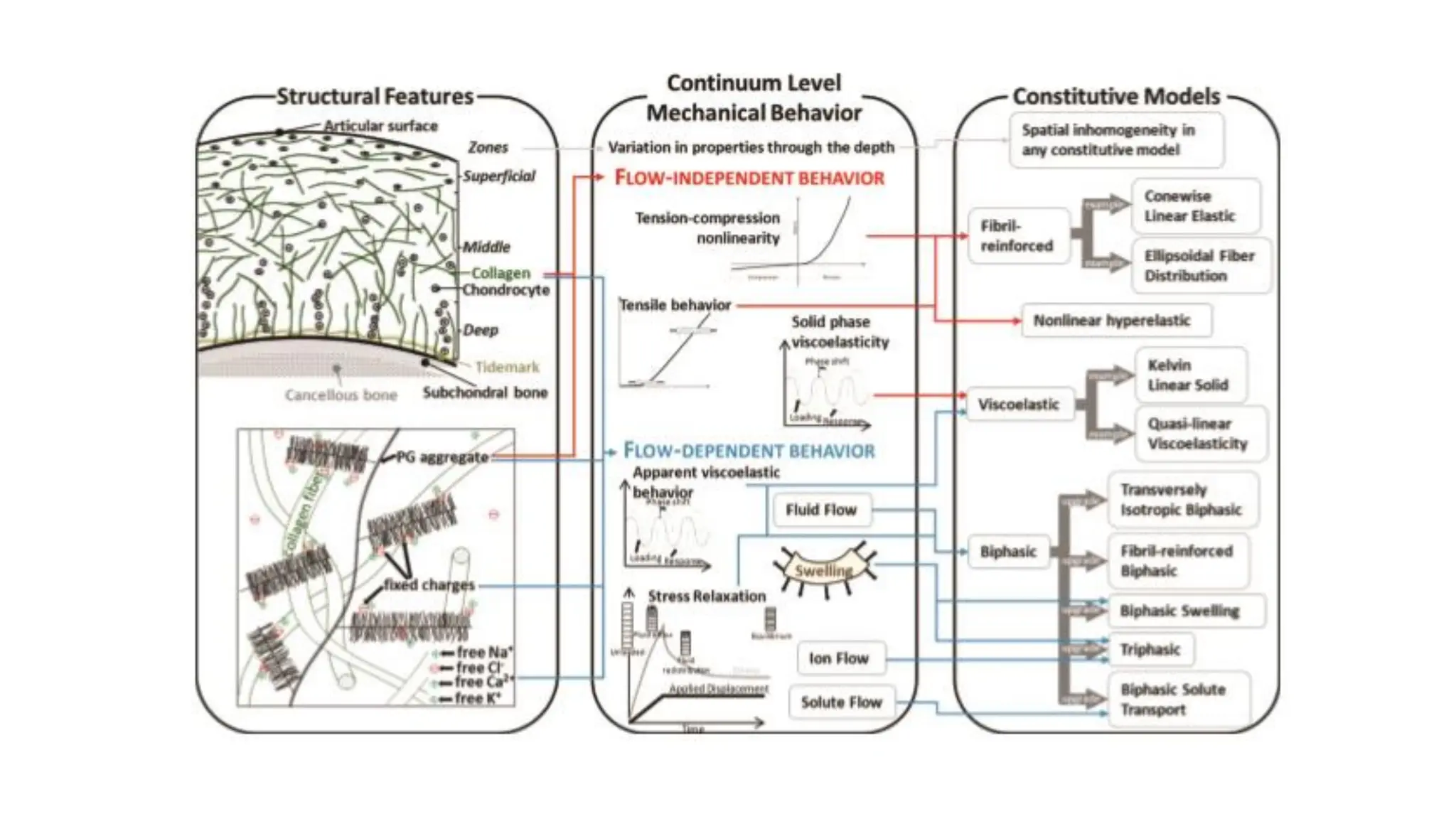
Structureand composition vary with depth
Material properties also vary with depth
Material behavior varies within the same joint,
and spatially within each joint within each part
Problems in cartilage models
30.
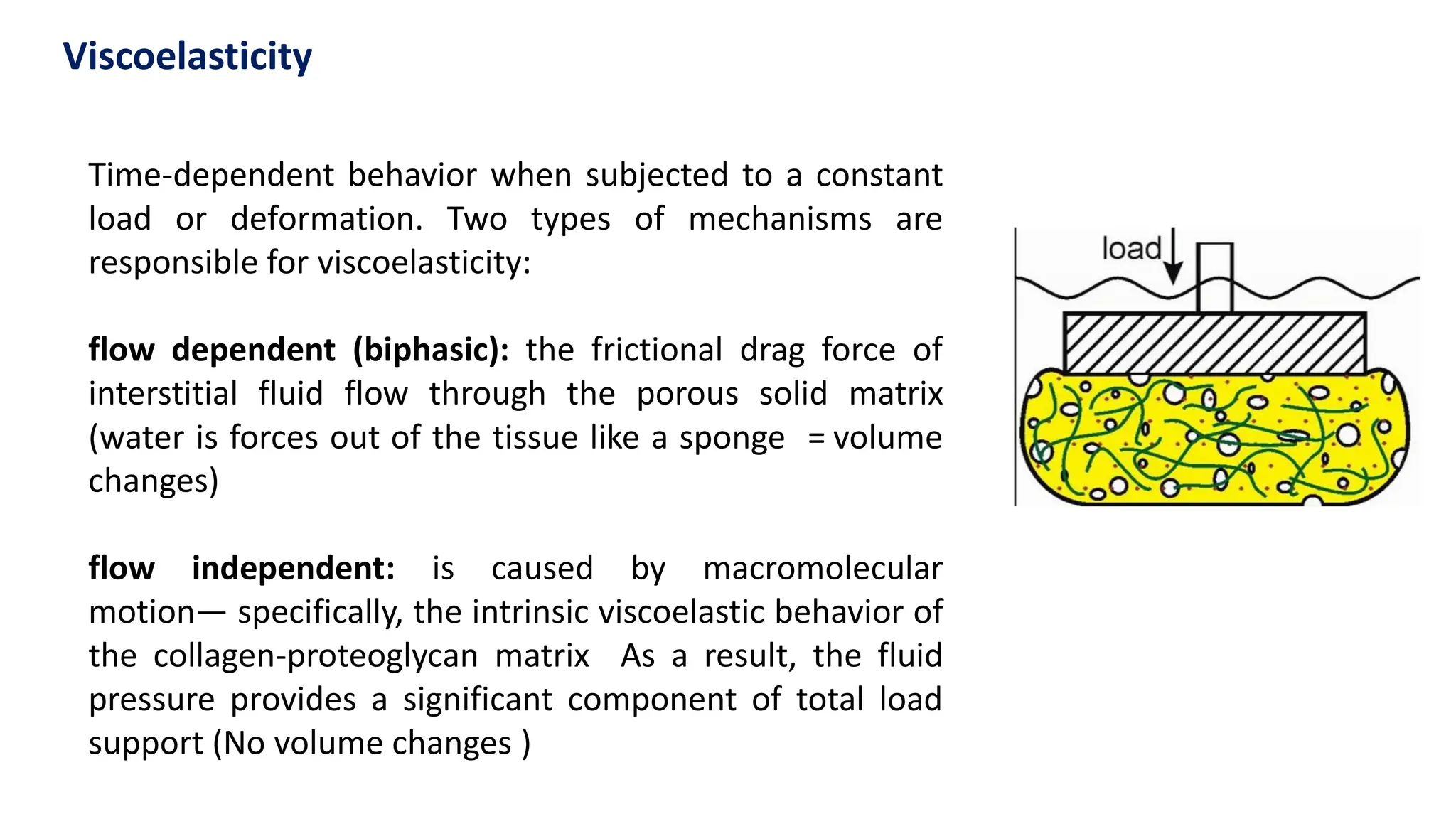
Time-dependent behavior whensubjected to a constant
load or deformation. Two types of mechanisms are
responsible for viscoelasticity:
flow dependent (biphasic): the frictional drag force of
interstitial fluid flow through the porous solid matrix
(water is forces out of the tissue like a sponge = volume
changes)
flow independent: is caused by macromolecular
motion— specifically, the intrinsic viscoelastic behavior of
the collagen-proteoglycan matrix As a result, the fluid
pressure provides a significant component of total load
support (No volume changes )
Viscoelasticity
31.
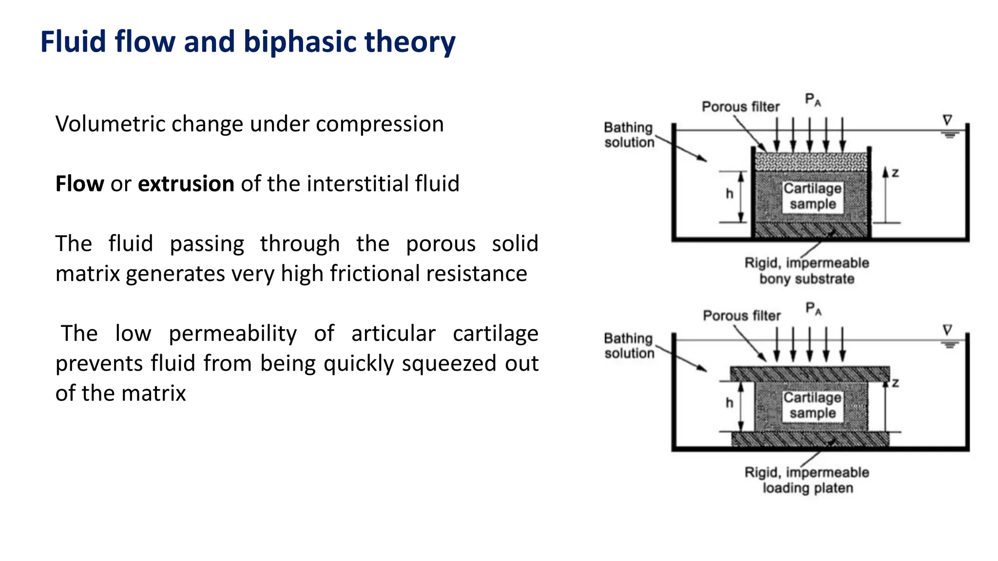
Fluid flow andbiphasic theory
Volumetric change under compression
Flow or extrusion of the interstitial fluid
The fluid passing through the porous solid
matrix generates very high frictional resistance
The low permeability of articular cartilage
prevents fluid from being quickly squeezed out
of the matrix
32.
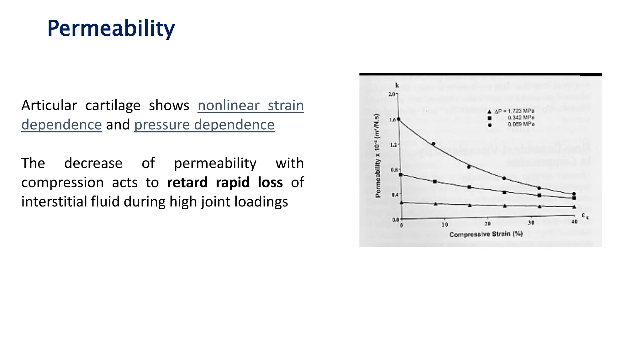
Permeability
Articular cartilage showsnonlinear strain
dependence and pressure dependence
The decrease of permeability with
compression acts to retard rapid loss of
interstitial fluid during high joint loadings
33.
Confined compression test
Copiousexudation of fluid at start
but the rate of exudation decreases
over time from points A to B to C
Compression force
34.
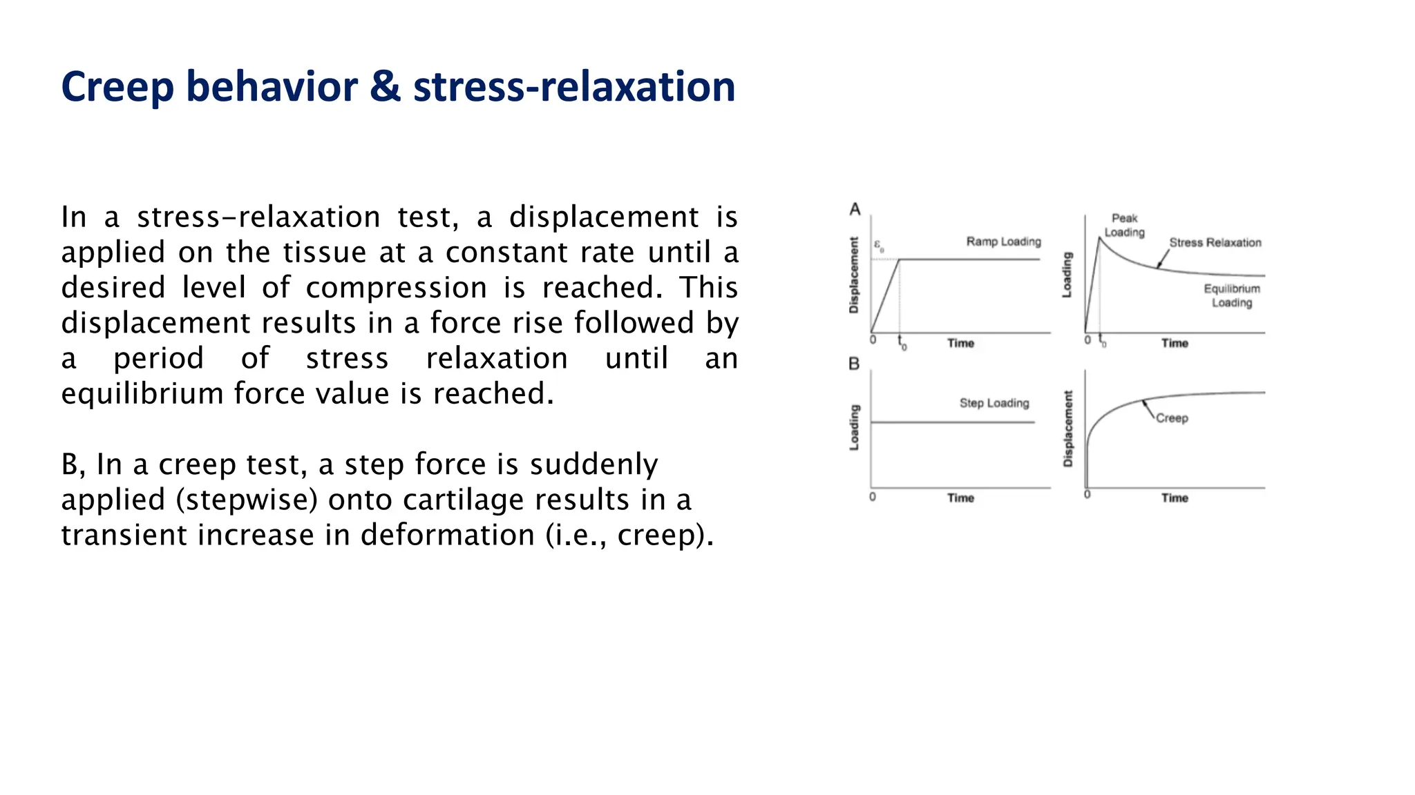
Creep behavior &stress-relaxation
In a stress-relaxation test, a displacement is
applied on the tissue at a constant rate until a
desired level of compression is reached. This
displacement results in a force rise followed by
a period of stress relaxation until an
equilibrium force value is reached.
B, In a creep test, a step force is suddenly
applied (stepwise) onto cartilage results in a
transient increase in deformation (i.e., creep).
35.
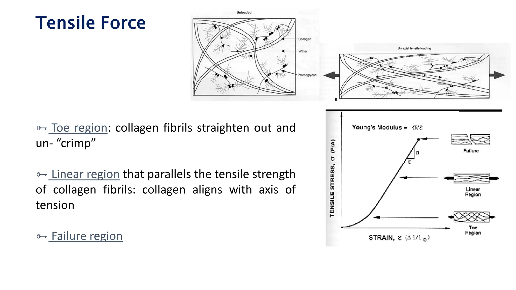
Tensile Force
Toeregion: collagen fibrils straighten out and
un- “crimp”
Linear region that parallels the tensile strength
of collagen fibrils: collagen aligns with axis of
tension
Failure region
36.
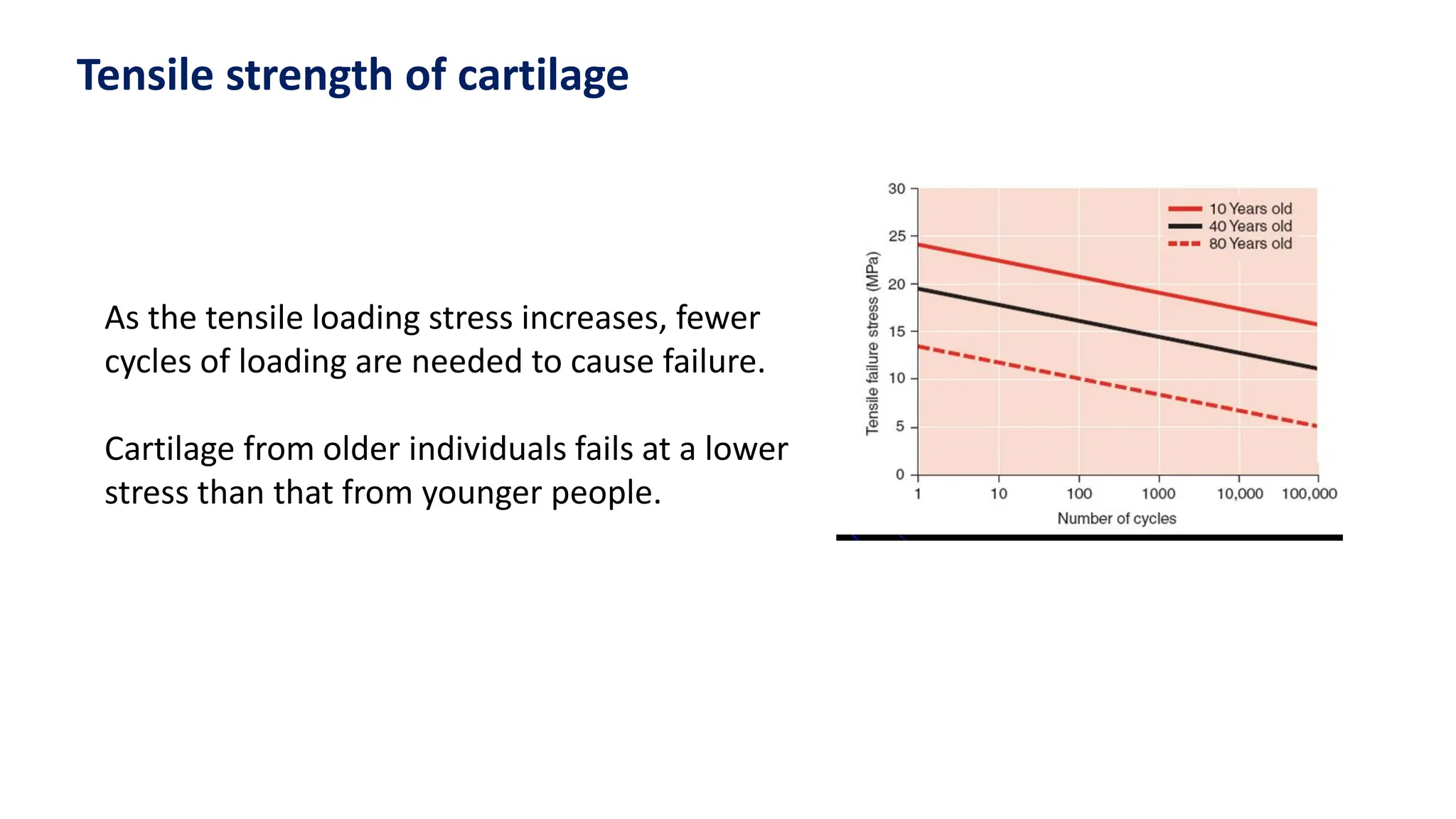
As the tensileloading stress increases, fewer
cycles of loading are needed to cause failure.
Cartilage from older individuals fails at a lower
stress than that from younger people.
Tensile strength of cartilage
37.
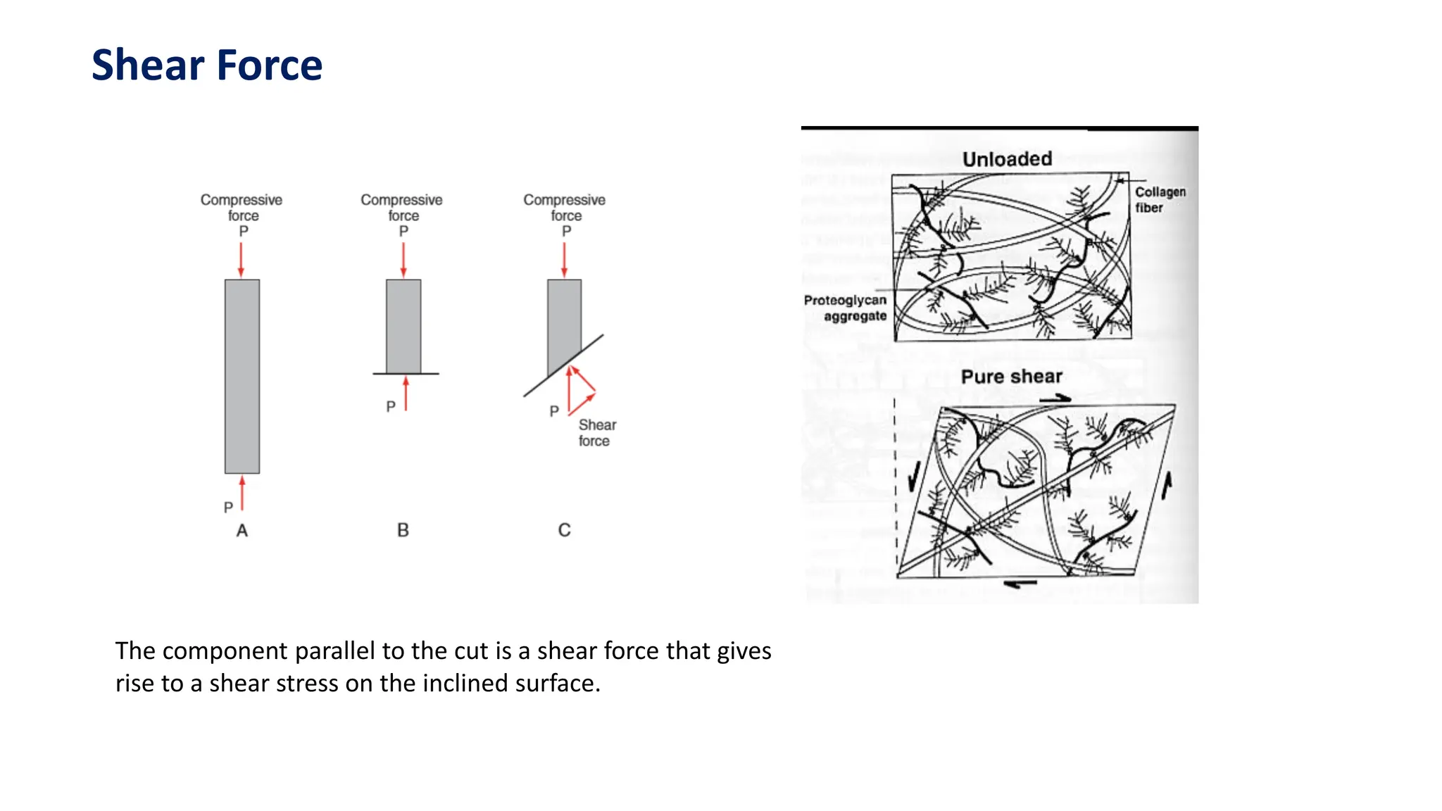
Shear Force
The componentparallel to the cut is a shear force that gives
rise to a shear stress on the inclined surface.
38.
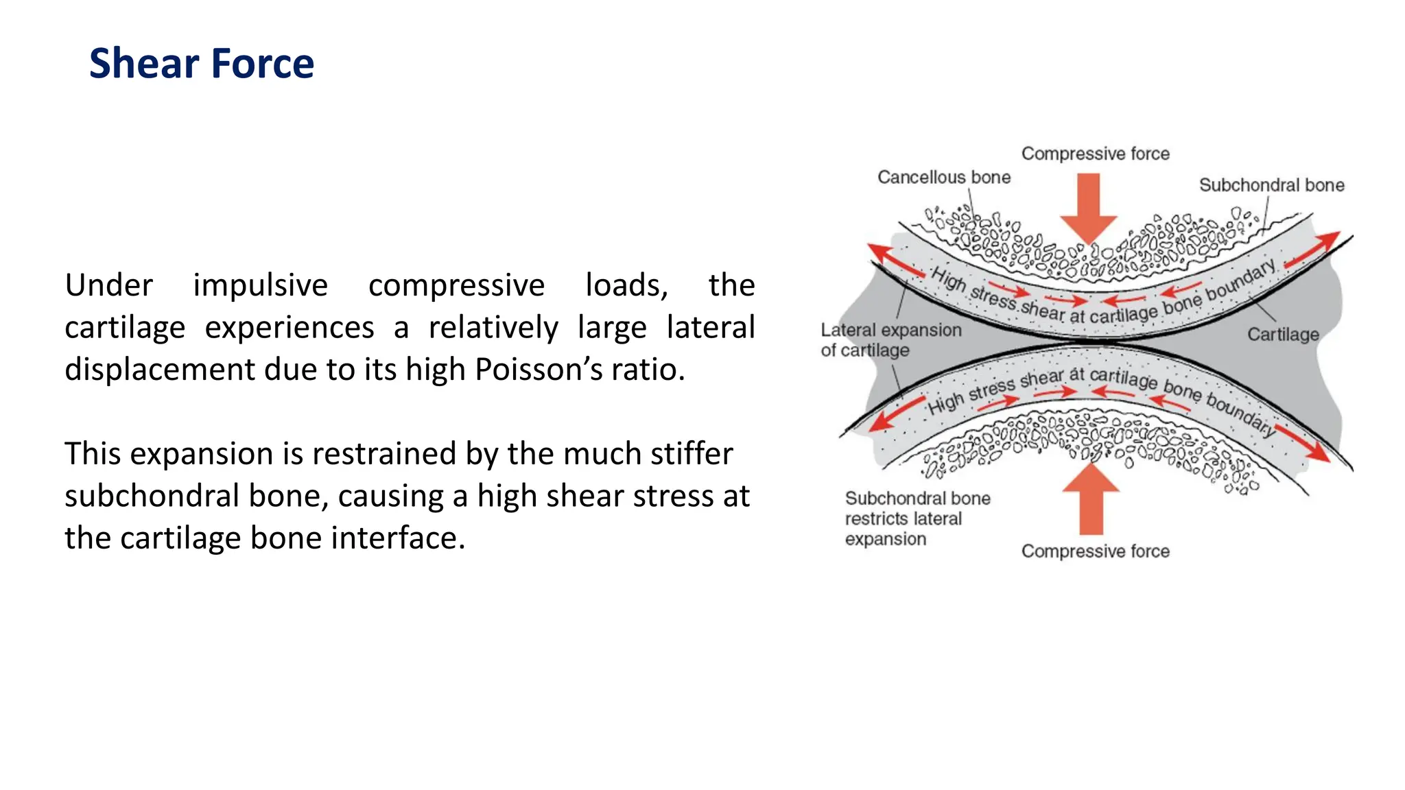
Under impulsive compressiveloads, the
cartilage experiences a relatively large lateral
displacement due to its high Poisson’s ratio.
This expansion is restrained by the much stiffer
subchondral bone, causing a high shear stress at
the cartilage bone interface.
Shear Force
39.
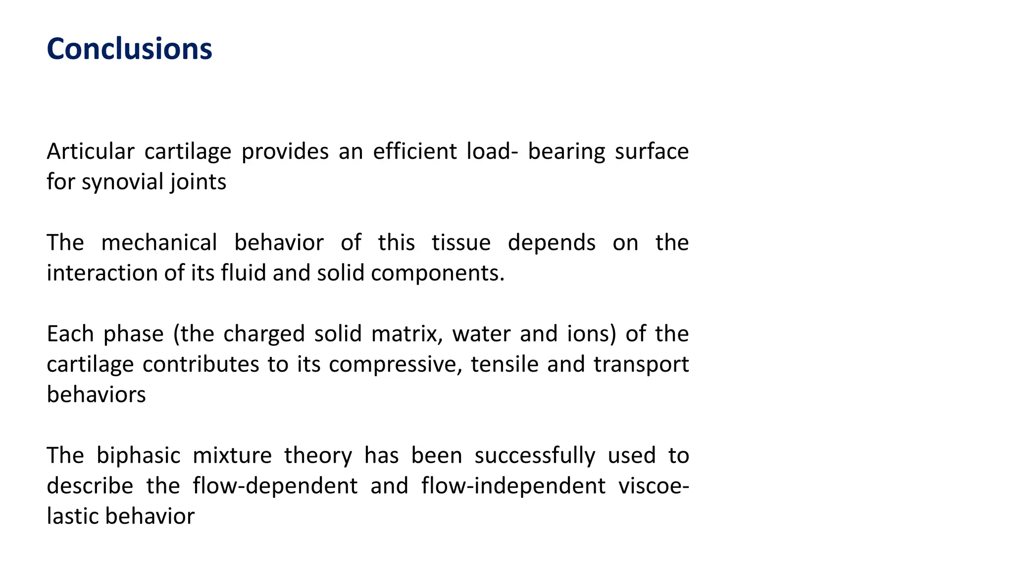
Articular cartilage providesan efficient load- bearing surface
for synovial joints
The mechanical behavior of this tissue depends on the
interaction of its fluid and solid components.
Each phase (the charged solid matrix, water and ions) of the
cartilage contributes to its compressive, tensile and transport
behaviors
The biphasic mixture theory has been successfully used to
describe the flow-dependent and flow-independent viscoe-
lastic behavior
Conclusions


![Cartilage_[Autosaved].pptx](https://cdn.slidesharecdn.com/ss_thumbnails/cartilageautosaved-230825071732-ce10acc9-thumbnail.jpg?width=640&height=640&fit=bounds)


