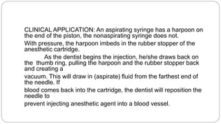
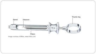
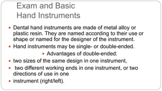
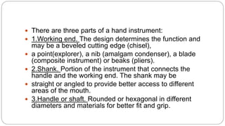
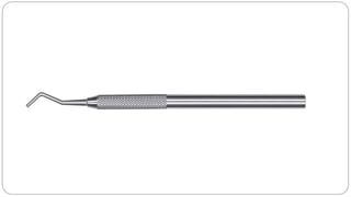
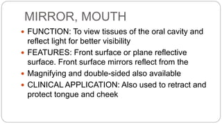
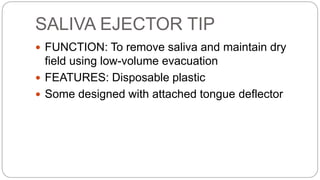
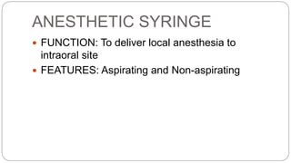
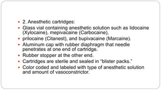
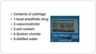
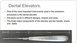
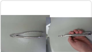
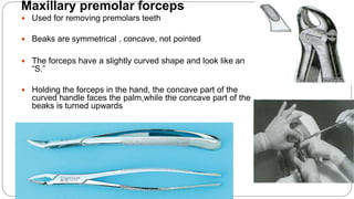
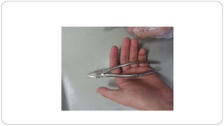
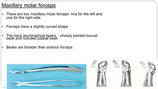
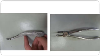
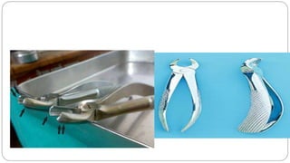
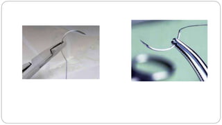
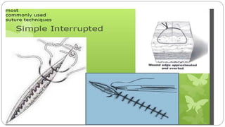
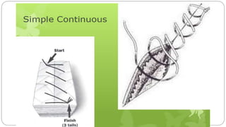
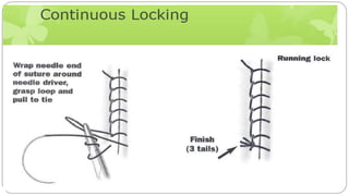
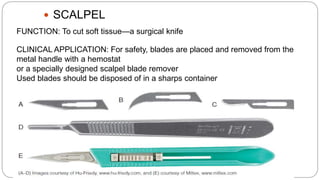
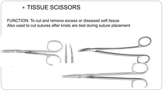
BY: Amanj kaify
Hawler Medical University
College of Dentistry
ARMAMENTARIUM FOR
BASIC ORAL SURGERY
BASIC EXTRACTION SET-UP
 PURPOSE: To provide instrumentation
for surgical removal of tooth/teeth.Oral
Surgery
 1. Local anesthesia syringe, needles,
and cartridges
 2. Sterile gauze
 3. Surgical aspirating tip
 4. Cotton pliers
 5. Mouth mirror
 6. Periosteal elevator
 7. Straight elevators
 8. Surgical curette
 9. Hemostat
 10. Extraction forceps (selected for
EXTRACTION/ALVEOPLASTY/GINGIVOPLASTY
SET-UP
 PURPOSE: To provide instrumentation for surgically removing multiple
teeth, reshaping boneand gingiva, and placing sutures.Oral Surgery
 1. Local anesthesia set-up 2. Tissue retractor
 3. Scalpel(s) 4. Mouth prop
 5. Sterile gauze 6. Surgical aspirating tip
 7. Cotton pliers 8. Mouth mirror
 9. Periosteal elevator 10. Straight elevators
 11. Tissue retractor 12. Surgical curette
 13. Bone file 14. Extraction forceps (selected for
 specific tooth/teeth)
 15. Rongeur 16. Tissue scissor
 17. Needle holder 18. Hemostat
 19. Suture
Exam and Basic
Hand Instruments
 Dental hand instruments are made of metal alloy or
plastic resin. They are named according to their use or
shape or named for the designer of the instrument.
 Hand instruments may be single- or double-ended.
 Advantages of double-ended:
 two sizes of the same design in one instrument,
 two different working ends in one instrument, or two
directions of use in one
 instrument (right/left).
 There are three parts of a hand instrument:
 1.Working end. The design determines the function and
may be a beveled cutting edge (chisel),
 a point(explorer), a nib (amalgam condenser), a blade
(composite instrument) or beaks (pliers).
 2.Shank. Portion of the instrument that connects the
handle and the working end. The shank may be
 straight or angled to provide better access to different
areas of the mouth.
 3.Handle or shaft. Rounded or hexagonal in different
diameters and materials for better fit and grip.
MIRROR, MOUTH
 FUNCTION: To view tissues of the oral cavity and
reflect light for better visibility
 FEATURES: Front surface or plane reflective
surface. Front surface mirrors reflect from the
 Magnifying and double-sided also available
 CLINICAL APPLICATION: Also used to retract and
protect tongue and cheek
EXPLORER
 FUNCTION: To examine tooth surfaces for caries, calculus, or defects
using sense of touch (tactile)
 FEATURES: Thin, sharp working end comes in different designs
 May be single- or double-ended (different design on each side)
 CLINICAL APPLICATION: Also used to:
 Check fit of margins of restorations
 Evaluate root surfaces and
 furcation area in periodontal
 exam (11/12)
 Remove excess material from restoration or preparation
 Remove excess cement
COTTON PLIERS
 COTTON PLIERS
 FUNCTION: To place and remove small objects from the
oral cavity (i.e., cotton pellets, root canal instruments,
wedges)
 FEATURES: Serrated or nonserrated beaks, locking or
nonlocking handles
 Also known as College pliers or dressing pliers
 CLINICAL APPLICATION: Also used to retrieve materials
from drawers and containers to avoid
 cross-contamination
SALIVA EJECTOR TIP
 FUNCTION: To remove saliva and maintain dry
field using low-volume evacuation
 FEATURES: Disposable plastic
 Some designed with attached tongue deflector
-Local Anesthetic instruments
ANESTHETIC SYRINGE
 FUNCTION: To deliver local anesthesia to
intraoral site
 FEATURES: Aspirating and Non-aspirating
CLINICAL APPLICATION: An aspirating syringe has a harpoon on
the end of the piston, the nonaspirating syringe does not.
With pressure, the harpoon imbeds in the rubber stopper of the
anesthetic cartridge.
As the dentist begins the injection, he/she draws back on
the thumb ring, pulling the harpoon and the rubber stopper back
and creating a
vacuum. This will draw in (aspirate) fluid from the farthest end of
the needle. If
blood comes back into the cartridge, the dentist will reposition the
needle to
prevent injecting anesthetic agent into a blood vessel.
Self-Aspirating Syringes
Disposable
CCLAD (Computer Controlled Local
Anesthetic Delivery
LOCAL ANESTHESIA
ACCESSORIES
 1. Anesthetic needles:
 Two lengths—1 (short) and 1 5/8 (long)
 Three gauges (diameter)—25 gauge, 27 gauge,
and 30 gauge
 Some manufacturers identify gauge by color-
coding caps
 Available with plastic or metal hubs
 Advantage of larger needles
 1)Less deflection
 2)Greater accuracy in injection
 3)Less chance for breakage
 4)More reliable aspiration
 5)No perceptual difference in patient discomfort
Large diameter
Medium diameter
Small diameter
 2. Anesthetic cartridges:
 Glass vial containing anesthetic solution such as lidocaine
(Xylocaine), mepivacaine (Carbocaine),
 prilocaine (Citanest), and bupivacaine (Marcaine).
 Aluminum cap with rubber diaphragm that needle
penetrates at one end of cartridge.
 Rubber stopper at the other end.
 Cartridges are sterile and sealed in “blister packs.”
 Color coded and labeled with type of anesthetic solution
and amount of vasoconstrictor.
 Contents of cartridge
 1-local anesthetic drug
 2-vasoconstrictor
 3-anti oxidant
 4-Sodium choride
 5-distilled water
 Topical anesthetic: Used prior to local anesthetic
injection to decrease discomfort
 One of the most important instruments used in the extraction
procedure is the dental elevator
 Elevators come in different designs, shapes and sizes
 The three major components of the elevator are the handle, shank,
and blade
Dental Elevators.
 The handle of the elevator is pear shaped
 In some situations, crossbar or T-bar handles are used.
 The shank of the elevator simply connects the handle to the working end, or blade,
of the elevator.
 The blade of the elevator is the working tip of the
 The blade has two surfaces: a convex and a concave one.
Types of elevators
 Straight elevator it is used to luxate the teeth

 The blade of the straight elevator has a concave surface on one side
 Come in different sizes
 Cryer’s elevator –
 they come in pairs
The triangle shaped elevator is most useful when a broken root
remains in the tooth socket and the adjacent socket is empty
( Commonly used in the mandibular arch)
In experts hands they can be used in the maxilla to luxate maxillary teeth
or roots
Winter’s cross-bar elevator
Coupland Chisel
Used to elevate and loosen the tooth from the periodontal
ligament.
Elevation is done to create space and prevent trauma to
adjacent teeth and tissues.
Available in sizes 1, 2 and 3 – working end gets larger with
increase in size number.
 Warwick-James elevator – this is a delicate elevator
 Available as straight and curved (paired)
 It is used to luxate the teeth in the maxilla
 Since the handle is flat , the amount of the force aginat the bone will
decrease ( reduce the incidence of the fracture)
 Crane pick elevator
 Crane pick elevator is a heavy instrument used to elevate the whole
roots or even teeth
 This elevator possess a sharp and thick , curved working that can be
used for removing root
 Root tip pick, or apexo elevator– this is a delicate
instrument
 The instrument is wedged between the root and the bone
 It is not used to push the roots
Apexo elevator
 Double-angled elevators (also refered as Apexo elevator)
 They are mainly used to remove root tips in both jaws
 Their handle is similar to that of the straight elevator.
 The shank has a double angle
 The blade has a sharp point which can easily remove small broken root
tips
Dental Forceps
Function
Removal of tooth from alveolar bone
Designed in various styles and configurations to adapt to variety of teeth for
which they are used
Components
Components of a dental extraction forceps consists of:
- handle
- Hinge
- beaks
1. Handle
 They are made of adequate size
 They are serrated
 For the maxillary teeth, the forceps are held with palm under the
forceps
 For the mandibular teeth, the palm is kept on top of the forceps
 Handles can be straight or curved
 This provides the operator with a sense of "better fit"
2. Hinge
 Like the shank of an elevator
 The hinge transfers and concentrates the forces applied
to the beaks.
 The American type of forceps has a hinge that is directed in a
horizontal direction with the handles of the forcep
 While The English type of forceps has a hinge that is directed
vertically to the handles of the forecep
3. Beaks
 The beaks of the forcep are concave on their inner
aspect and shaped to fit around the root of the tooth
 The beak is designed to adapt to the tooth root at the
junction of the crown and root
Individual Forceps
Upper anterior forceps
 Used for extracting upper incisors & canines
 Beaks are symmetrical & are placed in the same line as the handles
 beaks are concave and not pointed
 Beaks are shorter than the handles, so that load arm is shorter than
the working arm
Maxillary premolar forceps
 Used for removing premolars teeth
 Beaks are symmetrical , concave, not pointed
 The forceps have a slightly curved shape and look like an
“S.”
 Holding the forceps in the hand, the concave part of the
curved handle faces the palm,while the concave part of the
beaks is turned upwards
Maxillary molar forceps
 There are two maxillary molar forceps: one for the left and
one for the right side
 Forceps have a slightly curved shape
 The have asymmetrical beaks : sharply pointed buccal
beak and rounded palatal beak
 Beaks are broader than anterior forceps
 Maxillary Third Molar Forceps.
 It is the longest forceps, due to the posterior position of the third molar
 The beaks are offset from the handle in a bayonet fashion
 The beaks of forceps are concave and smooth (without pointed ends)
 The forceps can be used for extraction of both the left and right maxillry third
molar and maxillary second molar (conical roots)
 Maxillary cowhorn forceps.
 They are particularly useful for maxillary molars whose crowns are severely decayed.
 The sharply pointed beaks may reach deeper into the trifurcation
 The major disadvantage is that they crush alveolar bone, and when used on intact
teeth without due caution, fracture of large amounts of buccal alveolar bone may
occur.
 Maxillary cow horn forceps has a bayonet design, and are commonly used
to extract the maxillary third molar , as well as , it can be used to extraxt the
maxillary first and secnd molar
 Maxillary cow horn forceps have unidentical beaks, one has pointed tip and
the other has bifid pointed beaks
 With experinced hands, the surgeon can extract the maxillary third molar by
engaging the furcation area between the buccal roots by one pointed tip of
the bifid beak so that other tips engane the trifurcation area from the distal
area and from the palatal sides
 Or , as it recommened by the manufacture, the dentist can usually extract
the third molar by engaging the single pointed beak on the furcation area
between the buccal roots and the other bifid pointed tip engages the palatal
root
 If Maxillary cow horn forceps is used to extract the maxillary first and second
molar, the dentist should engage the single pointed beak on the furcation
area between the buccal roots and the other bifid pointed beak should
engages the palatal root ( strong apical force is important to engage the
Upper root forceps
 Designed for removing maxillary roots
 The handles of the root tip forceps are straight, while the
beaks are narrow and offset from the handle in a bayonet
fashion
 Beaks closely approximate each other and they meat at the
ends
 Beaks are narrow to fit to the circumference of the root &
provide firm grip
Lower anterior forceps
 Lower anterior forceps have identical , short closed beaks
 Beaks are narrower than lower molar forceps, similar to upper root forceps
 Beaks are at right angles to the handles
 They can also be used as lower root forceps
Mandibular premolar forcep
 The mandibular premolar forcep have identical long and broad open beaks
 The mandibular premolar forcep and can be use to extract the mandibular
canine
Lower molar forceps
 Beaks are at right angles to the handles
 Beaks are symmetrically pointed & the sharp pointed tips engage the
bifurcation at the buccal & lingual surfaces
 Beaks are more broader & stout
 Mandibular Third Molar Forceps.
 These have straight handles, while the beaks, are curved at a
right angle compared to the handles.
The beaks are a little longer compared to the previous forceps
 Because this tooth varies in the shape and size and because there is usually no root
bifurcation, the ends of the beaks of the forceps are concave without a pointed
design.( most useful for the third molar with have fused conical roots)
 Other types of the lower third forcep, have bilateral pointed tips in the center to adapt
into the bifuraction area if it is present between the roots
 Lower cowhorns forceps
 They have two heavy beaks with a very sharp tips that can fit into the root bifurcation
 These forceps are often used when the crown of the tooth is badly broken down.
 They often cause the tooth to split in two the roots can be removed separately with
elevators
 Mandibular Root Tip Forceps.
 The handles of the root tip forceps are straight,while the beaks are
curved at a right angle.
 Their ends are very narrow and meet at the tip when the forceps are
closed
 HEMOSTATS
FUNCTION: To securely hold small items, clamp blood vessels, and remove
small pieces of tooth
or bone
 NEEDLE HOLDERS
 FUNCTION: To hold suture needle
 FEATURES: Similar to hemostat but with a
concave area on inside of each beak to allow for
curve of suture needle
 To avoid needle breakage, place the needle
holder on the needle just beyond the suture
attachment point and at right angles to the curve
of the needle
 SUTURE
FUNCTION: To close incision site
“Stitches” hold tissues in place during healing
FEATURES: Suture material attached to sterile stainless steel needle
Different sizes and designs of needles
Suture may be absorbable—plain or chromic gut, polyglycolic acid (PGA,
Vicryl)
or nonabsorbable—silk, polyester, nylon, polypropylene
Sized by diameter of suture material: 3–0 (000), 4–0 (0000), 5–0 (00000) most
common sizes used in dentistry (smaller number larger diameter)
CLINICAL APPLICATION: Nonabsorbable sutures usually removed at 7–10 days
postsurgical visit
Placed with needle holder or hemostat
 SCALPEL
FUNCTION: To cut soft tissue—a surgical knife
CLINICAL APPLICATION: For safety, blades are placed and removed from the
metal handle with a hemostat
or a specially designed scalpel blade remover
Used blades should be disposed of in a sharps container
 SCALPEL BLADE REMOVER
To safely remove blade from scalpel
handle
 RONGEURS—SIDE-CUTTING and END-
CUTTINGTo cut and contour bone—removes sharp edges of alveolar crest after
extractions
for better contour of alveolar ridge; removes exostoses
 BONE CHISEL AND MALLET
FUNCTION: To remove bone for better contour of alveolar ridge; remove
exostoses, i.e., tori
 BONE FILE
FUNCTION: To smooth bone for better contour of alveolar ridge, often following
use of
rongeurs
FEATURES: Straight or curved working ends
Crosscut or straight cutting ridges
Double ended
 TISSUE SCISSORS
FUNCTION: To cut and remove excess or diseased soft tissue
Also used to cut sutures after knots are tied during suture placement
Armamentrium

Armamentrium

  • 1.
    BY: Amanj kaify HawlerMedical University College of Dentistry ARMAMENTARIUM FOR BASIC ORAL SURGERY
  • 2.
    BASIC EXTRACTION SET-UP PURPOSE: To provide instrumentation for surgical removal of tooth/teeth.Oral Surgery  1. Local anesthesia syringe, needles, and cartridges  2. Sterile gauze  3. Surgical aspirating tip  4. Cotton pliers  5. Mouth mirror  6. Periosteal elevator  7. Straight elevators  8. Surgical curette  9. Hemostat  10. Extraction forceps (selected for
  • 3.
    EXTRACTION/ALVEOPLASTY/GINGIVOPLASTY SET-UP  PURPOSE: Toprovide instrumentation for surgically removing multiple teeth, reshaping boneand gingiva, and placing sutures.Oral Surgery  1. Local anesthesia set-up 2. Tissue retractor  3. Scalpel(s) 4. Mouth prop  5. Sterile gauze 6. Surgical aspirating tip  7. Cotton pliers 8. Mouth mirror  9. Periosteal elevator 10. Straight elevators  11. Tissue retractor 12. Surgical curette  13. Bone file 14. Extraction forceps (selected for  specific tooth/teeth)  15. Rongeur 16. Tissue scissor  17. Needle holder 18. Hemostat  19. Suture
  • 4.
    Exam and Basic HandInstruments  Dental hand instruments are made of metal alloy or plastic resin. They are named according to their use or shape or named for the designer of the instrument.  Hand instruments may be single- or double-ended.  Advantages of double-ended:  two sizes of the same design in one instrument,  two different working ends in one instrument, or two directions of use in one  instrument (right/left).
  • 5.
     There arethree parts of a hand instrument:  1.Working end. The design determines the function and may be a beveled cutting edge (chisel),  a point(explorer), a nib (amalgam condenser), a blade (composite instrument) or beaks (pliers).  2.Shank. Portion of the instrument that connects the handle and the working end. The shank may be  straight or angled to provide better access to different areas of the mouth.  3.Handle or shaft. Rounded or hexagonal in different diameters and materials for better fit and grip.
  • 7.
    MIRROR, MOUTH  FUNCTION:To view tissues of the oral cavity and reflect light for better visibility  FEATURES: Front surface or plane reflective surface. Front surface mirrors reflect from the  Magnifying and double-sided also available  CLINICAL APPLICATION: Also used to retract and protect tongue and cheek
  • 9.
    EXPLORER  FUNCTION: Toexamine tooth surfaces for caries, calculus, or defects using sense of touch (tactile)  FEATURES: Thin, sharp working end comes in different designs  May be single- or double-ended (different design on each side)  CLINICAL APPLICATION: Also used to:  Check fit of margins of restorations  Evaluate root surfaces and  furcation area in periodontal  exam (11/12)  Remove excess material from restoration or preparation  Remove excess cement
  • 11.
    COTTON PLIERS  COTTONPLIERS  FUNCTION: To place and remove small objects from the oral cavity (i.e., cotton pellets, root canal instruments, wedges)  FEATURES: Serrated or nonserrated beaks, locking or nonlocking handles  Also known as College pliers or dressing pliers  CLINICAL APPLICATION: Also used to retrieve materials from drawers and containers to avoid  cross-contamination
  • 13.
    SALIVA EJECTOR TIP FUNCTION: To remove saliva and maintain dry field using low-volume evacuation  FEATURES: Disposable plastic  Some designed with attached tongue deflector
  • 15.
  • 16.
    ANESTHETIC SYRINGE  FUNCTION:To deliver local anesthesia to intraoral site  FEATURES: Aspirating and Non-aspirating
  • 17.
    CLINICAL APPLICATION: Anaspirating syringe has a harpoon on the end of the piston, the nonaspirating syringe does not. With pressure, the harpoon imbeds in the rubber stopper of the anesthetic cartridge. As the dentist begins the injection, he/she draws back on the thumb ring, pulling the harpoon and the rubber stopper back and creating a vacuum. This will draw in (aspirate) fluid from the farthest end of the needle. If blood comes back into the cartridge, the dentist will reposition the needle to prevent injecting anesthetic agent into a blood vessel.
  • 19.
  • 21.
  • 23.
    CCLAD (Computer ControlledLocal Anesthetic Delivery
  • 24.
    LOCAL ANESTHESIA ACCESSORIES  1.Anesthetic needles:  Two lengths—1 (short) and 1 5/8 (long)  Three gauges (diameter)—25 gauge, 27 gauge, and 30 gauge  Some manufacturers identify gauge by color- coding caps  Available with plastic or metal hubs
  • 26.
     Advantage oflarger needles  1)Less deflection  2)Greater accuracy in injection  3)Less chance for breakage  4)More reliable aspiration  5)No perceptual difference in patient discomfort
  • 28.
  • 29.
     2. Anestheticcartridges:  Glass vial containing anesthetic solution such as lidocaine (Xylocaine), mepivacaine (Carbocaine),  prilocaine (Citanest), and bupivacaine (Marcaine).  Aluminum cap with rubber diaphragm that needle penetrates at one end of cartridge.  Rubber stopper at the other end.  Cartridges are sterile and sealed in “blister packs.”  Color coded and labeled with type of anesthetic solution and amount of vasoconstrictor.
  • 30.
     Contents ofcartridge  1-local anesthetic drug  2-vasoconstrictor  3-anti oxidant  4-Sodium choride  5-distilled water
  • 31.
     Topical anesthetic:Used prior to local anesthetic injection to decrease discomfort
  • 32.
     One ofthe most important instruments used in the extraction procedure is the dental elevator  Elevators come in different designs, shapes and sizes  The three major components of the elevator are the handle, shank, and blade Dental Elevators.
  • 33.
     The handleof the elevator is pear shaped  In some situations, crossbar or T-bar handles are used.
  • 34.
     The shankof the elevator simply connects the handle to the working end, or blade, of the elevator.  The blade of the elevator is the working tip of the  The blade has two surfaces: a convex and a concave one.
  • 35.
    Types of elevators Straight elevator it is used to luxate the teeth   The blade of the straight elevator has a concave surface on one side  Come in different sizes
  • 37.
     Cryer’s elevator–  they come in pairs The triangle shaped elevator is most useful when a broken root remains in the tooth socket and the adjacent socket is empty ( Commonly used in the mandibular arch) In experts hands they can be used in the maxilla to luxate maxillary teeth or roots Winter’s cross-bar elevator
  • 39.
    Coupland Chisel Used toelevate and loosen the tooth from the periodontal ligament. Elevation is done to create space and prevent trauma to adjacent teeth and tissues. Available in sizes 1, 2 and 3 – working end gets larger with increase in size number.
  • 41.
     Warwick-James elevator– this is a delicate elevator  Available as straight and curved (paired)  It is used to luxate the teeth in the maxilla  Since the handle is flat , the amount of the force aginat the bone will decrease ( reduce the incidence of the fracture)
  • 42.
     Crane pickelevator  Crane pick elevator is a heavy instrument used to elevate the whole roots or even teeth  This elevator possess a sharp and thick , curved working that can be used for removing root
  • 43.
     Root tippick, or apexo elevator– this is a delicate instrument  The instrument is wedged between the root and the bone  It is not used to push the roots
  • 44.
  • 45.
     Double-angled elevators(also refered as Apexo elevator)  They are mainly used to remove root tips in both jaws  Their handle is similar to that of the straight elevator.  The shank has a double angle  The blade has a sharp point which can easily remove small broken root tips
  • 46.
    Dental Forceps Function Removal oftooth from alveolar bone Designed in various styles and configurations to adapt to variety of teeth for which they are used Components Components of a dental extraction forceps consists of: - handle - Hinge - beaks
  • 47.
    1. Handle  Theyare made of adequate size  They are serrated  For the maxillary teeth, the forceps are held with palm under the forceps
  • 48.
     For themandibular teeth, the palm is kept on top of the forceps  Handles can be straight or curved  This provides the operator with a sense of "better fit"
  • 49.
    2. Hinge  Likethe shank of an elevator  The hinge transfers and concentrates the forces applied to the beaks.
  • 50.
     The Americantype of forceps has a hinge that is directed in a horizontal direction with the handles of the forcep  While The English type of forceps has a hinge that is directed vertically to the handles of the forecep
  • 51.
    3. Beaks  Thebeaks of the forcep are concave on their inner aspect and shaped to fit around the root of the tooth  The beak is designed to adapt to the tooth root at the junction of the crown and root
  • 52.
    Individual Forceps Upper anteriorforceps  Used for extracting upper incisors & canines  Beaks are symmetrical & are placed in the same line as the handles  beaks are concave and not pointed  Beaks are shorter than the handles, so that load arm is shorter than the working arm
  • 54.
    Maxillary premolar forceps Used for removing premolars teeth  Beaks are symmetrical , concave, not pointed  The forceps have a slightly curved shape and look like an “S.”  Holding the forceps in the hand, the concave part of the curved handle faces the palm,while the concave part of the beaks is turned upwards
  • 56.
    Maxillary molar forceps There are two maxillary molar forceps: one for the left and one for the right side  Forceps have a slightly curved shape  The have asymmetrical beaks : sharply pointed buccal beak and rounded palatal beak  Beaks are broader than anterior forceps
  • 59.
     Maxillary ThirdMolar Forceps.  It is the longest forceps, due to the posterior position of the third molar  The beaks are offset from the handle in a bayonet fashion  The beaks of forceps are concave and smooth (without pointed ends)  The forceps can be used for extraction of both the left and right maxillry third molar and maxillary second molar (conical roots)
  • 60.
     Maxillary cowhornforceps.  They are particularly useful for maxillary molars whose crowns are severely decayed.  The sharply pointed beaks may reach deeper into the trifurcation  The major disadvantage is that they crush alveolar bone, and when used on intact teeth without due caution, fracture of large amounts of buccal alveolar bone may occur.
  • 61.
     Maxillary cowhorn forceps has a bayonet design, and are commonly used to extract the maxillary third molar , as well as , it can be used to extraxt the maxillary first and secnd molar  Maxillary cow horn forceps have unidentical beaks, one has pointed tip and the other has bifid pointed beaks  With experinced hands, the surgeon can extract the maxillary third molar by engaging the furcation area between the buccal roots by one pointed tip of the bifid beak so that other tips engane the trifurcation area from the distal area and from the palatal sides  Or , as it recommened by the manufacture, the dentist can usually extract the third molar by engaging the single pointed beak on the furcation area between the buccal roots and the other bifid pointed tip engages the palatal root  If Maxillary cow horn forceps is used to extract the maxillary first and second molar, the dentist should engage the single pointed beak on the furcation area between the buccal roots and the other bifid pointed beak should engages the palatal root ( strong apical force is important to engage the
  • 64.
    Upper root forceps Designed for removing maxillary roots  The handles of the root tip forceps are straight, while the beaks are narrow and offset from the handle in a bayonet fashion  Beaks closely approximate each other and they meat at the ends  Beaks are narrow to fit to the circumference of the root & provide firm grip
  • 66.
    Lower anterior forceps Lower anterior forceps have identical , short closed beaks  Beaks are narrower than lower molar forceps, similar to upper root forceps  Beaks are at right angles to the handles  They can also be used as lower root forceps
  • 67.
    Mandibular premolar forcep The mandibular premolar forcep have identical long and broad open beaks  The mandibular premolar forcep and can be use to extract the mandibular canine
  • 68.
    Lower molar forceps Beaks are at right angles to the handles  Beaks are symmetrically pointed & the sharp pointed tips engage the bifurcation at the buccal & lingual surfaces  Beaks are more broader & stout
  • 69.
     Mandibular ThirdMolar Forceps.  These have straight handles, while the beaks, are curved at a right angle compared to the handles. The beaks are a little longer compared to the previous forceps  Because this tooth varies in the shape and size and because there is usually no root bifurcation, the ends of the beaks of the forceps are concave without a pointed design.( most useful for the third molar with have fused conical roots)  Other types of the lower third forcep, have bilateral pointed tips in the center to adapt into the bifuraction area if it is present between the roots
  • 70.
     Lower cowhornsforceps  They have two heavy beaks with a very sharp tips that can fit into the root bifurcation  These forceps are often used when the crown of the tooth is badly broken down.  They often cause the tooth to split in two the roots can be removed separately with elevators
  • 71.
     Mandibular RootTip Forceps.  The handles of the root tip forceps are straight,while the beaks are curved at a right angle.  Their ends are very narrow and meet at the tip when the forceps are closed
  • 72.
     HEMOSTATS FUNCTION: Tosecurely hold small items, clamp blood vessels, and remove small pieces of tooth or bone
  • 73.
     NEEDLE HOLDERS FUNCTION: To hold suture needle  FEATURES: Similar to hemostat but with a concave area on inside of each beak to allow for curve of suture needle
  • 74.
     To avoidneedle breakage, place the needle holder on the needle just beyond the suture attachment point and at right angles to the curve of the needle
  • 75.
     SUTURE FUNCTION: Toclose incision site “Stitches” hold tissues in place during healing FEATURES: Suture material attached to sterile stainless steel needle Different sizes and designs of needles Suture may be absorbable—plain or chromic gut, polyglycolic acid (PGA, Vicryl) or nonabsorbable—silk, polyester, nylon, polypropylene Sized by diameter of suture material: 3–0 (000), 4–0 (0000), 5–0 (00000) most common sizes used in dentistry (smaller number larger diameter) CLINICAL APPLICATION: Nonabsorbable sutures usually removed at 7–10 days postsurgical visit Placed with needle holder or hemostat
  • 84.
     SCALPEL FUNCTION: Tocut soft tissue—a surgical knife CLINICAL APPLICATION: For safety, blades are placed and removed from the metal handle with a hemostat or a specially designed scalpel blade remover Used blades should be disposed of in a sharps container
  • 85.
     SCALPEL BLADEREMOVER To safely remove blade from scalpel handle
  • 86.
     RONGEURS—SIDE-CUTTING andEND- CUTTINGTo cut and contour bone—removes sharp edges of alveolar crest after extractions for better contour of alveolar ridge; removes exostoses
  • 87.
     BONE CHISELAND MALLET FUNCTION: To remove bone for better contour of alveolar ridge; remove exostoses, i.e., tori
  • 88.
     BONE FILE FUNCTION:To smooth bone for better contour of alveolar ridge, often following use of rongeurs FEATURES: Straight or curved working ends Crosscut or straight cutting ridges Double ended
  • 89.
     TISSUE SCISSORS FUNCTION:To cut and remove excess or diseased soft tissue Also used to cut sutures after knots are tied during suture placement