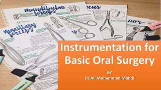
Instrumentation for Basic Oral Surgery Dr.Ali Mohammed AbuTrab
•Download as PPTX, PDF•
1 like•186 views

Instrumentation for Basic Oral Surgery Dr.Ali Mohammed AbuTrab

Recommended
Recommended
More Related Content
What's hot
What's hot (20)
Similar to Instrumentation for Basic Oral Surgery Dr.Ali Mohammed AbuTrab
Similar to Instrumentation for Basic Oral Surgery Dr.Ali Mohammed AbuTrab (20)
Recently uploaded
Recently uploaded (20)
Instrumentation for Basic Oral Surgery Dr.Ali Mohammed AbuTrab
- 2. • This lecture will introduce the basic and the main instruments that perform the procedures of oral surgery…….. • Instruments for Incising Tissue • Instruments for Elevating Mucoperiosteum • Instruments for Retraction of Soft Tissue • Instruments for Grasping Tissue • Instruments for Removing Bone • Instruments for Removing Pathologic Tissue • Instruments for Suturing Mucosa • Instruments for Extracting the Teeth (Dental elevators -Extraction forceps)
- 3. Instruments for Incising Tissue • Many surgical procedures begin with an incision. The primary instrument for making incisions is the scalpel, which is composed of a handle and a sterile, very sharp blade. blade handle the pen grasp for maximal control There are many types of blades and each type has its own uses.
- 4. • The most common blades that use in our field Shape Description Uses No.10 - A blade with a curved belly - The belly being the sharp edge Making skin incisions No.11 -A blade with straight and an angled edge with a pointed end Making stab incisions to insert drains No.12 -A blade with crescent shape and the inner side is sharp Useful for mucogingival procedures in which incisions are made on the posterior aspects of teeth or in the maxillary tuberosity area No.15 -The most frequently used scalpel blade for intraoral surgery - The blade is similar in shape to the larger No. 10 blade Used to make incisions around teeth and through soft tissue
- 5. Instruments for Elevating Mucoperiosteum • When an incision is made through the periosteum, ideally the periosteum should be reflected from the underlying cortical bone. No. 9 Molt periosteal elevator 1-This instrument has a sharp, pointed end and a broader, rounded end. 2-The pointed end is used to begin the periosteal reflection and to reflect dental papillae from between teeth (dissection). 3- The broad, rounded end is used to continue the elevation of the periosteum from bone.
- 6. Instruments for Retraction of Soft Tissue • Good access and vision are critical to performing excellent surgery. • Use to Retract the cheek, tongue, and mucoperiosteal flaps to provide access and visibility during surgery Shape Description Uses Austin retractor -right-angle retractor -These retractors can also be used to retract the cheek and a mucoperiosteal flap simultaneously. -Retractors are also used to help protect soft tissue from sharp cutting instruments Minnesota retractor -broad offset retractor Before the flap is created, the retractor is held loosely in the cheek. Once the flap is reflected, the retractor edge is placed on bone and is then used to retract the flap
- 7. Shape Description Uses Seldin retractor -Retractor may look similar to a periosteal elevator, the leading edge is not sharp but, instead, smooth; these instruments are not typically used to elevate the mucoperiosteum Used to retract oral soft tissue The Weider retractor -A broad, heart-shaped retractor that is serrated on one side It can more firmly engage the tongue and retract it medially and anteriorly . When this retractor is used, care must be taken not to position it so far posteriorly as to cause gagging or to push the tongue into the oropharynx A towel clip -When a biopsy procedure is to be performed on the posterior aspect of the tongue, the most positive way to control the tongue is by holding the anterior tongue with a towel clip.
- 8. Instruments for Grasping Tissue • Various oral surgical procedures require the surgeon to grasp soft tissue to incise it, to stop bleeding, or to pass a suture needle. • Adson forceps These are delicate forceps, with or without small teeth at the tips, that can be used to hold tissue gently while stabilizing it. When this instrument is used, care should be taken not to grasp the tissue too tightly to avoid crushing it. • Stillies forceps These forceps are usually 7 to 9 inches long and can easily grasp tissue in the posterior part of the mouth, still leaving enough of the instrument protruding beyond the lips for the surgeon to hold and control it With tooth Without tooth
- 9. • Allis tissue forceps -Use when removing larger amounts of tissue or doing biopsies. -forceps with locking handles and teeth that will firmly grip the tissue are necessary. -The Allis forceps should never be used on tissue that is to be left in the mouth because they cause a relatively large amount of tissue crushing. Comparison of Adson beaks (right) with Allis beaks (left) shows the differences in their designs and uses.
- 10. Instruments for Removing Bone Rongeurs Forceps • The instrument most commonly used for removing bone in dentoalveolar surgery (Alveoloplasty). • This instrument has sharp blades that are squeezed together by the handles, cutting or pinching through bone. • Rongeur forceps have a rebound mechanism incorporated so that when hand pressure is released, the instrument reopens (like spring). • Two major designs for rongeur forceps are : 1-a side-cutting forceps and 2-the side- and end-cutting forceps • Rongeurs can be used to remove large amounts of bone efficiently and quickly. Because a rongeur is a delicate instrument, the surgeon should not use it to remove large amounts of bone in single bites. Rather, smaller amounts of bone should be removed in multiple bites. The end-cutting forceps can be inserted into sockets for the removal of interradicular bone and can also be used to remove sharp edges of bone. Side-cutting rongeurs used for trimming and recontouring alveolar bone and gross tissue removal.
- 11. • Burr and Handpiece • Moderate-speed, high-torque handpieces with sharp carbide round burs remove cortical bone efficiently. • The handpiece must not exhaust air into the operative field, which would make it improper to use the typical high-speed air-turbine drills employed in routine restorative dentistry. The reason is that the air exhausted into the wound may be forced into deeper tissue planes and produce tissue emphysema, a dangerous occurrence. • Bone File • The bone file is usually a double-ended instrument with small and larger ends. The bone file cannot be used efficiently for removal of large amounts of bone; therefore it is used only for final smoothing.
- 12. Instruments for Removing Pathologic Tissue • The curette commonly used for oral surgery is an angled, double-ended instrument used to remove soft tissue from bony defects. • Its principal use is to remove granulomas or small cysts from periapical lesions, but the curette may also be used to remove small amounts of granulation tissue debris from a tooth socket. • Larger currettes are available for removing soft tissue from larger bony cavities such as cysts.
- 13. Instruments for Suturing Mucosa • Needle Holder an instrument with a locking handle and a short, blunt beak. For intraoral placement of sutures, a 7-inch (15- cm) needle holder is usually recommended. Hemostat: longer and thinner beak Needle holder: shorter and thicker The face of the hemostat has parallel grooves that do not allow a firm grip on the needle The face of the shorter beak of the needle holder is cross-hatched to ensure a positive grip on the needle
- 14. Suture Needle Shape Description Uses Triangular tip with the apex forming a cutting surface Used for tough tissue, such as skin (use of a tapered needle with skin causes excess trauma because of difficulty in penetration) Similar to a conventional cutting needle except the cutting edge faces down instead of up This may decrease the likelihood of sutures pulling through soft tissue (most used in oral surgery) Gradually taper to the point and cross- section reveals a round, smooth shaft Used for tissue that is easy to penetrate, such as bowel or blood vessels • Most sutures with the suture material swaged onto the base of the needle. • Shapes vary from a quarter circle to five-eighths of a circle, depending on how confined the operating field is. • Choice of needle should not alter the tissue to be sutured as little as possible and this depend on : -The tissue being sutured -Ease of access to the tissue
- 15. Suture Material They are classified according : • 1-Size :The size of suture relates to its diameter and is designated by a series of zeros. The diameter most commonly used in the suturing of oral mucosa is 3-0 (000). A larger-sized suture is 2-0, or 0. Smaller sizes are designated with more zeros, for example 4-0, 5-0, and 7- 0. Sutures of very fine size, such as 7-0, are usually used in conspicuous places on the skin— for example, the face—because properly placed smaller sutures usually cause less scarring. • 2-Resorbability: Sutures may be resorbable or nonresorbable. Nonresorbable suture materials include such types as silk, nylon, vinyl, and stainless steel. The most commonly used nonresorbable suture in the oral cavity is silk. Nylon, vinyl, and stainless steel are rarely used in the mouth. Resorbable sutures are primarily made of gut. Although the term catgut is often used to designate this type of suture, gut actually is derived from the serosal surface of sheep intestines. Plain catgut resorbs quickly in the oral cavity, rarely lasting longer than 3 to 5 days. chromic gut lasts longer than plain gut by up to 7 to 10 days. polyglycolic acid and polylactic acid materials are slowly resorbed, taking up to 4 weeks to do so. Such long- lasting resorbable sutures are rarely indicated for basic oral surgery.
- 16. • 3-Monofilament or polyfilament: Monofilament sutures are sutures such as plain and chromic gut, nylon, and stainless steel. Polyfilament sutures are braided sutures such as silk, polyglycolic acid, and polylactic acid. Sutures that are made of braided material are easier to handle and tie than monofilament sutures and rarely come untied. • One of the most commonly used sutures for the oral cavity is 3-0 black silk. The size 3-0 has the appropriate amount of strength; the polyfilament nature of the silk makes it straightforward to tie and well tolerated by the patient’s soft tissues. The color makes the suture easy to see when the patient returns for suture removal. Sutures that are holding mucosa together usually stay no longer than 5 to 7 days, so the wicking action is of little clinical importance. Many surgeons prefer 3-0 chromic suture to avoid the need to later remove it.
- 17. Scissors • Suture scissors usually have short cutting edges because their sole purpose is to cut sutures. They usually have long handles and thumb and finger rings. Scissors are held in the same manner as needle holders. Scissor shape Description The most commonly used suture scissors for oral surgery are Dean scissors. These have slightly curved handles and serrated blades that make cutting sutures easier. Iris scissors are small, sharp- pointed, delicate tools used for fine work. -These scissors are designed for cutting soft tissue. -They can have either sharp or blunt (rounded) tips.
- 18. Instruments for Extracting the Teeth A- Elevators • The three major components of the elevator are the handle, shank, and blade. • Types of Elevators: (1) the straight type, (2) the triangle or pennant-shaped type, and (3) the pick type.
- 19. Straight Elevators • The straight elevator is the most commonly used elevator to luxate teeth. The blade of the straight elevator has a concave surface on one side that is placed toward the tooth to be elevated. The small straight elevator is frequently used for beginning the luxation of an erupted tooth before application of the forceps. Larger straight elevators are used to displace roots from their sockets and to luxate teeth that are more widely spaced, or they are used once a smaller-sized straight elevator becomes less effective.
- 20. • The shape of the blade of the straight elevator can be angled from the shank, allowing this instrument to be used in the more posterior aspects of the mouth. Two examples of the angled-shank elevator with a blade similar to that of the straight elevator are the Miller elevator and the Potts elevator.
- 21. Coupland chisel It has similar shape of the luxator, but the tip is sharper and has straight-cut. Depending on the width of the working end, there are different sizes including: Size 1, 2 and 3. Coupland chisel can be used to elevate teeth (by leverage action) and separate teeth. It can also remove bone to create point of application.
- 22. The Triangle or Pennant-Shaped( Cryer Elevator ) • These elevators are provided in pairs: a left and a right. The triangular elevator is most useful when a broken root remains in the tooth socket and the adjacent socket is empty. • The tip of the triangular elevator is placed into the socket with the shank of the elevator resting on the buccal plate of bone. The elevator is then turned in a wheel-and-axle rotation, with the sharp tip of the elevator engaging the cementum of the remaining distal root; the elevator is then turned and the root is delivered.
- 23. The Pick Type Elevators • A- Crane pick: This instrument is used as a lever to elevate a broken root from the tooth socket. Usually it is necessary to drill a hole with a burr (purchase point) approximately 3 mm deep into the root just at the bony crest. The tip of the pick is then inserted into the hole, and, with the buccal plate of bone as a fulcrum, the root is elevated from the tooth socket. Occasionally the sharp point can be used without preparing a purchase point by engaging the cementum or the furcation of the tooth. • B- Apexo elevator: The root-tip pick is a delicate instrument that is used to tease small root tips from their sockets. It must be emphasized that this is a thin instrument and should not be used as a wheel and- axle or lever type of elevator such as the Cryer elevator or the Crane pick. The root-tip pick is used to tease the very small root end of a tooth by inserting the tip into the periodontal ligament space between the root tip and the socket wall. This instrument works best on roots left after a tooth has been well elevated.
- 24. B-Periotomes • Are instruments used to extract teeth while preserving the anatomy of the tooth’s socket. The general principle behind their use is to sever some of the periodontal ligaments of the tooth to facilitate its removal. There are varying types of periotomes with different blade shapes. • The tip of the periotome blade is inserted into the periodontal ligament space and advanced using pressure in the apical direction along the long axis of the tooth. It is advanced about 2 to 3 mm and then removed and reinserted into an adjacent accessible site. The process is continued around the tooth, gradually advancing the depth of the periotome tip while progressing apically. Once sufficient severance of periodontal ligaments has been accomplished, the tooth is removed by using a dental elevator, extraction forceps, or both, taking care to avoid excessive expansion or fracture of bone.
- 25. Extraction Forceps -Forceps are used to lift elevator-luxated teeth from their sockets rather than to pull teeth from their sockets. - The handles have a serrated surface to allow a positive grip and to prevent slippage. British forceps type American forceps type The usual American type of forceps has a hinge in a horizontal direction. The British preference is for a vertical hinge and a corresponding vertically positioned handle. The beaks of the extraction forceps are the source of the greatest variation among forceps. The beaks are designed to adapt to the tooth root near the junction of the crown and root. It must be remembered that the beaks of the forceps are designed to be adapted to the root structure of the tooth and not to the crown of the tooth.
- 26. Maxillary Forceps Single rooted teeth (incisors- canine and second premolar) American type British type Beaks that are found on the same level as the handles characterize these forceps, and the beaks are concave and not pointed .
- 27. Maxillary Forceps Upper premolars The forceps used for premolars have a slightly curved shape and look like an “S.” Holding the forceps in the hand, the concave part of the curved part of the handle faces the palm, while the concave part of the beaks is turned upwards. The ends of the beaks of the forceps are concave and are not pointed. These forceps ma also be used for extraction of the six anterior teeth of the upper jaw. British type American type No. 150 Forceps
- 28. Maxillary Forceps Upper Molars (6-7) • Maxillary Molar Forceps, for the First and Second Molar. There are two of these forceps: one for the left and one for the right side. Just like the previously mentioned forceps, they have a slightly curved shape that looks like an “S” .The buccal beak of each forceps has a pointed design, which fits into the buccal bifurcation of the two buccal roots, while the palatal beak is concave and fits into the convex surface of the palatal root. Upper Molar (Left) Upper Molar (Light)
- 29. Maxillary Forceps Upper Molars(8) • Maxillary Third Molar Forceps. These forceps have a slightly curved shape, just like the aforementioned forceps, and are the longest forceps, due to the posterior position of the third molar. Because this tooth varies in shape and size, the beaks of the forceps are concave and smooth (without pointed ends), so that these forceps may be used for extraction of both the left and right third molar of the upper jaw.
- 30. Maxillary Forceps Cowhorns Forceps • Maxillary Cowhorn Molar Forceps. The upper cowhorn forceps are a variation of the maxillary molar forceps. The beaks of this type of forceps have sharply pointed ends, which fit into the trifurcation of the roots of the molars. They are primarily used for extraction of teeth with severely decayed crowns, because when they are used to extract intact teeth, they may fracture the buccal alveolar bone due to the large amount of force they generate.
- 31. Maxillary Root Tip Forceps Maxillary Root Tip Forceps • Maxillary Root Tip Forceps. The handles of the root tip forceps are straight, while the beaks are narrow and angle-shaped. The ends of the beaks are concave and without a pointed design.
- 32. Mandibular Forceps Anterior And Premolars Teeth Forceps • Mandibular Universal Forceps or No. 151 Forceps. Unlike the maxillary forceps, the beaks and handles of these forceps face the same direction, creating an arch. When the forceps are held in the hand, the concave part of the arch of the handles faces the palm, while the beaks obviously face downward. The ends of the beaks are concave, without pointed-ends. The no. 151 forceps are used for extraction of the six anterior teeth and the four premolars of the lower jaw. Mandibular Universal Forceps or No. 151 Forceps British pattern
- 33. Mandibular Forceps Molars Forceps • These forceps are used for both sides of the jaw and have straight handles while the beaks are curved at approximately a right angle compared to the handles. Both beaks of the forceps have pointed ends, which fit into the bifurcation of the roots buccally and lingually .These forceps are used for the removal of both the first and second molar of the right and left side of the lower jaw. American pattern British pattern
- 34. Mandibular Forceps Third Molar Forceps • . These forceps also have straight handles, while the beaks, just like those of the first and second molar forceps, are curved at a right angle compared to the handles. The beaks are a little longer compared to the previous forceps, due to the posterior position of the third molar in the dental arch. Because this tooth varies in size and shape and because there is usually no root bifurcation, the ends of the beaks of the forceps are concave without a pointed design
- 35. Mandibular Forceps Cowhorn Molar Forceps • The lower cowhorn forceps or no. 23 forceps are a variation of the mandibular molar forceps. In comparison to the standard forceps, the beaks have a semicircular shape with sharply pointed ends so that they can fit into the bifurcation of the roots and firmly grasp the tooth. wing to the function of these forceps, tooth extraction may be achieved quite easily as long as the roots are not curved. With the beaks of the forceps grasping the crown of the molar and the sharp ends fitting into the root bifurcation, the surgeon squeezes the handles and, using small buccolingual movements, slides the tooth out of the socket. Also, the cowhorn forceps are very useful for sectioning roots of posterior teeth in the lower jaw, when their crowns are severely decayed. After grasping the roots, the teeth are easily sectioned after applying pressure at the bifurcation point.
- 36. Mandibular Forceps Root Tip Forceps • The handles of the root tip forceps are straight , while the beaks are curved at a right angle. Their ends are very narrow and meet at the tip when the forceps are closed.
- 37. References: • James R. Hupp , Edward Ellis , Myron R. Tucker ;”CONTEMPORARY ORAL AND MAXILLOFACIAL SURGERY, SEVENTH EDITION “; PART II- Principles of Exodontia, chapter7,Instrumentation for Basic Oral Surgery. • Fragiskos D. Fragiskos; “Oral Surgery”;chapter 4 Equipment, Instruments,and Materials. • Seth Delpachitra,Anton Sklavos,Ricky Kumar; ”Principles of Dentoalveolar Extractions”
- 38. Any Question????
- 39. Thank you