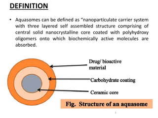
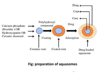
AQUASOMES
PRESENTED BY,
NIMMI ROY
1ST YEAE MPHARM
DEPT OF PHARMACEUTICS
SRINIVAS COLLEGE OF
PHARMACY
1
CONTENTS
• Introduction
• Bodies of water
• Principle
• Preparation
• Evaluation
• Applications
• References
2
DEFINITION
• Aquasomes can be defined as “nanoparticulate carrier system
with three layered self assembled structure comprising of
central solid nanocrystalline core coated with polyhydroxy
oligomers onto which biochemically active molecules are
absorbed.
3
INTRODUCTION
• Aquasomes are first discovered by NIR KOSSOVSKY.
• Consists of solid crystalline core , carbohydrate core and
active drug.
• It is spherical in shape and size of 60-30nm.
• Aquasomes are called as “Bodies of water” and their water
property protect and preserve fragile biological molecules.
4
BODIES OF WATER
• Water like properties.
• Protect and preserve fragile biological molecules.
• Maintain conformational integrity as well as high degree of
surface exposure in targeting of bio-active molecules/
5
PRINCIPLE OF SELF ASSEMBLY
• Assembly of micromolecule is governed by 3 process.
Interaction between charged groups.
Hydrogen bonding and dehydration.
Structural stability.
6
Interaction between charged groups
• Interaction of charged group such as amino, carbonyl,
sulphate, phosphate groupsfacilitate the long range approach
of self assembling subunits.
• Charged groups also play role in stabilizing teritiary structure
of folded proteins.
• Example of ion pairs-carboxylated/phosphate group boundto
ionized arginine/lysine side chain of protein.
7
Hydrogen bonding and Dehydration effect
• Hydrogen bond are formed between hydrogen atom attached
to an electronegative donor atom and an electronegative or
basic acceptor.
• Hydrogen bond help in base pair matching and stabilization
of secondary protein structure.
• Molecules that form hydrogen bonds are hydrophilicand these
molecule confer significant degree of organization to the
surronding water molecules.
8
Structural stability
• The structural stability of protein in the biological
environment is determined by the interaction between
charged groups and hydrogen bond largely external to the
molecule and vander walls forces largely internal to the
molecule.
• Vander walls forces are largely responsible for the hardness or
softness of the molecule.The vander walls interaction amoung
hydrophilic side chains promotes stability of compact helical
structures.
9
PREPARATION
• Simple and straight forward approach with minimum solvent
usuage.
• No homogenization steps.
• 3 steps of preparation by using the principle of assembly:-
1. Preparation of the core-
i. Co precipitation
ii. Self-precipitation
iii. Sonication
iv. PAMAM
2.Carbohydrate coating of the core
3.Immobilization of drug molecule
10
PREPARATION OF THE CORE
• Co-precipitation
11
• Self-precipitation
12
• Sonication
13
• PAMAM
 Carboxilic acid terminated half generation
poly(amidoamine)(PAMAM) used.
 Forms amorphus hydroxyapatite cores with a
mixture of calcium phosphate.
14
CARBOHYDRATE COATING OF CORE
• Coating material – Cellulose , citrate , pyrodoxal-5-phosphate ,
sucrose and trehalose
• Adsorption method-
• Lactose coated by adsorption method by direct incubation and by
nonsolvent addition.
15
IMMOBILIZATION OF DRUG MOLECULE
• The drug can be loaded by partial adsorption.
16
17
Fig: preparation of aquasomes
EVALUATION
18
EVALUATION OF CERAMIC CORE
Structure analysis-
Interpretated by fourier transformed infrared spectroscopy
using hydroxypatite powder in the wave no range of 4000-400
cm⁻¹.
Phase analysis of core-
Cores exposed to Cu K-α radiation in a wide angle X-ray
diffractometer.
Particle morphology-
Determined by using transmission electron microscopy one
drop of aqueous dispersion is placed over a 400-mesh carbon-
coated copper grid followed by negative staining with
phosphotungstic acid and placed at the accelerataing voltage.
19
EVALUATION OF SUGAR COATING
• Colorimetric analysis-(Anthrone method)
20
• Concanavalin –A-Induced aggregation
21
EVALUATION OF DRUG LOADED AQUASOMES
Size and shape-
• Morphological examination- transmission electron
microscope.
• Mean particle size and size distribution-A photon correlation
spectroscopy using a Autosizer II C apparatus and SEM.
• Chemical composition and crystalline structure – X-ray
powder diffractometry.
Glass transition temperature-
• Carbohydrates and protein studied by using DSC analyzer.
In-process stability studies-
• Stability and integrity of protein during the formulation of the
aquasomes determined by using sodium dodecyl
sulfatepolyacrylamide gel electrophoresis(SDS-PAGE)
22
In vitro drug release studies-
• To study the release pattern of drug
23
 Drug loading efficiency-
• Ensure the amount of drug which is bound on the surface of
aquasomes.
 Hb loading capacity-(Drabkin’s method)
• Estimated by the difference between the control
sample(HbaA solution)and the free hemoglobin contained in
all fractions without nanoparticles.
 Antigen loading efficiency-
24
APPLICATIONS
• Oxygen carrier
• For immunotherapy
• For oral route
• For immunopotentiation
• Anti thrombic activity
• Antigen delivery
• Delivery of poorly soluble drugs
• Enzyme delivery
• Insulin delivery
• Vaccine delivery
25
CONCLUSION
• Aquasomes appears to be promising carriers for
delivery of broad range of conformational sensitive
molecule better biological activity as it contain
carbohydrate coating over creamic core.Molecular
plasticizers,carbohydrates prevent destructive carrier
intraction and helps to preserve spatial qualities and
the crystalline natural core provide structural
stability and overall integrity. This strategy may
benificially extended to novel drug delivery of other
bioactive molecule.
26
REFERENCES
1. Jain N.K. Advances in controlled drug delivery system , 317-
328.
2. wani S.U, Yerawar A.N. International journal of pharmacy
and technology 2015; 2:1.
3. Gulati monica, Singh sachin. Potential applications of
aquasomes for therapeutic delivery of proteins and peptides,
4. www.pharmainfonet .com/ aquasomes.
27
28

Aquasomes evaluation

  • 1.
    AQUASOMES PRESENTED BY, NIMMI ROY 1STYEAE MPHARM DEPT OF PHARMACEUTICS SRINIVAS COLLEGE OF PHARMACY 1
  • 2.
    CONTENTS • Introduction • Bodiesof water • Principle • Preparation • Evaluation • Applications • References 2
  • 3.
    DEFINITION • Aquasomes canbe defined as “nanoparticulate carrier system with three layered self assembled structure comprising of central solid nanocrystalline core coated with polyhydroxy oligomers onto which biochemically active molecules are absorbed. 3
  • 4.
    INTRODUCTION • Aquasomes arefirst discovered by NIR KOSSOVSKY. • Consists of solid crystalline core , carbohydrate core and active drug. • It is spherical in shape and size of 60-30nm. • Aquasomes are called as “Bodies of water” and their water property protect and preserve fragile biological molecules. 4
  • 5.
    BODIES OF WATER •Water like properties. • Protect and preserve fragile biological molecules. • Maintain conformational integrity as well as high degree of surface exposure in targeting of bio-active molecules/ 5
  • 6.
    PRINCIPLE OF SELFASSEMBLY • Assembly of micromolecule is governed by 3 process. Interaction between charged groups. Hydrogen bonding and dehydration. Structural stability. 6
  • 7.
    Interaction between chargedgroups • Interaction of charged group such as amino, carbonyl, sulphate, phosphate groupsfacilitate the long range approach of self assembling subunits. • Charged groups also play role in stabilizing teritiary structure of folded proteins. • Example of ion pairs-carboxylated/phosphate group boundto ionized arginine/lysine side chain of protein. 7
  • 8.
    Hydrogen bonding andDehydration effect • Hydrogen bond are formed between hydrogen atom attached to an electronegative donor atom and an electronegative or basic acceptor. • Hydrogen bond help in base pair matching and stabilization of secondary protein structure. • Molecules that form hydrogen bonds are hydrophilicand these molecule confer significant degree of organization to the surronding water molecules. 8
  • 9.
    Structural stability • Thestructural stability of protein in the biological environment is determined by the interaction between charged groups and hydrogen bond largely external to the molecule and vander walls forces largely internal to the molecule. • Vander walls forces are largely responsible for the hardness or softness of the molecule.The vander walls interaction amoung hydrophilic side chains promotes stability of compact helical structures. 9
  • 10.
    PREPARATION • Simple andstraight forward approach with minimum solvent usuage. • No homogenization steps. • 3 steps of preparation by using the principle of assembly:- 1. Preparation of the core- i. Co precipitation ii. Self-precipitation iii. Sonication iv. PAMAM 2.Carbohydrate coating of the core 3.Immobilization of drug molecule 10
  • 11.
    PREPARATION OF THECORE • Co-precipitation 11
  • 12.
  • 13.
  • 14.
    • PAMAM  Carboxilicacid terminated half generation poly(amidoamine)(PAMAM) used.  Forms amorphus hydroxyapatite cores with a mixture of calcium phosphate. 14
  • 15.
    CARBOHYDRATE COATING OFCORE • Coating material – Cellulose , citrate , pyrodoxal-5-phosphate , sucrose and trehalose • Adsorption method- • Lactose coated by adsorption method by direct incubation and by nonsolvent addition. 15
  • 16.
    IMMOBILIZATION OF DRUGMOLECULE • The drug can be loaded by partial adsorption. 16
  • 17.
  • 18.
  • 19.
    EVALUATION OF CERAMICCORE Structure analysis- Interpretated by fourier transformed infrared spectroscopy using hydroxypatite powder in the wave no range of 4000-400 cm⁻¹. Phase analysis of core- Cores exposed to Cu K-α radiation in a wide angle X-ray diffractometer. Particle morphology- Determined by using transmission electron microscopy one drop of aqueous dispersion is placed over a 400-mesh carbon- coated copper grid followed by negative staining with phosphotungstic acid and placed at the accelerataing voltage. 19
  • 20.
    EVALUATION OF SUGARCOATING • Colorimetric analysis-(Anthrone method) 20
  • 21.
  • 22.
    EVALUATION OF DRUGLOADED AQUASOMES Size and shape- • Morphological examination- transmission electron microscope. • Mean particle size and size distribution-A photon correlation spectroscopy using a Autosizer II C apparatus and SEM. • Chemical composition and crystalline structure – X-ray powder diffractometry. Glass transition temperature- • Carbohydrates and protein studied by using DSC analyzer. In-process stability studies- • Stability and integrity of protein during the formulation of the aquasomes determined by using sodium dodecyl sulfatepolyacrylamide gel electrophoresis(SDS-PAGE) 22
  • 23.
    In vitro drugrelease studies- • To study the release pattern of drug 23
  • 24.
     Drug loadingefficiency- • Ensure the amount of drug which is bound on the surface of aquasomes.  Hb loading capacity-(Drabkin’s method) • Estimated by the difference between the control sample(HbaA solution)and the free hemoglobin contained in all fractions without nanoparticles.  Antigen loading efficiency- 24
  • 25.
    APPLICATIONS • Oxygen carrier •For immunotherapy • For oral route • For immunopotentiation • Anti thrombic activity • Antigen delivery • Delivery of poorly soluble drugs • Enzyme delivery • Insulin delivery • Vaccine delivery 25
  • 26.
    CONCLUSION • Aquasomes appearsto be promising carriers for delivery of broad range of conformational sensitive molecule better biological activity as it contain carbohydrate coating over creamic core.Molecular plasticizers,carbohydrates prevent destructive carrier intraction and helps to preserve spatial qualities and the crystalline natural core provide structural stability and overall integrity. This strategy may benificially extended to novel drug delivery of other bioactive molecule. 26
  • 27.
    REFERENCES 1. Jain N.K.Advances in controlled drug delivery system , 317- 328. 2. wani S.U, Yerawar A.N. International journal of pharmacy and technology 2015; 2:1. 3. Gulati monica, Singh sachin. Potential applications of aquasomes for therapeutic delivery of proteins and peptides, 4. www.pharmainfonet .com/ aquasomes. 27
  • 28.