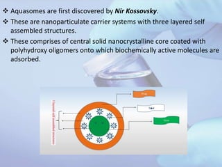
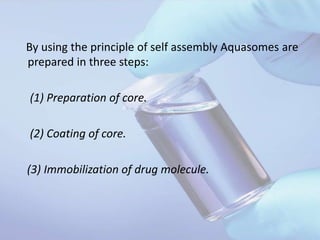
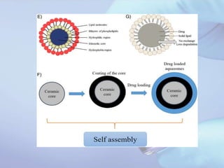
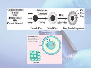
2. Aquasomes are first discovered by Nir Kossovsky.
These are nanoparticulate carrier systems with three layered self
assembled structures.
These comprises of central solid nanocrystalline core coated with
polyhydroxy oligomers onto which biochemically active molecules are
adsorbed.
3. Aquasomes are also called as “bodies of water” and their water like
properties protect and preserve fragile biological molecules.
This property of maintaining conformational integrity as well as high
degree of surface exposure made it as a successful carrier system for
bioactive molecules like peptide, protein, hormones, antigens and
genes to specific sites .
They are assemblies of simple polymers, complex lipid mixtures with
diameter ranging between 30 to 500 nm.
As these are solid or glassy particles dispersed in an aqueous
environment, they exhibit the physical properties of colloids and their
mechanism of action is controlled by their surface chemistry.
4. Aquasomes deliver their contents through combination of
specific targeting, slow and sustained release process.
5. METHOD OF PREPARATION OF
AQUASOMES:
By using the “principle of self assembly’’, the
aquasomes are prepared in three steps i.e.,
1. Preparation of core,
2. Coating of core, and
3. Immobilization of drug molecule
6. Principle of self assembly :
In aqueous biological environments , the assembly of
macro molecule is governed by three process.
(1) Interaction between charged group.
(2) Hydrogen bonding and dehydration effect.
(3) structural stability.
7. (1) Interaction between charged
group :
• Most of the Biological product are charged due to intrinsic
chemical group or absorbed ion from the biological
environment.
• Interaction of charged group such as amino, carbonyl, sulphate,
phosphate groups facilitate the long range approach of self
assembling sub units.
• Charged groups also play role in stabilizing tertiary structure of
folded proteins.
• Example of ion pairs -carboxylated /phosphate group bound to
ionized arginine / lysine side chain of protein.
8. (2)Hydrogen bonding and dehydration
effect :
Hydrogen bond are formed between hydrogen atom attached to an
electronegative donor atom (Ex oxygen, Nitrogen,) and an
electronegative or basic acceptor (Ex carbonyl oxygen).
Hydrogen bond help in base pair matching and stabilization of
secondary protein structure such as a helix and b sheets.
Molecule that form hydrogen bonds are hydrophilic and these
molecules confer significant degree of organization to the
surrounding water molecule.
In the case of hydrophobic molecules, which do not form hydrogen
bonds, their tendency to repel water helps to organize the moiety to
surrounding environment. The organized water decreases level of
entropy and is thermodynamically unfavorable, molecules dehydrate
and get self-assembled.
9. (3) Structural Stability :
• The structural stability of Protein in the biological environment is
determined by the interaction between charged groups and hydrogen
bond largely external to the molecule and vander walls forces largely
internal to the molecule.
• Vander walls forces are largely responsible for the hardness or
softness of the molecule.
• The vander walls interaction among hydrophilic side chains
promotes stability of compact helical structures
10. By using the principle of self assembly Aquasomes are
prepared in three steps:
(1) Preparation of core.
(2) Coating of core.
(3) Immobilization of drug molecule.
11. (1) Preparation of core :
This stage mainly depends on the selection of material for core along
with its physical and chemical properties.
This can be fabricated by the :
Sonication
Colloidal precipitation
For the core material, ceramic material widely used, as they are
structurally to be known.
Commonly used ceramic core are tin oxide and calcium phosphate,
carbon ceramics (diamonds)
12. Example:
synthesis of nanocrystalline tin oxide core material.
This can be prepared by
• Direct current reactive.
• Magnetron sputtering.
3 inch diameter target of highly purified Tin is
sputtered in High pressure gas mixture of argon and oxygen.
The ultra fine particle form in gas phase are collect on copper tube
cool at 700K with liquid nitrogen.
13. Synthesis of nano crystalbrushite (calciumphosphate dihydrate)
This can be prepared by :
• colloidal dispersion
• Sonication
• By reaction of disodium hydrogen phosphate and calcium
chloride.
• They measure b/w 50-150nm. And exhibit clean and reactive
surface.
14. Selection of core material
Ex: tin oxide/carbon ceramics(diamonds)/bruhite(calcium phosphate dihydrate)
Fabrication of core
Techniques used for fabrication
Colloidal precipitation and sonication
Magneteron sputtering
Plasma condensation
Centrifugation of precipitated core
washing
Washing of sedimented ppt with excess of water to remove sodium chloride
formed during reaction
filteration
Resuspend the ppt in distilled water and pass through a fine membrane filter to
collect particles of desired size
Ceramic core
15. (2) Carbohydrate Coating
• The second step involves coating by carbohydrate on the surface of
ceramic cores.
• There are number of processes to enable the carbohydrate
(polyhydroxy oligomers) coating to adsorb epitaxially on to the
surface of the nanocrystalline ceramic cores.
• Process generally entail addition of poly hydroxy oligomer into an
aqueous dispersion of cores under sonication.
• Lyophilization (to promote the irreversible adsorption of
carbohydrate on the surface of ceramic core).
• Excess of carbohydrate is removed by stir cell ultrafilteration.
16. Commonly used coating material,
• Cellobiose
• Citrate
• Sucrose
• Trehalose
• Pyridoxal -5- phosphate.
(3) Immobilization of drug
The surface modified Nano crystalline core provide the solid
phase for subsequent non denaturing self assembly for a broad
range of biological active molecule.
• Drug can be loaded by partial adsorption
17.
18.
19. Characterization of ceramic core
Size distribution:
For morphological characterization and size distribution
analysis, scanning electron microscopy (SEM) and
transmission electron microscopy (TEM) are generally
used.
Core, coated core, as well as drug-loaded aquasomes
are analyzed by these techniques.
Mean particle size and zeta potential of the particles
can also be determined by using “photon correlation
spectroscopy’’.
20. Characterization of coated core :
• Coating of sugar over the ceramic core can be confirmed by
i. concanavalin A-induced aggregation method (determines the
amount of sugar coated over core) or
ii. anthrone method (Determines the residual sugar unbound or
residual sugar remaining after coating).
iii. Furthermore, the adsorption of sugar over the core can also
be confirmed by measurement of zeta potential.
21. Characterization of drug-loaded aquasomes
Drug payload
i. The drug loading can be determined by incubating the basic aquasomes
formulation (i.e., without drug) in a known concentration of the drug
solution for 24 hours at 4°C. The supernatant is then separated by high-
speed centrifugation for 1 hour at low temperature in a refrigerated
centrifuge. The drug remaining in the supernatant liquid after loading can
be estimated by any suitable method of analysis.
In vitro drug release studies
i. The in vitro release kinetics of the loaded drug is determined to study the
release pattern of drug from the aquasomes by incubating a known
quantity of drug-loaded aquasomes in a buffer of suitable pH at 37 °C with
continuous stirring.
ii. Samples are withdrawn periodically and centrifuged at high speed for
certain lengths of time. Equal volumes of medium must be replaced after
each withdrawal. The supernatants are then analyzed for the amount of
drug released by any suitable method .
22. Reference :
• Jain N.K Advances in controlled drug delivery system, 317-328
• Potential Applications of Aquasomes for Therapeutic Delivery
of Proteins and Peptides, Gulati monica , singh sachin
• www.pharmainfonet.com/aquasomes.